

The DOI matches its Crossref record; 271 of the 272 references it cites were found in a registry or an index.
Short-chain fatty acids, secondary bile acids and indoles: gut microbial metabolites with effects on enteroendocrine cell function and their potential as therapies for metabolic disease
Open accessAbstract
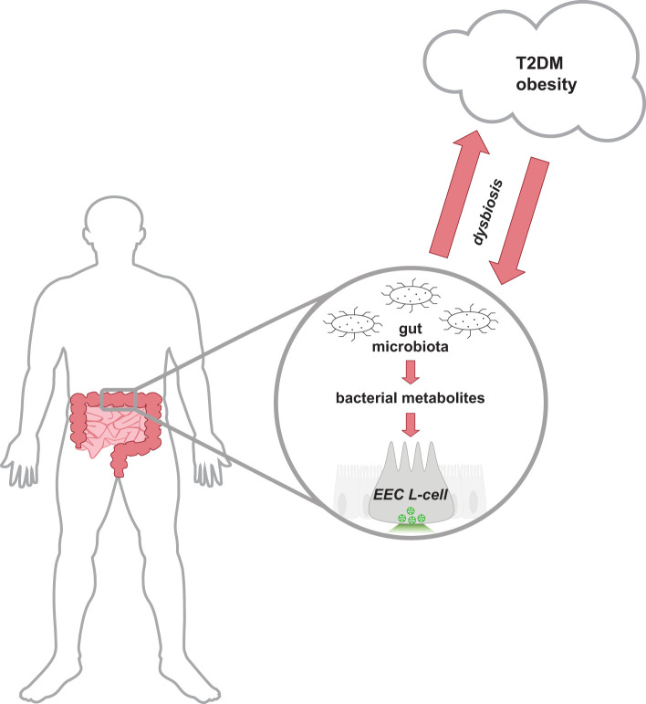
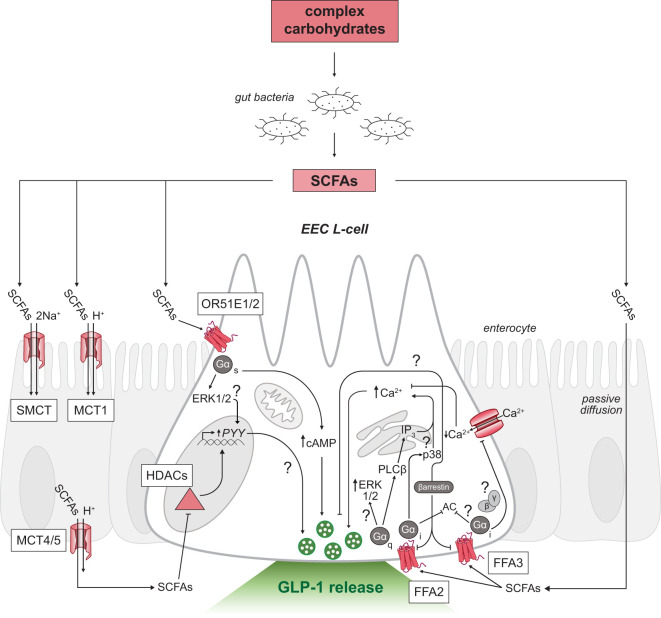
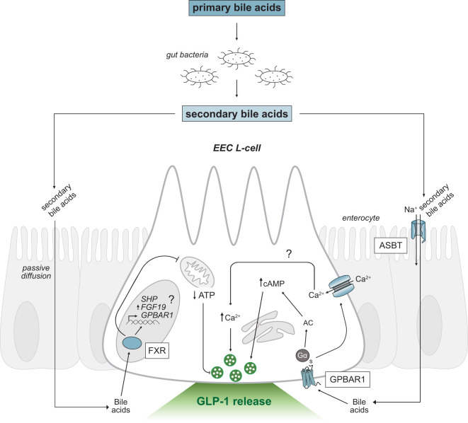
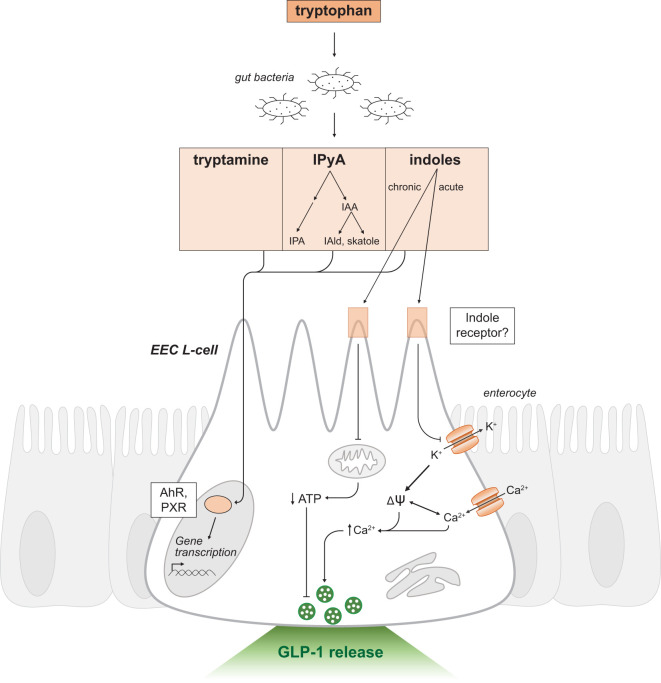
The gastrointestinal tract hosts the largest ecosystem of microorganisms in the body. The metabolism of ingested nutrients by gut bacteria produces novel chemical mediators that can influence chemosensory cells lining the gastrointestinal tract. Specifically, hormone-releasing enteroendocrine cells which express a host of receptors activated by these bacterial metabolites. This review will focus on the activation mechanisms of glucagon-like peptide-1 releasing enteroendocrine cells by the three main bacterial metabolites produced in the gut: short-chain fatty acids, secondary bile acids and indoles. Given the importance of enteroendocrine cells in regulating glucose homeostasis and food intake, we will also discuss therapies based on these bacterial metabolites used in the treatment of metabolic diseases such as diabetes and obesity. Elucidating the mechanisms gut bacteria can influence cellular function in the host will advance our understanding of this fundamental symbiotic relationship and unlock the potential of harnessing these pathways to improve human health.
Overview
The claims and statistics of this paper haven’t been checked yet.
Run the check — free, no account needed. It reads the open-access text and checks the sources it cites.
Integrity signals
Registry
Cites
Statistics
Figures
Verification
Claims the paper makes
The claims and statistics of this paper haven’t been checked yet.
Run the check — free, no account needed. It reads the open-access text and checks the sources it cites.
Claims other papers make about this one
Each is checked against this paper’s own text: does it say what the citing paper says it does?
In the citing paper
“the gut microbiota-derived metabolites, including SCFAs, secondary BAs, and indoles, have been demonstrated to modulate gut hormone secretion from EECs.”
From Microbiota–gut–brain axis and its therapeutic applications in neurodegenerative diseases (2024) · cites this paper as [547]
Self-check
Percentages, sums and reported differences re-computed from the paper’s own numbers.
The self-check needs the paper’s full text.
Citations
Cites
An obesity-associated gut microbiome with increased capacity for energy harvest
Turnbaugh, P. J., Ley, R. E., Mahowald, M. A., Magrini, V., Mardis, E. R., & Gordon, J. I. (2006) · Nature · doi:10.1038/nature05414 · Cited by 12,444
Human gut microbes associated with obesity
Ley, R. E., Turnbaugh, P. J., Klein, S., & Gordon, J. I. (2006) · Nature · doi:10.1038/4441022a · Cited by 8,785
Changes of Intestinal Functions in Liver Cirrhosis
Fukui, H., & Wiest, R. (2016) · Inflammatory Intestinal Diseases · doi:10.1159/000444436 · Cited by 6,820
A metagenome-wide association study of gut microbiota in type 2 diabetes
Qin, J., Li, Y., Cai, Z., Li, S., Zhu, J., Zhang, F., Liang, S., Zhang, W., Guan, Y., Shen, D., et al. (2012) · Nature · doi:10.1038/nature11450 · Cited by 6,762 · cited 1×
“In earlier studies, obesity and insulin resistance were associated with an increased abundance of Bacillota and concomitant decrease of Bacteroidota in both animal (7, 168), and human studies (6, 167, 169).”
BackgroundThe gut microbiota as an environmental factor that regulates fat storage
Bäckhed, F., Ding, H., Wang, T., Hooper, L. V., Koh, G. Y., Nagy, A., Semenkovich, C. F., & Gordon, J. I. (2004) · Proceedings of the National Academy of Sciences · doi:10.1073/pnas.0407076101 · Cited by 6,245 · cited 1×
“Dysbiosis of host-gut microbiota equilibrium may precede metabolic disease as similar shifts in intestinal gut bacteria composition can disrupt nutrient and energy metabolism (9).”
BackgroundObesity alters gut microbial ecology
Ley, R. E., Bäckhed, F., Turnbaugh, P., Lozupone, C. A., Knight, R. D., & Gordon, J. I. (2005) · Proceedings of the National Academy of Sciences · doi:10.1073/pnas.0504978102 · Cited by 6,176 · cited 1×
“In earlier studies, obesity and insulin resistance were associated with an increased abundance of Bacillota and concomitant decrease of Bacteroidota in both animal (7, 168), and human studies (6, 167, 169).”
BackgroundRevised Estimates for the Number of Human and Bacteria Cells in the Body
Sender, R., Fuchs, S., & Milo, R. (2016) · PLOS Biology · doi:10.1371/journal.pbio.1002533 · Cited by 5,425 · cited 1×
“Interestingly, the distal small intestine and colon harbor the greatest density of PYY and GLP-1 releasing L-cells (41), paralleling the distribution of gut bacteria (42).”
BackgroundHost-Bacterial Mutualism in the Human Intestine
Bäckhed, F., Ley, R. E., Sonnenburg, J. L., Peterson, D. A., & Gordon, J. I. (2005) · Science · doi:10.1126/science.1104816 · Cited by 5,277 · cited 1×
“In earlier studies, obesity and insulin resistance were associated with an increased abundance of Bacillota and concomitant decrease of Bacteroidota in both animal (7, 168), and human studies (6, 167, 169).”
Backgroundden Besten, G., van Eunen, K., Groen, A. K., Venema, K., Reijngoud, D.-J., & Bakker, B. M. (2013) · Journal of Lipid Research · doi:10.1194/jlr.r036012 · Cited by 5,048 · cited 2×
“The absorbed butyrate is largely utilized by colonocytes for energy, and the remaining absorbed SCFAs are transported through portal blood circulation back to the liver where SCFAs are primarily converted to glucose stores (4).”
BackgroundBiology of Incretins: GLP-1 and GIP
Baggio, L. L., & Drucker, D. J. (2007) · Gastroenterology · doi:10.1053/j.gastro.2007.03.054 · Cited by 3,639
Cited by
Sorted by citations
Microbiota–gut–brain axis and its therapeutic applications in neurodegenerative diseases
Loh, J. S., Mak, W. Q., Tan, L. K. S., Ng, C. X., Chan, H. H., Yeow, S. H., Foo, J. B., Ong, Y. S., How, C. W., & Khaw, K. Y. (2024) · Signal Transduction and Targeted Therapy · doi:10.1038/s41392-024-01743-1 · Cited by 1,193
Li, Z. H. (2024) · Gut · doi:10.1136/gutjnl-2024-331880 · Cited by 79
Gut dysbiosis in patients with chronic pain: a systematic review and meta-analysis
Goudman, L., Demuyser, T., Pilitsis, J. G., Billot, M., Manuel Roulaud, B., Rigoard, P., & Moens, M. (2024) · Frontiers in Immunology · doi:10.3389/fimmu.2024.1342833 · Cited by 82
Jameson, K. G., Kazmi, S. A., Ohara, T. E., Son, C., Yu, K. B., Mazdeyasnan, D., Leshan, E., Vuong, H. E., Paramo, J., Lopez-Romero, A., et al. (2024) · iScience · doi:10.1016/j.isci.2024.111699 · Cited by 88
“57 Moreover, EECs are enriched for FFAR2 and TGR5 compared to other epithelial cell subtypes (Figures S7B–S7C).”
Microbiota in cancer: current understandings and future perspectives
Yao, Y., Zhu, Y., Chen, K., Chen, J., Li, Y., Li, D., & Wei, P. (2026) · Signal Transduction and Targeted Therapy · doi:10.1038/s41392-025-02335-3 · Cited by 56
Musazadeh, V., Assadian, K., Rajabi, F., Faghfouri, A. H., Soleymani, Y., Kavyani, Z., & Najafiyan, B. (2024) · Pharmacological Research · doi:10.1016/j.phrs.2024.107398 · Cited by 49
Abildinova, G. Z., Benberin, V. V., Vochshenkova, T. A., Afshar, A., Mussin, N. M., Kaliyev, A. A., Zhussupova, Z., & Tamadon, A. (2024) · Frontiers in Microbiology · doi:10.3389/fmicb.2024.1463958 · Cited by 44
Barathan, M., Ng, S. L., Lokanathan, Y., Ng, M. H., & Law, J. X. (2024) · Vaccines · doi:10.3390/vaccines12111282 · Cited by 41
Enteroendocrine cells regulate intestinal homeostasis and epithelial function
Nwako, J. G., & McCauley, H. A. (2024) · Molecular and Cellular Endocrinology · doi:10.1016/j.mce.2024.112339 · Cited by 49
Li, L., Li, R., Tian, Q., Luo, Y., Li, R., Lin, X., Ou, Y., Guo, T., Chen, X., Pan, A., et al. (2024) · Cell Reports Medicine · doi:10.1016/j.xcrm.2024.101801 · Cited by 46
Therapeutic targeting of obesity-induced neuroinflammation and neurodegeneration
Zeng, J., Cheong, L. Y. T., & Lo, C. H. (2025) · Frontiers in Endocrinology · doi:10.3389/fendo.2024.1456948 · Cited by 35
Mapping pesticide-induced metabolic alterations in human gut bacteria
Chen, L., Yan, H., Di, S., Guo, C., Zhang, H., Zhang, S., Gold, A., Wang, Y., Hu, M., Wu, D., et al. (2025) · Nature Communications · doi:10.1038/s41467-025-59747-6 · Cited by 39
Stanimirov, B., Đanić, M., Pavlović, N., Zaklan, D., Lazarević, S., Mikov, M., & Stankov, K. (2025) · International Journal of Molecular Sciences · doi:10.3390/ijms262412167 · Cited by 39
López-Almada, G., Mejía-León, M. E., & Salazar‐López, N. J. (2024) · Foods · doi:10.3390/foods13223529 · Cited by 36
GLP‐1 agonists and the gut microbiome: A bidirectional relationship
Kamath, S., Chan, N. S. L., & Joyce, P. (2026) · British Journal of Clinical Pharmacology · doi:10.1002/bcp.70487 · Cited by 35
“20,60,61 These interactions extend beyond glycaemic control, embedding GLP-1 within a broader host – microbe metabolic network that governs insulinotropic responses, energy balance and neuroendocrine regulation.”
Zhao, Y., Zhao, M., Zhang, Y., Fu, Z., Jin, T., Song, J., Huang, Y., Zhao, C., & Wang, M. (2023) · Journal of Ethnopharmacology · doi:10.1016/j.jep.2023.117383 · Cited by 30
Ma, J., Wang, J., Wan, Y., Wang, S., & Jiang, C. (2024) · Journal of Ethnopharmacology · doi:10.1016/j.jep.2024.118815 · Cited by 28
Rzeczycki, P., Pęciak, O., Plust, M., & Droździk, M. (2025) · International Journal of Molecular Sciences · doi:10.3390/ijms262411897 · Cited by 30
“Short-chain fatty acids, secondary bile acids, and tryptophan catabolites have emerged as key mediators of microbiota–transporter interactions [37,38].”
Unraveling the Pathophysiology of Irritable Bowel Syndrome: Mechanisms and Insights
Aggeletopoulou, I., Papantoniou, K., Pastras, P., & Triantos, C. K. (2025) · International Journal of Molecular Sciences · doi:10.3390/ijms262110598 · Cited by 25
Fortunato, I. M., Pereira, Q. C., Oliveira, F. D. S., Alvarez, M. C., dos Santos, T. W., & Ribeiro, M. L. (2024) · International Journal of Molecular Sciences · doi:10.3390/ijms25031803 · Cited by 24
Xie, Y.-H., Li, X., Meng, Q., Li, J., Wang, X., Zhu, L., Wang, W., & Li, X. (2024) · Microbiology Spectrum · doi:10.1128/spectrum.00291-24 · Cited by 21
Harnessing Bacterial Extracellular Vesicle Immune Effects for Cancer Therapy
Karaman, İ., Pathak, A., Bayık, D., & Watson, D. C. (2024) · Pathogens and Immunity · doi:10.20411/pai.v9i1.657 · Cited by 20
“For instance, acetate produced by Bifidobacterium bifidum [25] and the short-chain fatty acid butyrate [26] were shown to enhance anti-tumor immunity by increasing tumor-infiltrating IFNγ secreting CD8+ T cells.”
BackgroundEffect of Gut Dysbiosis on Onset of GI Cancers
Kumari, S., Srilatha, M., & Nagaraju, G. P. (2024) · Cancers · doi:10.3390/cancers17010090 · Cited by 19
Maternal Nutrition, Toxicants, and Epigenetic Programming of Obesity Across Generations
Alum, E., Aloh, H., Obasi, D., Okoroh, P., Aniokete, U., & Emeruwa, A. (2025) · Diabetes, Metabolic Syndrome and Obesity · doi:10.2147/dmso.s579409 · Cited by 14
“156 For example, butyrate-mediated HDAC inhibition in fetal liver promotes histone acetylation at lipid oxidation genes, improving metabolic efficiency.”
Lu, C., Liu, D., Wu, Q., Zeng, J., Xiong, Y., & Luo, T. L. (2024) · npj Biofilms and Microbiomes · doi:10.1038/s41522-024-00585-7 · Cited by 15
“Gutmicrobial metabolites,suchastrimethylamineN-oxide,asourpreviousreport, acceleratestheprogressionofatherosclerosis 15 , whereas short-chain fatty acids and secondary bile acids (SBAs) confer cardioprotective effects 16,17 .”
BackgroundMicic, D. (2023) · Acta Endocrinologica (Bucharest) · doi:10.4183/aeb.2023.234 · Cited by 13
Zhu, L., Lv, B., Gao, Y., & Qin, D. (2025) · Archives of Biochemistry and Biophysics · doi:10.1016/j.abb.2025.110341 · Cited by 11
Fang, H., Li, H., Chen, Y., Liu, X., Zhao, J., Ross, R. P., Stanton, C., Chen, W., & Yang, B. K. (2024) · Food Bioscience · doi:10.1016/j.fbio.2024.104058 · Cited by 10
Aliuş, C., Breazu, A., Panţu, C., Toader, C., Șerban, M., Covache-Busuioc, R.-A., Munteanu, O., & Dumitru, A. V. (2025) · International Journal of Molecular Sciences · doi:10.3390/ijms262210959 · Cited by 12
Tao, W., Yu, Y., Tan, D., Huang, X., Huang, J., Lin, C., & Yu, R. (2025) · Frontiers in Cellular and Infection Microbiology · doi:10.3389/fcimb.2025.1603442 · Cited by 9
Modvig, I. M., Smits, M. M., Galsgaard, K. D., Hjørne, A. P., Drzazga, A. K., ROSENKILDE, M. M., & Holst, J. J. (2024) · Nutrition and Diabetes · doi:10.1038/s41387-024-00303-4 · Cited by 9
Xue, X., Zhou, H., Gao, J., Li, X., Wang, J., Bai, W., Bai, Y., Fan, L., Chang, H., & Shi, S. (2024) · Heliyon · doi:10.1016/j.heliyon.2024.e38339 · Cited by 10
Du, Z., Luo, Z., Huang, Y., Zhou, T., Ma, L., Wu, D., Yao, X., Shen, L., Yu, S., Yong, K., et al. (2024) · Frontiers in Microbiology · doi:10.3389/fmicb.2024.1373402 · Cited by 8
Qu, Z., Wu, Y., Jiang, Y., Fan, J., Cao, L., Dong, Y., Fang, S., & Gu, S. (2025) · Nutrients · doi:10.3390/nu17172886 · Cited by 8
“More importantly, microbially derived SCFAs, secondary bile acids, and indole derivatives can activate enteroendocrine cells, promoting the secretion of glucagon-like peptide-1 (GLP-1) and thereby influencing host neuroendocrine regulation and energy metabolism [12].”
Gut microbiome dysbiosis in PCOS: from pathogenesis to microbiome-targeted therapies
Du, X., Su, H., Huang, Y., Liu, J., Li, Q., Yang, X., Tao, X., & Li, R. (2026) · Frontiers in Endocrinology · doi:10.3389/fendo.2026.1747766 · Cited by 10
“Certain Clostridium species produce indole metabolites that activate pancreatic glucagon-like peptide-1 (GLP-1) secretion, enhancing glucose-stimulated insulin release (25).”
Magnesium and LongevityPreprint
Chambers, P. W. (2024) · Qeios · doi:10.32388/n1scbr.3 · Cited by 8
Oso, T. A., Ahmed, M. M., Okesanya, O. J., Adebayo, U. O., Obadeyi, K. B., Othman, Z. K., & Lucero‐Prisno, D. E. (2025) · Journal of Alzheimer s Disease · doi:10.1177/13872877251407700 · Cited by 9
Research progress on intestinal microbiota regulating cognitive function through the gut-brain axis
Lin, K., Peng, F., He, K., Qian, Z., Mei, X., Su, Z., Wujimaiti, Y., Xia, X., & Zhang, T. (2024) · Neurological Sciences · doi:10.1007/s10072-024-07525-5 · Cited by 8
“Upon the presence of carbohydrates, triglycerides, and proteins in the intestine, EECs release substances that regulate gastrointestinal functions, including motility, secretion, and food intake[38–40].”
Aswinanand, B., Haridevamuthu, B., Guru, A., & Arockiaraj, J. (2025) · Antonie van Leeuwenhoek · doi:10.1007/s10482-025-02097-6 · Cited by 6
“…Bile Acids (BAs), Indole Derivatives (IDs), Choline Metabolites (CMs), and Bacterial Vitamins (BVs) that prevent pathogenic invasion, maintain intestinal homeostasis, and contribute to immunoregulation and metabolic activities (Arpaia et al. 2013; Masse and Lu 2023; Neves et al. 2015) (Table 1).”
Lewandowski, R. (2026) · FEMS Microbiology Letters · doi:10.1093/femsle/fnag035 · Cited by 6
Kacena, C. (2024) · Nutrition Reviews · doi:10.1093/nutrit/nuae221 · Cited by 7
Zhang, F. X., Tian, J., Li, J., Zhang, T., & Wang, X. (2025) · Fish & Shellfish Immunology · doi:10.1016/j.fsi.2025.110683 · Cited by 5
Moustakli, E., Messini, C. I., Potiris, A., Zikopoulos, A., Arkoulis, I., Kozonis, A., Karampitsakos, T., Machairoudias, P., Machairiotis, N., Antsaklis, P. G., et al. (2026) · Antioxidants · doi:10.3390/antiox15020174 · Cited by 5
“Collectively, microbiome-derived metabolites contribute to an antioxidant environment that supports mitochondrial resilience within reproductive tissues [24,28,39,52].”
Microbiota-Derived Metabolites in the Epigenetic Regulation of Redox Homeostasis
Mester, P., Steinmann, S. M., Mehler, S., Müller, M., & Gülow, K. (2026) · Antioxidants · doi:10.3390/antiox15070897 · Cited by 6
“Clinical phenotypes associated with dysbiosis may therefore arise in part from a reorganization of the metabolite landscape rather than from compositional changes alone [14,171].”
Scaldaferri, F., D’Onofrio, A. M., Chiera, E., Gomez-Nguyen, A., Ferrajoli, G. F., Vincenzo, F. D., Petito, V., Laterza, L., Pugliese, D., Napolitano, D., et al. (2025) · Microorganisms · doi:10.3390/microorganisms13061208 · Cited by 6
“The metabolic pathway includes the microbial production of metabolites—such as short-chain fatty acids (SCFAs) (e.g., butyrate and propionate), tryptophan metabolites, and bile acids—that influence both intestinal homeostasis and CNS signaling [9].”
Magnesium and LongevityPreprint
Chambers, P. W. (2024) · Qeios · doi:10.32388/n1scbr.2 · Cited by 5
Biomarkers in wearables, ingestible, and implantable sensors for health monitoring
Ghosh, G., & Sharma, R. (2025) · Progress in molecular biology and translational science · doi:10.1016/bs.pmbts.2025.05.015 · Cited by 4
Design and Characterization of a Generalist Biosensor for Indole Derivatives
Pham, C., Stogios, P. J., Savchenko, A. V., & Mahadevan, R. (2024) · ACS Synthetic Biology · doi:10.1021/acssynbio.3c00736 · Cited by 4
Yang, W., Yin, L., Xie, Q., Tan, M., Liu, Y., Xu, J., & Wu, J. (2026) · Frontiers in Nutrition · doi:10.3389/fnut.2025.1699600 · Cited by 3
Homs, E., Vives, M., Torrell, H., Castillo, D. D., Sánchez, A., & Sabench, F. (2026) · Cirugía Española · doi:10.1016/j.ciresp.2026.800297 · Cited by 3
Freschi, M. L., Künstner, A., Huber, G., Stölting, I., Busch, H. S., Hirose, M., & Raasch, W. (2024) · Frontiers in Pharmacology · doi:10.3389/fphar.2024.1453989 · Cited by 3
Feng, L., Feng, J., He, L., Chen, F., Feng, X., & Wang, S. (2025) · Applied Microbiology · doi:10.3390/applmicrobiol5010029 · Cited by 3
Liang, M., Li, L., & Fan, J. (2025) · Clinical Reviews in Allergy & Immunology · doi:10.1007/s12016-025-09100-y · Cited by 2
“These metabolites profoundly influence host metabolism and maintain immune homeostasis by modulating immune cell functions [277, 286, 287].”
The Role of the Gut Microbiome Dysbiosis in Metabolic Dysfunction: A Mini Review
Shafik, A. N., Fahim, V. F., Iskander, F. A., Elsayegh, H. A., Serag, H., & Sallam, H. S. (2025) · Healthcare · doi:10.3390/healthcare13233029 · Cited by 5
“These secondary bile acids are exclusively produced by intestinal bacteria, and their levels are influenced by the composition of the gut microbiota [115].”
Targeting the Gut Microbiota with Pro- and Postbiotics: Emerging Strategies against Alopecia
Singh, V. B., Hussain, M. S., Das, J., Chellappan, D. K., Chaudhary, P., Sridhar, S. B., Panigrahy, U. P., Ramzan, M., Mondal, M., & Ashique, S. (2026) · Probiotics and Antimicrobial Proteins · doi:10.1007/s12602-026-10918-x · Cited by 2
Macrophage-associated kinase signaling in atherosclerosis – a systematic review
Müller, J. S., & van der Vorst, E. P. C. (2026) · Cell Communication and Signaling · doi:10.1186/s12964-026-02829-9 · Cited by 2
Bautista, J., & López-Cortés, A. (2026) · Frontiers in Immunology · doi:10.3389/fimmu.2026.1887873 · Cited by 2
Zhou, L., Zhang, C.-L., Jiang, K., Cheng, H., Xiong, W.-W., & Zhu, J. (2024) · Biological and Pharmaceutical Bulletin · doi:10.1248/bpb.b24-00052 · Cited by 2
Stepanova, N. (2024) · Eurasian Journal of Medicine and Oncology · doi:10.14744/ejmo.2024.53968 · Cited by 2
“[19, 20, 24, 40] 23, 38, 40] Conversely, the decrease in butyrate-producing bacteria observed in T2DM leads to the disruption of the integrity of the gut barrier and “leaky gut,” and it is also associated with chronic low-grade inflammation.”
BackgroundMetabolic crosstalk: Exploring the dynamic interactions between gut microbiota and metabolism
Akash, M. S. H., & Rehman, K. (2025) · Elsevier eBooks · doi:10.1016/b978-0-443-36312-2.00017-6 · Cited by 1
Gut and oral microbiome profiles in patients with obesity and ischemic heart disease
Starodubova, A. V., Leonov, G. E., Shaposhnikova, N. N., Varaeva, Y. R., Николаевна, Л. Е., Fotin, D. P., Kirichenko, T. V., Ekta, M. B., Markina, Y. V., Игоревич, К. С., et al. (2025) · Frontiers in Cellular and Infection Microbiology · doi:10.3389/fcimb.2025.1695279 · Cited by 1
Chen, B., Gg, C., Leng, X., Li, Q., Wu, W., Wu, W., Liu, Z., Zhong, Z., Zheng, X., Kang, W., et al. (2026) · Advanced Science · doi:10.1002/advs.202519949 · Cited by 2
“…modulating enteroendocrine and enterochromaffin cells through microbial metabolites such as secondary bile acids, indole derivatives, and SCFAs, which regulate the secretion of neuropeptides and neurotransmitters, including glucagon-like peptide-1 (GLP-1) and 5-hydroxytryptamine (5-HT) [ 363–365 ].”
Kulkarni, C., Washimkar, K. R., Yadav, S., Porwal, K., Sadhukhan, S., Sharma, S., Rajput, S., Hurkadale, P. J., Gayen, J. R., Mugale, M. N., et al. (2026) · Journal of Nutrition · doi:10.1016/j.tjnut.2026.101509 · Cited by 1
Zhou, J., Xin, X., & Wang, Y. (2026) · The Journal of Nutritional Biochemistry · doi:10.1016/j.jnutbio.2026.110361 · Cited by 1
Peptidomics: A New Dimension in Microbiome Research
Hilpert, K. (2026) · Protein and Peptide Letters · doi:10.2174/0109298665436241260327111926 · Cited by 1
Liu, S., Wang, M., Sun, X., Jia, Z., & Huang, K. (2026) · Metabolites · doi:10.3390/metabo16040259 · Cited by 1
The Gut Microbiota-Metabolite-Host Axis in Cardiometabolic Diseases: Bridging Bench to Bedside
Chen, L., & Yang, D. (2026) · Cardiovascular Drugs and Therapy · doi:10.1007/s10557-026-07875-6 · Cited by 1
“This enhances insulin secretion [93 ].”
Ganamurali, N., & Sabarathinam, S. (2026) · Chronobiology International · doi:10.1080/07420528.2026.2660826 · Cited by 1
“When this dialogue is functioning properly, it maintains metabolic and immune health, but any break in communication can lead to inflammatory or metabolic disorders (Bonaz et al. 2018; Overby and Ferguson 2021; Masse and Lu 2023).”
Gupta, J., Chakraborty, A., Bhattacharya, D., Nag, M., Lahiri, D., Elayaperumal, S., Gill, H. S., Rajeev, M., Pandit, S., Sharma, S., et al. (2026) · SN Comprehensive Clinical Medicine · doi:10.1007/s42399-026-02402-y · Cited by 1
Han, X., Guo, X. L., & Qiu, J. (2026) · Frontiers in Immunology · doi:10.3389/fimmu.2026.1762013 · Cited by 1
“These metabolites not only maintain intestinal homeostasis but also regulate the reproductive tract microenvironment and immune responses at distant sites (62, 63).”
Ezzi, M. Y. (2026) · Metabolites · doi:10.3390/metabo16060371 · Cited by 1
“Furthermore, it has also been observed that perioperative administration of probiotics showed better results in patients undergoing gastrointestinal surgery [42].”
Li, M., Sun, P., Zhou, X., Yang, X., & Li, W. (2026) · Nutritional Neuroscience · doi:10.1080/1028415x.2026.2686736 · Cited by 1
Amruthavarshini, S. N., Kumari, N., & Anand, S. (2026) · Molecular Neurobiology · doi:10.1007/s12035-026-06015-3 · Cited by 1
“…prebiotic strategies can positively change the gut physiology or GM by encouraging the production of butyrate, indoles, and secondary bile acid profiles that further regulate gut immunity and mucosal homeostasis, which are linked to positive effects on the CNS and its functions [122, 123].”
Tang, H., Zhang, Y., Wang, Y., Zhou, Z., Sun, R., Chen, R., Yang, L., Shao, M., & Liao, J. (2026) · Frontiers in Reproductive Health · doi:10.3389/frph.2026.1845581 · Cited by 1
“Tryptophan metabolites, especially indole derivatives, can regulate the activity and antioxidant capacity of respiratory chain complexes, and the kynurenine pathway can also act as a metabolic regulator to modulate mitophagy and biogenesis (24, 32).”
Almutawif, Y. A., & Eid, H. M. A. (2026) · Archives of Microbiology · doi:10.1007/s00203-026-05051-w · Cited by 1
“These alterations spread to the metabolite layer, affecting SCFA pools, bile acid composition, and indole derivatives (Masse and Lu 2023, Fan et al. 2022).”
Cömert, T. K., Yavuz, A., & KURAN, Ö. (2026) · International Journal of Molecular Sciences · doi:10.3390/ijms27146488 · Cited by 1
“Experimental studies further suggest that maternal polyphenol intake may attenuate gut dysbiosis and support intestinal homeostasis and immune regulation [96,97].”
Cornelli, U., & Casella, C. (2026) · Toxics · doi:10.3390/toxics14070634 · Cited by 1
“Additionally, MPs interact with the metabolism of bile acids, altering the balance of primary to secondary bile acids and influencing FXR/TGR5 signalling [21].”
Li, L., Han, Y., Yu, M., Yang, H., Tareen, A. M., Arain, M. A., Wang, Y., & Yin, S. (2026) · Comprehensive physiology · doi:10.1002/cph4.70233 · Cited by 1
Bacterial indole‐3‐butyric acid promotes fish PUFA synthesis
Li, R., Yuan, B., Yi, X., Lin, J., Huang, B., Yin, E., Zhou, J., Min, Q., Lv, M., Zhang, B., et al. (2026) · iMetaOmics. · doi:10.1002/imo2.70136 · Cited by 1
“SCFAs generated through microbial fermentation not only fuel intestinal epithelial cells but also modulate signaling pathways involving PPARs and G ‐ protein ‐ coupled receptors, thereby influencing host insulin sensitivity and lipid metabolic gene expression [10 – 12].”
Kang, P., Zheng, Y., Song, S., Gao, Y., Song, Y., Wang, H., Jiang, T., & Xiang, X. (2026) · Frontiers in Nutrition · doi:10.3389/fnut.2026.1851659 · Cited by 1
Magnesium and LongevityPreprint
Chambers, P. W. (2024) · Qeios · doi:10.32388/n1scbr · Cited by 4
Alteration of gut microbiota composition in patients with prediabetes and type 2 diabetes mellitus
Александровна, М. О., Алексеевич, Т. А., Олеговна, Щ. А., Martirosian, N. S., Кузина, И. А., Валерьевна, Г. Е., Эдуардовна, Т. М., Акрамовна, Э. Э., & Petunina, N. A. (2025) · Meditsinskiy sovet = Medical Council · doi:10.21518/ms2025-032
Akash, M. S. H., & Rehman, K. (2025) · Elsevier eBooks · doi:10.1016/b978-0-443-36312-2.00018-8
Suzuki, E., Deleray, V., Zemlin, J., Kousha, A., Nonoguchi, H. A., Sun, D., Tsai, C., Zuffa, S., Kvitne, K. E., Dorrestein, P. C., et al. (2026) · Gut Microbes Reports · doi:10.1080/29933935.2026.2705707
Tao, W., Xu, L., Yang, R., Sun, C., Yu, A., Xie, Y., Liu, Z., Zhang, X., Guo, B., Fu, S., et al. (2025) · SSRN Electronic Journal · doi:10.2139/ssrn.5912765
Qu, Y., Huo, Y., Wang, Y., Mu, Y., Yu, X., Tian, L., Guo, J., Yang, X., & Chen, Y. (2026) · Food Safety and Health · doi:10.1002/fsh3.70093
“Studies have shown that short‐chain fatty acids (SCFAs) may signal through cell surface G‐protein coupled receptors (Masse and Lu 2023), thereby exerting potent anti‐inflammatory effects and maintaining immune homeostasis (Cui et al. 2025).”
Jia, S., Lin, S., Liu, Q., Wang, S., Chen, X., & Sun, N. (2026) · Journal of Agricultural and Food Chemistry · doi:10.1021/acs.jafc.6c01144
Advanced methods and technological advances in personalized microbiome research
Rana, S., Joshi, V., Kumar, P., Kumar, M., & Sahoo, G. C. (2026) · Elsevier eBooks · doi:10.1016/b978-0-443-40483-2.00002-9
Parhi, P., Banerjee, A., & Thatoi, H. (2026) · Journal of Chemotherapy · doi:10.1080/1120009x.2026.2653339
Tao, W., Xu, L., Yang, R., Sun, C., Yu, A., Xie, Y., Liu, Z., Zhang, X., Guo, B., Fu, S., et al. (2026) · Animal nutrition · doi:10.1016/j.aninu.2026.04.001
Microbiome influence on cancer
Khan, F., Ahmed, S., & Tayyaba, U. (2026) · Elsevier eBooks · doi:10.1016/b978-0-443-40551-8.00005-6
The Leptin Renaissance: Orchestrating the Next Endocrine Paradigm
Junaid, M., Zeming, H., & Zhu, Y. (2026) · Journal of Cellular Biochemistry · doi:10.1002/jcb.70091
Liu, S., Tian, L., Chen, W., Geng, J., He, Z., & Zhou, J. (2026) · Nutrients · doi:10.3390/nu18111778
“The transmission of signals through enteroendocrine cells by microbial products and metabolites has been demonstrated to regulate neurotransmitter secretion [19].”
Nowacka, A., Śniegocki, M., & Ziółkowska, E. (2026) · International Journal of Molecular Sciences · doi:10.3390/ijms27115109
Su, Z., Huang, Z., Huang, Y. Q., Guo, J., Wang, Q., Xu, S., Kang, Y., Wang, Z., Shi, Y., & Jia, B. (2026) · Pharmacological Research · doi:10.1016/j.phrs.2026.108318 · Cited by 1
Oriquat, G., Abdulsahib, W. K., Mustafa, W. W., Gajjar, T. B., Hanumanthayya, M., Shukla, S. K., Panigrahi, R., Bainsal, N., & Khaitova, S. (2026) · PROTOPLASMA · doi:10.1007/s00709-026-02235-5 · Cited by 1
Zhang, S. Z., Tu, J., Hong, W., Liu, J., Xue, C., & Dong, N. (2026) · Applied and Environmental Microbiology · doi:10.1128/aem.02412-25
Microbiome-Derived Effectors and Convergent Host Pathways in Organ Injury and Fibrosis
D'Alessandro, V., Fujimoto, H., D’Alessandro‐Gabazza, C. N., Toda, M., Shah, R., Nishihama, K., Hataji, O., Gabazza, E. C., Leckband, D. E., Cann, I., et al. (2026) · Frontiers in Bioscience-Landmark · doi:10.31083/fbl51062
Gut microbial metabolites in cutaneous inflammation: shared mechanisms and therapeutic opportunities
Hu, B., Du, L., Chu, D., Kou, E., Zhao, H., Dong, B., Wang, B., & Zhu, Y. (2026) · Frontiers in Immunology · doi:10.3389/fimmu.2026.1850282
“Secondary bile acids such as LCA and DCA act through TGR5 and FXR in skin cells (86).”
InfluentialHuang, Q., Liang, Z., Zeng, Y., Yao, C., He, J. S., Cao, Y., & Chen, Y. (2026) · Food Frontiers · doi:10.1002/fft2.70337
Metadata
- Authors
- Masse, K. E., & Lu, V. B.
- Affiliations
- Western University (CA)
- Corresponding author
- Lu, Van B.
- ORCID
- Lu, Van B.
- Venue
- Frontiers in Endocrinology
- Publisher
- Frontiers Media SA
- Published
- 2023-07-25
- Volume
- 14
- Pages
- 1169624-1169624
- ISSN
- 1664-2392 (ISSN-L)
- PMC
- PMC10407565
- OpenAlex
- W4385251361
- Cited by
- 120
- References
- 272
- Citations by year
- 2026: 66 · 2025: 23 · 2024: 29 · 2023: 2
- FWCI
- 3.16
- Type
- journal article
- Language
- en
- Open access
- gold
- doi.org
- Licence
- Locations
- Frontiers in Endocrinology (publishedVersion, cc-by)
- PubMed Central (publishedVersion, cc-by)
- PubMed (publishedVersion)
- DOAJ (DOAJ: Directory of Open Access Journals) (submittedVersion, cc-by-sa)
- Funders
- Natural Sciences and Engineering Research Council of Canada
- Topics
- Life Sciences › Biochemistry, Genetics and Molecular Biology › Molecular Biology › Gut microbiota and health
- Health Sciences › Medicine › Gastroenterology › Gastrointestinal motility and disorders
- Health Sciences › Medicine › Endocrinology, Diabetes and Metabolism › Diabetes Treatment and Management
- Keywords
- short-chain fatty acids, secondary bile acids, indole, gut microbiota metabolites, enteroendocrine cells, glucagon-like peptide-1, glucose homeostasis, food intake regulation, diabetes mellitus, obesity, metabolic disease treatment, gut microbiota, host-microbe symbiosis
- MeSH terms
- Gastrointestinal Microbiome, Bacteria, Bile Acids and Salts, Fatty Acids, Volatile, Humans, Indoles, Metabolic Diseases, Ecosystem, Enteroendocrine Cells
About these checks
Registry, retraction, Cites and statistics results are computed, not judged, with one exception: a printed reference that no index lists is read once by a model before it counts as a citation error. Claims are extracted by a model and kept only when their quote appears verbatim in the text; each cited source is then checked, with a model’s help, for whether it says what the claim relies on. Those verdicts can be wrong — read the claim’s quote and the cited source before relying on one.
Statistics and arithmetic are recomputed, not judged.
Confidence bands are shown only for fields calibrated on our annotated test set. None is calibrated yet, so no band is shown.
What counts as a claim ›How we measure confidence ›How we check this ›
Are you an author, or do you see a problem with this page? Request a review
