

The DOI matches its Crossref record; 3 of the 3 references it cites were found in a registry or an index.
PANDORA v2.0: Benchmarking peptide-MHC II models and software improvements
Open accessPublished version of a preprint
Abstract
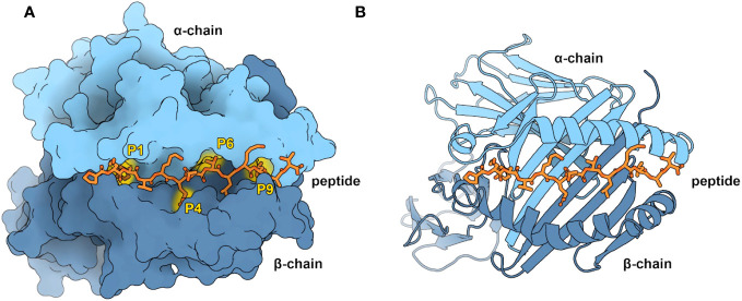
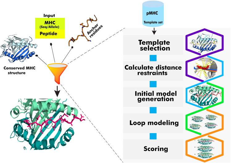
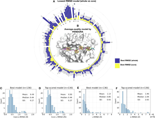
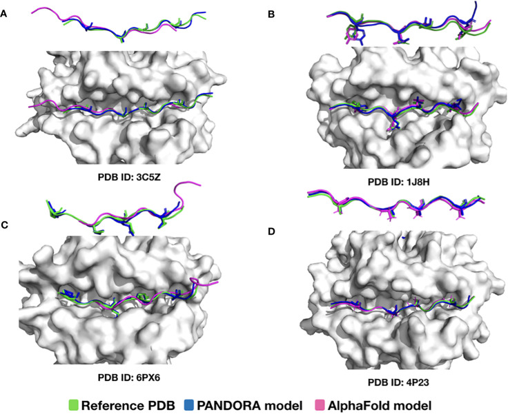
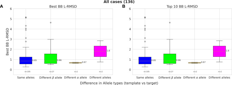
T-cell specificity to differentiate between self and non-self relies on T-cell receptor (TCR) recognition of peptides presented by the Major Histocompatibility Complex (MHC). Investigations into the three-dimensional (3D) structures of peptide:MHC (pMHC) complexes have provided valuable insights of MHC functions. Given the limited availability of experimental pMHC structures and considerable diversity of peptides and MHC alleles, it calls for the development of efficient and reliable computational approaches for modeling pMHC structures. Here we present an update of PANDORA and the systematic evaluation of its performance in modelling 3D structures of pMHC class II complexes (pMHC-II), which play a key role in the cancer immune response. PANDORA is a modelling software that can build low-energy models in a few minutes by restraining peptide residues inside the MHC-II binding groove. We benchmarked PANDORA on 136 experimentally determined pMHC-II structures covering 44 unique αβ chain pairs. Our pipeline achieves a median backbone Ligand-Root Mean Squared Deviation (L-RMSD) of 0.42 Å on the binding core and 0.88 Å on the whole peptide for the benchmark dataset. We incorporated software improvements to make PANDORA a pan-allele framework and improved the user interface and software quality. Its computational efficiency allows enriching the wealth of pMHC binding affinity and mass spectrometry data with 3D models. These models can be used as a starting point for molecular dynamics simulations or structure-boosted deep learning algorithms to identify MHC-binding peptides. PANDORA is available as a Python package through Conda or as a source installation at https://github.com/X-lab-3D/PANDORA .
Overview
The claims and statistics of this paper haven’t been checked yet.
Run the check — free, no account needed. It reads the open-access text and checks the sources it cites.
Integrity signals
Registry
Cites
Statistics
Figures
Verification
Claims the paper makes
The claims and statistics of this paper haven’t been checked yet.
Run the check — free, no account needed. It reads the open-access text and checks the sources it cites.
Self-check
Percentages, sums and reported differences re-computed from the paper’s own numbers.
The self-check needs the paper’s full text.
Citations
Cites
Highly accurate protein structure prediction with AlphaFold
Jumper, J., Evans, R., Pritzel, A., Green, T., Figurnov, M., Ronneberger, O., Tunyasuvunakool, K., Bates, R., Žídek, A., Potapenko, A., et al. (2021) · Nature · doi:10.1038/s41586-021-03819-2 · Cited by 43,764
Towards a systems understanding of MHC class I and MHC class II antigen presentation
Neefjes, J., Jongsma, M. L. M., Paul, P., & Bakke, O. (2011) · Nature Reviews Immunology · doi:10.1038/nri3084 · Cited by 2,044
MHC-II neoantigens shape tumour immunity and response to immunotherapy
Alspach, E., Lussier, D. M., Miceli, A. P., Kizhvatov, I., DuPage, M., Luoma, A. M., Meng, W., Lichti, C. F., Esaulova, E., Vomund, A. N., et al. (2019) · Nature · doi:10.1038/s41586-019-1671-8 · Cited by 846
Research works printed but not linked: 49
- Janeway, 2001, Immunobiology: the immune system in health and disease, 732
- Blum, 2013, Pathways of antigen processing, Annu Rev Immunol, 31, 10.1146/annurev-immunol-032712-095910 · DOI
- Madden, 1995, The three-dimensional structure of peptide-MHC complexes, Annu Rev Immunol, 13, 587, 10.1146/annurev.iy.13.040195.003103 · DOI
- Abualrous, 2023, MHC-II dynamics are maintained in HLA-DR allotypes to ensure catalyzed peptide exchange, Nat Chem Biol, 19, 1, 10.1038/s41589-023-01316-3 · DOI
- Thomas, 2017, Structure of the TAPBPR–MHC I complex defines the mechanism of peptide loading and editing, Science, 358, 10.1126/science.aao6001 · DOI
- Zhang, 1998, Structural principles that govern the peptide-binding motifs of class I MHC molecules, JMB, 281, 10.1006/jmbi.1998.1982 · DOI
- Sinigaglia, 1994, Defining rules for the peptide-MHC class II interaction, Curr Opin Immunol, 6, 10.1016/0952-7915(94)90033-7 · DOI
- Ferrante, 2007, Cooperativity of hydrophobic anchor interactions: evidence for epitope selection by MHC class II as a folding process, J Immunol, 178, 10.4049/jimmunol.178.11.7181 · DOI
- Murthy, 1997, The class II MHC protein HLA-DR1 in complex with an endogenous peptide: implications for the structural basis of the specificity of peptide binding, Structure, 5, 10.1016/S0969-2126(97)00288-8 · DOI
- Wucherpfennig, 2001, Insights into autoimmunity gained from structural analysis of MHC–peptide complexes, Curr Opin Immunol, 13, 10.1016/S0952-7915(01)00274-6 · DOI
- Madura, 2015, Structural basis for ineffective T-cell responses to MHC anchor residue-improved “heteroclitic” peptides: Molecular immunology, Eur J Immunol, 45, 10.1002/eji.201445114 · DOI
- Smith, 2021, Structurally silent peptide anchor modifications allosterically modulate T cell recognition in a receptor-dependent manner, Proc Natl Acad Sci USA, 118, e2018125118, 10.1073/pnas.2018125118 · DOI
- Saotome, 2023, Structural analysis of cancer-relevant TCR-CD3 and peptide-MHC complexes by cryoEM, Nat Commun, 14, 2401, 10.1038/s41467-023-37532-7 · DOI
- Sušac, 2022, Structure of a fully assembled tumor-specific T cell receptor ligated by pMHC, Cell, 185, 3201, 10.1016/j.cell.2022.07.010 · DOI
- Sahin, 2017, Personalized RNA mutanome vaccines mobilize poly-specific therapeutic immunity against cancer, Nature, 547, 10.1038/nature23003 · DOI · Paper page
- Ott, 2017, An immunogenic personal neoantigen vaccine for patients with melanoma, Nature, 547, 10.1038/nature22991 · DOI · Paper page
- Kreiter, 2015, Mutant MHC class II epitopes drive therapeutic immune responses to cancer, Nature, 520, 10.1038/nature14426 · DOI · Paper page
- Tomasello, 2020, The Protein Imager: a full-featured online molecular viewer interface with server-side HQ-rendering capabilities, Bioinformatics, 36, 10.1093/bioinformatics/btaa009 · DOI
- Jardetzky, 1996, Crystallographic analysis of endogenous peptides associated with HLA-DR1 suggests a common, polyproline II-like conformation for bound peptides, Proc Natl Acad Sci USA, 93, 10.1073/pnas.93.2.734 · DOI
- Wieczorek, 2017, Major histocompatibility complex (MHC) class I and MHC class II proteins: conformational plasticity in antigen presentation, Front Immunol, 8, 10.3389/fimmu.2017.00292 · DOI
- Hammer, 1994, Precise prediction of major histocompatibility complex class II-peptide interaction based on peptide side chain scanning, J Exp Med, 180, 10.1084/jem.180.6.2353 · DOI
- Stern, 1994, Crystal structure of the human class II MHC protein HLA-DR1 complexed with an influenza virus peptide, Nature, 368, 10.1038/368215a0 · DOI
- Brown, 1993, Three-dimensional structure of the human class II histocompatibility antigen HLA-DR1, Nature, 364, 10.1038/364033a0 · DOI
- Kulski, 2022, Human leukocyte antigen super-locus: nexus of genomic supergenes, SNPs, indels, transcripts, and haplotypes, Hum Genome Var, 9, 49, 10.1038/s41439-022-00226-5 · DOI
- Barker, 2023, The IPD-IMGT/HLA database, Nucleic Acids Res, 51, 10.1093/nar/gkac1011 · DOI
- Berman, 2000, The protein data bank, Nucleic Acids Res, 28, 10.1093/nar/28.1.235 · DOI · Paper page
- Patronov, 2011, Peptide binding prediction for the human class II MHC allele HLA-DP2: a molecular docking approach, BMC Struct Biol, 11, 32, 10.1186/1472-6807-11-32 · DOI
- Patronov, 2014, Histidine Hydrogen Bonding in MHC at pH 5 and pH 7 Modeled by Molecular Docking and Molecular Dynamics Simulations, CAD, 10, 10.2174/15734099113096660050 · DOI
- Bordner, 2010, Towards universal structure-based prediction of class II MHC epitopes for diverse allotypes, PloS One, 5, 10.1371/journal.pone.0014383 · DOI
- Tong, 2004, Modeling the structure of bound peptide ligands to major histocompatibility complex, Protein Sci, 13, 10.1110/ps.04631204 · DOI
- Khan, 2010, pDOCK: a new technique for rapid and accurate docking of peptide ligands to Major Histocompatibility Complexes, Immunome Res, 6, 10.1186/1745-7580-6-S1-S2 · DOI
- Atanasova, 2013, EpiDOCK: a molecular docking-based tool for MHC class II binding prediction, PEDS, 26, 10.1093/protein/gzt018 · DOI
- Racle, 2019, Robust prediction of HLA class II epitopes by deep motif deconvolution of immunopeptidomes, Nat Biotechnol, 37, 10.1038/s41587-019-0289-6 · DOI
- Liu, 2022, DeepSeqPanII: an interpretable recurrent neural network model with attention mechanism for peptide-HLA class II binding prediction, IEEE/ACM Trans Comput Biol Bioinf., 19, 10.1109/TCBB.2021.3074927 · DOI
- Reynisson, 2020, NetMHCpan-4.1 and NetMHCIIpan-4.0: improved predictions of MHC antigen presentation by concurrent motif deconvolution and integration of MS MHC eluted ligand data, NAR, 48, 10.1093/nar/gkaa379 · DOI · Paper page
- Marzella, 2022, PANDORA: A fast, anchor-restrained modelling protocol for peptide: MHC complexes, Front Immunol, 13, 10.3389/fimmu.2022.878762 · DOI
- Marzella, 2023, The PANDORA software for anchor-restrained peptide:MHC modeling, Methods Mol Biol, 2673, 10.1007/978-1-0716-3239-0_18 · DOI
- Ehrenmann, 2010, IMGT/3Dstructure-DB and IMGT/DomainGapAlign: a database and a tool for immunoglobulins or antibodies, T cell receptors, MHC, IgSF and MhcSF, Nucleic Acids Res, 38, 10.1093/nar/gkp946 · DOI
- Webb, 2016, Comparative protein structure modeling using MODELLER, Curr Protoc Bioinf, 54, 1, 10.1002/prot.21804 · DOI
- Lensink, 2007, Docking and scoring protein complexes: CAPRI 3rd Edition, Proteins, 69, 10.1002/prot.21804 · DOI
- Mirdita, 2022, ColabFold: making protein folding accessible to all, Nat Methods, 19, 10.1038/s41592-022-01488-1 · DOI · Paper page
- Evans, 2021, Protein complex prediction with AlphaFold-Multimer, Bioinformatics;, 4, 10.1101/2021.10.04.463034 · DOI · Paper page
- Carson, 1997, T cell receptor recognition of MHC class II–bound peptide flanking residues enhances immunogenicity and results in altered TCR V region usage, Immunity, 7, 10.1016/S1074-7613(00)80360-X · DOI
- Vignali, 1994, Amino acid residues that flank core peptide epitopes and the extracellular domains of CD4 modulate differential signaling through the T cell receptor, JEM, 179, 10.1084/jem.179.6.1945 · DOI
- Muller, 1996, Activation of T cells by the ragged tail of MHC class II-presented peptides of the measles virus fusion protein, Int Immunol, 8, 10.1093/intimm/8.4.445 · DOI
- Zavala-Ruiz, 2004, A hairpin turn in a class II MHC-bound peptide orients residues outside the binding groove for T cell recognition, Proc Natl Acad Sci USA, 101, 10.1073/pnas.0403371101 · DOI
- Levy, 2023, Large-scale immunopeptidome analysis reveals recurrent posttranslational splicing of cancer- and immune-associated genes, MCP, 22, 100519, 10.1016/j.mcpro.2023.100519 · DOI
- Bloodworth, 2022, Rosetta FlexPepDock to predict peptide-MHC binding: An approach for non-canonical amino acids, PloS One, 17, 10.1371/journal.pone.0275759 · DOI
- Sandalova, 2022, Structural aspects of chemical modifications in the MHC-restricted immunopeptidome; Implications for immune recognition, Front Chem, 10, 10.3389/fchem.2022.861609 · DOI
Metadata
- Authors
- Parizi, F. M., Marzella, D. F., Ramakrishnan, G., ‘t Hoen, P. A. C., Karimi-Jafari, M. H., & Xue, L. C.
- Affiliations
- University of Tehran (IR)
- Radboud University Medical Center (NL)
- Corresponding author
- Xue, Li C.
- Venue
- Frontiers in Immunology
- Publisher
- Frontiers Media SA
- Published
- 2023-12-08
- Volume
- 14
- Pages
- 1285899-1285899
- ISSN
- 1664-3224 (ISSN-L)
- PMC
- PMC10739464
- OpenAlex
- W4389479390
- Cited by
- 14
- References
- 3
- Citations by year
- 2026: 3 · 2025: 4 · 2024: 6 · 2023: 1
- FWCI
- 1.24
- Type
- journal article
- Language
- en
- Open access
- gold
- doi.org
- Licence
- Locations
- Frontiers in Immunology (publishedVersion, cc-by)
- bioRxiv (Cold Spring Harbor Laboratory) (acceptedVersion)
- PubMed Central (publishedVersion, cc-by)
- PubMed (publishedVersion)
- Radboud Repository (Radboud University) (submittedVersion)
- DOAJ (DOAJ: Directory of Open Access Journals) (submittedVersion, cc-by-sa)
- Funders
- Radboud Universitair Medisch Centrum
- European Regional Development Fund
- Topics
- Life Sciences › Biochemistry, Genetics and Molecular Biology › Molecular Biology › vaccines and immunoinformatics approaches
- Life Sciences › Biochemistry, Genetics and Molecular Biology › Molecular Biology › Protein Structure and Dynamics
- Physical Sciences › Chemistry › Organic Chemistry › Chemical Synthesis and Analysis
- Keywords
- Pandora, peptide-MHC class II complexes, T cell receptor recognition, benchmarking, cancer immune response
- MeSH terms
- Histocompatibility Antigens, Major Histocompatibility Complex, Peptides, Software, Benchmarking
About these checks
Registry, retraction, Cites and statistics results are computed, not judged, with one exception: a printed reference that no index lists is read once by a model before it counts as a citation error. Claims are extracted by a model and kept only when their quote appears verbatim in the text; each cited source is then checked, with a model’s help, for whether it says what the claim relies on. Those verdicts can be wrong — read the claim’s quote and the cited source before relying on one.
Statistics and arithmetic are recomputed, not judged.
Confidence bands are shown only for fields calibrated on our annotated test set. None is calibrated yet, so no band is shown.
What counts as a claim ›How we measure confidence ›How we check this ›
Are you an author, or do you see a problem with this page? Request a review
