

The DOI matches its Crossref record; 278 of the 281 references it cites were found in a registry or an index.
Imaging of cerebrovascular pathology in animal models of Alzheimer's disease
Open accessAbstract
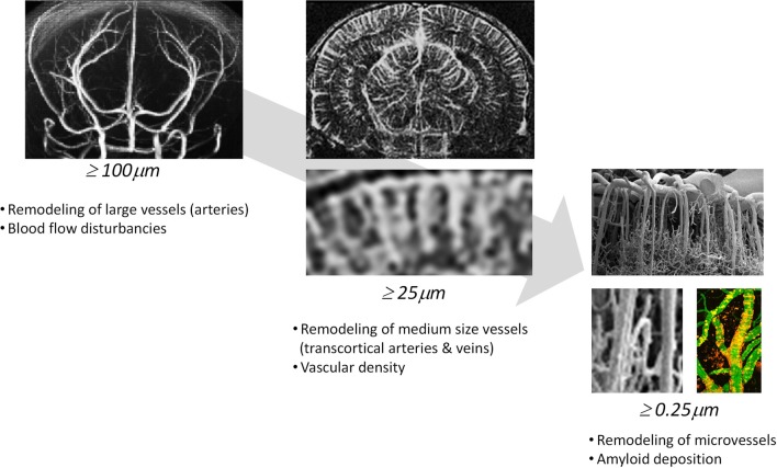
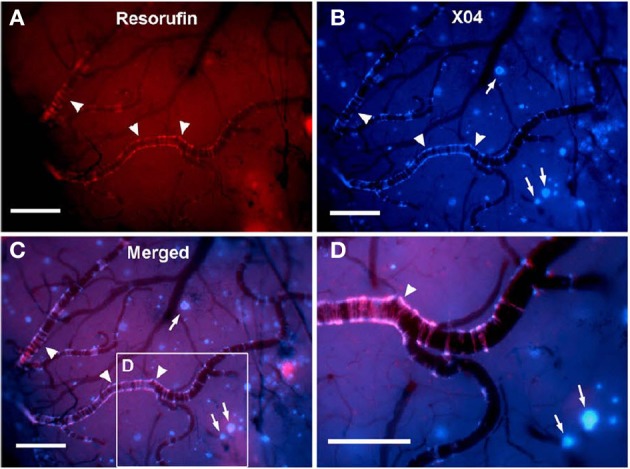
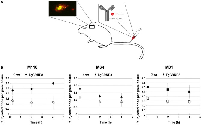
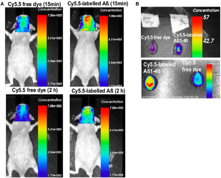
In Alzheimer's disease (AD), vascular pathology may interact with neurodegeneration and thus aggravate cognitive decline. As the relationship between these two processes is poorly understood, research has been increasingly focused on understanding the link between cerebrovascular alterations and AD. This has at last been spurred by the engineering of transgenic animals, which display pathological features of AD and develop cerebral amyloid angiopathy to various degrees. Transgenic models are versatile for investigating the role of amyloid deposition and vascular dysfunction, and for evaluating novel therapeutic concepts. In addition, research has benefited from the development of novel imaging techniques, which are capable of characterizing vascular pathology in vivo. They provide vascular structural read-outs and have the ability to assess the functional consequences of vascular dysfunction as well as to visualize and monitor the molecular processes underlying these pathological alterations. This article focusses on recent in vivo small animal imaging studies addressing vascular aspects related to AD. With the technical advances of imaging modalities such as magnetic resonance, nuclear and microscopic imaging, molecular, functional and structural information related to vascular pathology can now be visualized in vivo in small rodents. Imaging vascular and parenchymal amyloid-β (Aβ) deposition as well as Aβ transport pathways have been shown to be useful to characterize their dynamics and to elucidate their role in the development of cerebral amyloid angiopathy and AD. Structural and functional imaging read-outs have been employed to describe the deleterious affects of Aβ on vessel morphology, hemodynamics and vascular integrity. More recent imaging studies have also addressed how inflammatory processes partake in the pathogenesis of the disease. Moreover, imaging can be pivotal in the search for novel therapies targeting the vasculature.
Overview
The claims and statistics of this paper haven’t been checked yet.
Run the check — free, no account needed. It reads the open-access text and checks the sources it cites.
Integrity signals
Registry
Cites
Statistics
Figures
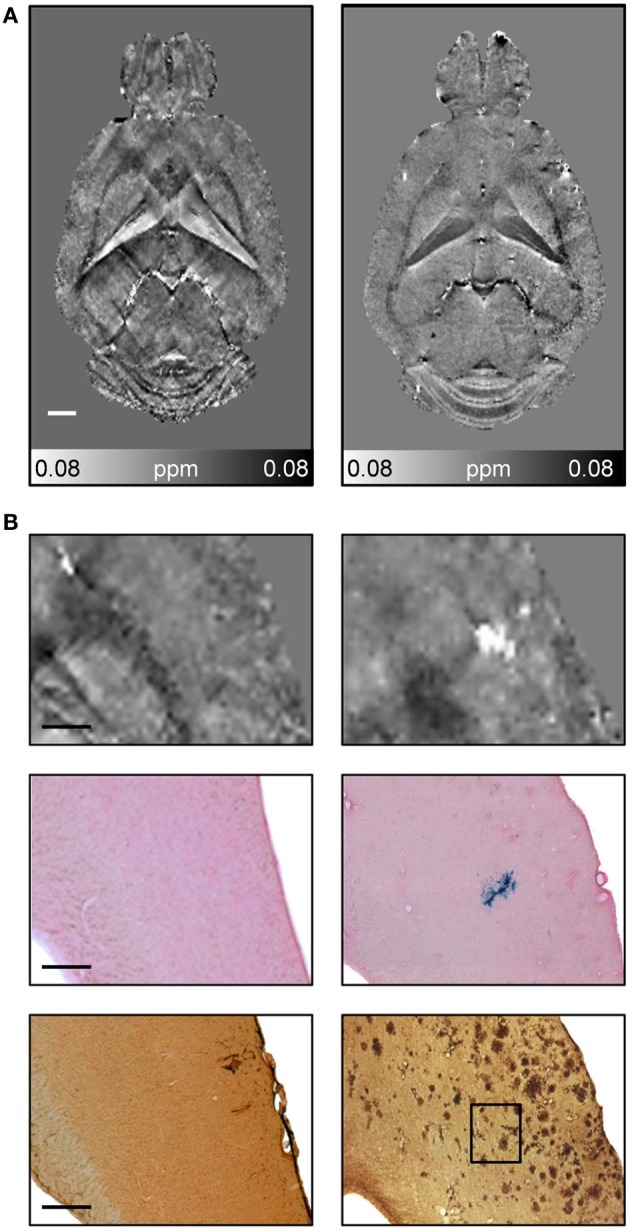
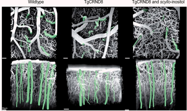
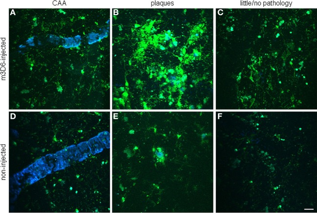
Verification
Claims the paper makes
The claims and statistics of this paper haven’t been checked yet.
Run the check — free, no account needed. It reads the open-access text and checks the sources it cites.
Claims other papers make about this one
Each is checked against this paper’s own text: does it say what the citing paper says it does?
The check wasn’t confident either way, so it gives no verdict — never disputed.
In the citing paper
“Fixation does alter the magnitude of water diffusion in the tissue, but measures of the directionality of diffusion are less affected [44][45][46].”
From Diffusion tensor imaging reveals whole-brain microstructural changes in the P301L mouse model of tauopathy. (2020) · cites this paper as [46]
Experimental — accuracy not yet measured
Self-check
Percentages, sums and reported differences re-computed from the paper’s own numbers.
The self-check needs the paper’s full text.
Citations
Cites
The Amyloid Hypothesis of Alzheimer’s Disease: Progress and Problems on the Road to Therapeutics
Hardy, J., & Selkoe, D. J. (2002) · Science · doi:10.1126/science.1072994 · Cited by 13,864
Neurophysiological investigation of the basis of the fMRI signal
Logothetis, N. K., Pauls, J., Augath, M., Trinath, T., & Oeltermann, A. (2001) · Nature · doi:10.1038/35084005 · Cited by 6,551
Correlative Memory Deficits, Aβ Elevation, and Amyloid Plaques in Transgenic Mice
Hsiao, K., Chapman, P., Nilsen, S., Eckman, C., Harigaya, Y., Younkin, S., Yang, F., & Cole, G. (1996) · Science · doi:10.1126/science.274.5284.99 · Cited by 4,210
Triple-Transgenic Model of Alzheimer’s Disease with Plaques and Tangles
Oddo, S., Caccamo, A., Shepherd, J. D., Murphy, M. P., Golde, T. E., Kayed, R., Metherate, R., Mattson, M. P., Akbari, Y., & LaFerla, F. M. (2003) · Neuron · doi:10.1016/s0896-6273(03)00434-3 · Cited by 4,126
Clinical and biomarker changes in dominantly inherited Alzheimer's disease
Bateman, R. J., Xiong, C., Benzinger, T. L. S., Fagan, A. M., Goate, A., Fox, N. C., Marcus, D. S., Cairns, N. J., Xie, X., Blazey, T. M., et al. (2012) · New England Journal of Medicine · doi:10.1056/nejmoa1202753 · Cited by 3,814
Ogawa, S., Tank, D. W., Menon, R., Ellermann, J. M., Kim, S. G., Merkle, H., & Ugurbil, K. (1992) · Proceedings of the National Academy of Sciences · doi:10.1073/pnas.89.13.5951 · Cited by 3,681
Oakley, H., Cole, S. L., Logan, S., Maus, E., Shao, P., Craft, J., Guillozet-Bongaarts, A., Ohno, M., Disterhoft, J., Van Eldik, L., et al. (2006) · The Journal of Neuroscience · doi:10.1523/jneurosci.1202-06.2006 · Cited by 3,530 · cited 13×
“Yes Richards et al., 2003; Woolley and Ballard, 2005; Weidensteiner et al., 2009 APPPS1 (APPPS1-21) hAPP751 K670N/M671L/V717I mThy1 X PS1 L166P knock-in Swe 2–4 mo 8 mo + Yes Yes Radde et al., 2006; Rupp et al., 2011; Montagne et al., 2012; Vom Berg et al., 2012 APPxPS1-Ki (APPSLPS1KI) hAPP751 K670N/N671L mThy1 PS1 M2…”
BackgroundInfluentialAlzheimer-type neuropathology in transgenic mice overexpressing V717F β-amyloid precursor protein
Games, D., Adams, D., Alessandrini, R., Barbour, R., Borthelette, P., Blackwell, C., Carr, T., Clemens, J., Donaldson, T., Gillespie, F., et al. (1995) · Nature · doi:10.1038/373523a0 · Cited by 2,516
Jarrett, J. T., Berger, E. P., & Lansbury, P. T. (1993) · Biochemistry · doi:10.1021/bi00069a001 · Cited by 2,022
Tau Suppression in a Neurodegenerative Mouse Model Improves Memory Function
SantaCruz, K., Lewis, J., Spires, T., Paulson, J., Kotilinek, L., Ingelsson, M., Guimaraes, A., DeTure, M., Ramsden, M., McGowan, E., et al. (2005) · Science · doi:10.1126/science.1113694 · Cited by 2,014
Research works printed but not linked: 3
- Beckmann, 2000, High resolution magnetic resonance angiography non-invasively reveals mouse strain differences in the cerebrovascular anatomy in vivo, Magn. Reson. Med, 44, 252, 10.1002/1522-2594(200008)44:2252::AID-MRM123.0.CO;2-G · DOI
- Jensen, 2000, MR imaging of microvasculature, Magn. Reson. Med, 44, 224, 10.1002/1522-2594(200008)44:2224::AID-MRM93.0.CO;2-M · DOI
- Tropres, 2001, Vessel size imaging, Magn. Reson. Med, 45, 397, 10.1002/1522-2594(200103)45:3397::AID-MRM10523.0.CO;2-3 · DOI
Cited by
Sorted by citations
Cerebrovascular Smooth Muscle Cells as the Drivers of Intramural Periarterial Drainage of the Brain
Aldea, R., Weller, R. O., Wilcock, D. M., Carare, R. O., & Richardson, G. W. (2019) · Frontiers in Aging Neuroscience · doi:10.3389/fnagi.2019.00001 · Cited by 340
Cerebrovascular pathology during the progression of experimental Alzheimer's disease
Giannoni, P., Arango-Lievano, M., das Neves, I., Rousset, M.-C., Baranger, K., Rivera, S., Jeanneteau, F. D., Claeysen, S., & Marchi, N. (2016) · Neurobiology of Disease · doi:10.1016/j.nbd.2016.01.001 · Cited by 152
Causes and consequences of baseline cerebral blood flow reductions in Alzheimer’s disease
Bracko, O., Hernández, J. C. C., Park, L., Nishimura, N., & Schaffer, C. B. (2021) · Journal of Cerebral Blood Flow & Metabolism · doi:10.1177/0271678x20982383 · Cited by 127
Coles, J. A., Myburgh, E., Brewer, J. M., & McMenamin, P. G. (2017) · Progress in Neurobiology · doi:10.1016/j.pneurobio.2017.05.002 · Cited by 123
Fard, M. T., & Stough, C. K. K. (2019) · Frontiers in Aging Neuroscience · doi:10.3389/fnagi.2019.00056 · Cited by 124
An Integrated View on Vascular Dysfunction in Alzheimer’s Disease
Klohs, J. (2019) · Neurodegenerative Diseases · doi:10.1159/000505625 · Cited by 111
Self-citationStarosolski, Z. A., Villamizar, C., Rendon, D. A., Paldino, M. J., Milewicz, D. M., Ghaghada, K. B., & Annapragada, A. (2015) · Scientific Reports · doi:10.1038/srep10178 · Cited by 112
Cerebral small vessel disease and Alzheimer’s disease
Cai, Z., Wang, C., He, W., Tu, H., Tang, Z., Xiao, M., & Yan, L. (2015) · Clinical Interventions in Aging · doi:10.2147/cia.s90871 · Cited by 102
Shen, Y., Hua, L., Yeh, C., Shen, L., Ying, M., Zhang, Z., Liu, G., Li, S., Chen, S., Chen, X., et al. (2020) · Theranostics · doi:10.7150/thno.44152 · Cited by 101
Bracko, O., Vinarcsik, L. K., Cruz Hernández, J. C., Ruiz-Uribe, N. E., Haft-Javaherian, M., Falkenhain, K., Ramanauskaite, E. M., Ali, M., Mohapatra, A., Swallow, M. A., et al. (2020) · Scientific Reports · doi:10.1038/s41598-020-65908-y · Cited by 85
Lourenço, C. F., Ledo, A., Barbosa, R. M., & Laranjinha, J. A. N. (2017) · Experimental Neurology · doi:10.1016/j.expneurol.2017.01.013 · Cited by 83
Microvascular Alterations in Alzheimer's Disease
Steinman, J., Sun, H., & Feng, Z. (2021) · Frontiers in Cellular Neuroscience · doi:10.3389/fncel.2020.618986 · Cited by 85
Kim, S. H., Ahn, J. H., Yang, H. J., Lee, P., Koh, G. Y., & Jeong, Y. (2020) · Acta Neuropathologica Communications · doi:10.1186/s40478-020-01042-0 · Cited by 79
Multi-scale optoacoustic molecular imaging of brain diseases
Razansky, D., Klohs, J., & Ni, R. (2021) · European Journal of Nuclear Medicine and Molecular Imaging · doi:10.1007/s00259-021-05207-4 · Cited by 77
Self-citationKasza, Á., Penke, B., Frank, Z., Bozsó, Z., Szegedi, V., Hunya, Á. G., Németh, K., Kozma, G., & Fülöp, L. (2017) · Molecules · doi:10.3390/molecules22112007 · Cited by 78
Endothelial Nitric Oxide Synthase–Deficient Mice
Liao, F., Lin, G., Chen, X., Chen, L., Zheng, W., Raghow, R., Zhou, F.-M., Shih, A. Y., & Tan, X.-L. (2021) · American Journal Of Pathology · doi:10.1016/j.ajpath.2021.02.022 · Cited by 68
Annexin A1 restores cerebrovascular integrity concomitant with reduced amyloid-β and tau pathology
Ries, M., Watts, H. R., Mota, B. C., Lopez, M. Y., Donat, C. K., Baxan, N., Pickering, J. A., Chau, T. W., Semmler, A., Gurung, B., et al. (2021) · Brain · doi:10.1093/brain/awab050 · Cited by 66
Ni, R., Rudin, M., & Klohs, J. (2018) · Photoacoustics · doi:10.1016/j.pacs.2018.04.001 · Cited by 53
Self-citationVascular cognitive impairment: Modeling a critical neurologic disease in vitro and in vivo
Helman, A. M., & Murphy, M. P. (2015) · Biochimica et Biophysica Acta (BBA) - Molecular Basis of Disease · doi:10.1016/j.bbadis.2015.12.009 · Cited by 55
Niendorf, T., Pohlmann, A., Reimann, H. M., Waiczies, H., Peper, E. S., Huelnhagen, T., Seeliger, E., Schreiber, A., Kettritz, R., Strobel, K., et al. (2015) · Frontiers in Pharmacology · doi:10.3389/fphar.2015.00255 · Cited by 48
Jandke, S., Garz, C., Schwanke, D., Sendtner, M., Heinze, H., Carare, R. O., & Schreiber, S. (2018) · Brain Pathology · doi:10.1111/bpa.12629 · Cited by 46
Petrushina, I., Hovakimyan, A., Harahap-Carrillo, I. S., Davtyan, H., Antonyan, T., Chailyan, G., Kazarian, K., Antonenko, M., Jullienne, A., Hamer, M. M., et al. (2020) · Neurobiology of Disease · doi:10.1016/j.nbd.2020.104823 · Cited by 48
Kniewallner, K. M., Foidl, B. M., & Humpel, C. (2018) · Scientific Reports · doi:10.1038/s41598-018-33768-2 · Cited by 48
Endothelins in inflammatory neurological diseases
D’Orléans-Juste, P., Ndunge, O. B. A., Desbiens, L., Tanowitz, H. B., & Desruisseaux, M. S. (2018) · Pharmacology & Therapeutics · doi:10.1016/j.pharmthera.2018.10.001 · Cited by 46
ELKAHLOUN, A. G., Hafko, R., & Saavedra, J. M. (2016) · Alzheimer s Research & Therapy · doi:10.1186/s13195-015-0167-5 · Cited by 45
Li, H.-C., Chen, P., Cheng, H.-F., Kuo, Y., & Huang, C. (2019) · IEEE Transactions on Biomedical Engineering · doi:10.1109/tbme.2019.2904702 · Cited by 44
Escrig, A., Canal, C., Sanchís, P., Fernández‐Gayol, O., Montilla, A., Comes, G., Molinero, A., Giralt, M., Giménez‐Llort, L., Becker‐Pauly, C., et al. (2019) · Brain Behavior and Immunity · doi:10.1016/j.bbi.2019.08.005 · Cited by 43
Uekawa, K., Hasegawa, Y., Senju, S., Nakagata, N., Ma, M., Nakagawa, T., Koibuchi, N., & Kim‐Mitsuyama, S. (2016) · Journal of Alzheimer s Disease · doi:10.3233/jad-150642 · Cited by 40
Lee, S.-C., Park, H.-H., Kim, S., Koh, S., Han, S.-H., & Yoon, M. (2019) · Analytical Chemistry · doi:10.1021/acs.analchem.8b03735 · Cited by 40
Bourassa, P., Tremblay, C., Schneider, J. A., Bennett, D. A., & Calon, F. (2020) · Neuropathology and Applied Neurobiology · doi:10.1111/nan.12599 · Cited by 38
Lana, D., Ugolini, F., Nosi, D., Wenk, G. L., & Giovannini, M. G. (2017) · Frontiers in Aging Neuroscience · doi:10.3389/fnagi.2017.00296 · Cited by 39
Wilhelmus, M. M. M., de Jager, M., Smit, A. B., van der Loo, R. J., & Drukarch, B. (2016) · Scientific Reports · doi:10.1038/srep20569 · Cited by 32
Ni, R., Kindler, D., Waag, R., Rouault, M., Ravikumar, P., Nitsch, R. M., Rudin, M., Camici, G. G., Liberale, L., Kulic, L., et al. (2019) · Frontiers in Aging Neuroscience · doi:10.3389/fnagi.2019.00027 · Cited by 31
Self-citationColeman, R. A., Liang, C., Patel, R., Ali, S., & Mukherjee, J. (2017) · Molecular Imaging · doi:10.1177/1536012117704557 · Cited by 29
Reuter, B., Venus, A., Heiler, P. M., Schad, L. R., Ebert, A., Hennerici, M. G., Grudzenski, S., & Fatar, M. (2016) · Frontiers in Aging Neuroscience · doi:10.3389/fnagi.2016.00170 · Cited by 28
Massalimova, A., Ni, R., Nitsch, R. M., Reisert, M., von Elverfeldt, D., & Klohs, J. (2020) · Neurodegenerative Diseases · doi:10.1159/000515754 · Cited by 28
Self-citationNeumann, U., Machauer, R., & Shimshek, D. R. (2019) · British Journal of Pharmacology · doi:10.1111/bph.14582 · Cited by 27
Self-citationA guide to using functional magnetic resonance imaging to study Alzheimer's disease in animal models
Asaad, M., & Lee, J. H. (2018) · Disease Models & Mechanisms · doi:10.1242/dmm.031724 · Cited by 26
A close look at brain dynamics: Cells and vessels seen by in vivo two-photon microscopy
Fumagalli, S., Ortolano, F., & Simoni, M. G. D. (2014) · Progress in Neurobiology · doi:10.1016/j.pneurobio.2014.06.005 · Cited by 27
Fibrin-targeting immunotherapy for dementia
Kantor, A. B., Akassoglou, K., & Stavenhagen, J. B. (2023) · The Journal of Prevention of Alzheimer's Disease · doi:10.14283/jpad.2023.105 · Cited by 24
Phosphorylated Aβ peptides in human Down syndrome brain and different Alzheimer’s-like mouse models
Kumar, S., Lemere, C. A., & Walter, J. (2020) · Acta Neuropathologica Communications · doi:10.1186/s40478-020-00959-w · Cited by 24
Hankeová, S., Šalplachta, J., Zikmund, T., Kavková, M., Hul, N. V., Břínek, A., Smekalova, V., Laznovsky, J., Dawit, F., Jaroš, J., et al. (2021) · eLife · doi:10.7554/elife.60916 · Cited by 24
Sharp, P. S., Ameen-Ali, K. E., Boorman, L., Harris, S., Wharton, S. B., Howarth, C., Shabir, O., Redgrave, P., & Berwick, J. (2019) · Journal of Cerebral Blood Flow & Metabolism · doi:10.1177/0271678x19890830 · Cited by 25
Beckmann, N., Doelemeyer, A., Zurbruegg, S., Bigot, K., Theil, D. J., Frieauff, W., Kolly, C., Moulin, P., Neddermann, D., Kreutzer, R., et al. (2016) · Neurobiology of Aging · doi:10.1016/j.neurobiolaging.2016.05.009 · Cited by 24
Self-citationLin, H., Wang, Z., Liao, Y., Yu, Z., Xu, H., Qin, T. G., Tang, J., Yang, X., Chen, S., Chen, X., et al. (2024) · EBioMedicine · doi:10.1016/j.ebiom.2024.105355 · Cited by 23
Dynamic changes in vascular size and density in transgenic mice with Alzheimer’s disease
Xu, X., Meng, T., Wen, Q., Tao, M., Wang, P., Zhong, K. L., & Shen, Y. (2020) · Aging · doi:10.18632/aging.103672 · Cited by 21
Ielacqua, G. D., Schlegel, F., Füchtemeier, M., Xandry, J., Rudin, M., & Klohs, J. (2016) · Frontiers in Aging Neuroscience · doi:10.3389/fnagi.2015.00241 · Cited by 17
Self-citationSkaaraas, G. H. E. S., Melbye, C., Puchades, M. A., Leung, D. S. Y., Jacobsen, Ø., Rao, S. B., Ottersen, O. P., Leergaard, T. B., & Torp, R. (2021) · Journal of Alzheimer s Disease · doi:10.3233/jad-210571 · Cited by 16
Maliszewska‐Cyna, E., Vecchio, L. M., Thomason, L. A. M., Oore, J., Steinman, J., Joo, I. L., Dorr, A. E., McLaurin, J., Sled, J. G., Stefanovic, B., et al. (2020) · NeuroImage · doi:10.1016/j.neuroimage.2020.117269 · Cited by 17
Mouse models of neurodegenerative disease: preclinical imaging and neurovascular component
Albanese, S., Greco, A., Auletta, L., & Mancini, M. (2018) · Brain Imaging and Behavior · doi:10.1007/s11682-017-9770-3 · Cited by 16
Deshpande, A., Elliott, J. M., Kari, N., Jiang, B., Michel, P., Toosizadeh, N., Fahadan, P. T., Kidwell, C. S., Wintermark, M., & Laksari, K. (2022) · Journal of Neuroimaging · doi:10.1111/jon.13023 · Cited by 15
Hill, L. K., Hoang, D. M., Chiriboga, L. A., Wısnıewskı, T., Sadowski, M. J., & Wadghiri, Y. Z. (2020) · Frontiers in Aging Neuroscience · doi:10.3389/fnagi.2020.585218 · Cited by 18
Tataryn, N. M., Singh, V., Dyke, J. P., Berk-Rauch, H., Clausen, D. M., Aronowitz, E., Norris, E. H., Strickland, S., & Ahn, H. J. (2021) · Neurobiology of Aging · doi:10.1016/j.neurobiolaging.2021.07.015 · Cited by 15
Xiong, Y., Zhang, Z., He, L., Ma, Y., Han, H., Zhao, X., & Guo, H. (2019) · Magnetic Resonance in Medicine · doi:10.1002/mrm.27855 · Cited by 12
BOLD amplitude correlates of preclinical Alzheimer’s disease
Hrybouski, S., Das, S. R., Xie, L., Brown, C., Flamporis, M., Lane, J., Nasrallah, I. M., Detre, J. A., Yushkevich, P. A., & Wolk, D. A. (2025) · Neurobiology of Aging · doi:10.1016/j.neurobiolaging.2025.03.007 · Cited by 13
Kindler, D., Maschio, C., Ni, R., Zerbi, V., Razansky, D., & Klohs, J. (2021) · Journal of Cerebral Blood Flow & Metabolism · doi:10.1177/0271678x211062274 · Cited by 11
Self-citationKindler, D., Maschio, C., Ni, R., Zerbi, V., Razansky, D., & Klohs, J. (2022) · Zurich Open Repository and Archive (University of Zurich) · doi:10.3929/ethz-b-000520246 · Cited by 10
Self-citationNi, R., Chen, Z., Shi, G., Villois, A., Zhou, Q., Arosio, P., Nitsch, R. M., Nilsson, K. P. R., Klohs, J., & Razansky, D. (2020) · bioRxiv (Cold Spring Harbor Laboratory) · doi:10.1101/2020.02.01.929844 · Cited by 10
Self-citationCell ageing: a flourishing field for neurodegenerative diseases
Brites, D. M. T. O. (2015) · AIMS molecular science · doi:10.3934/molsci.2015.3.225 · Cited by 10
Models of cerebral amyloid angiopathy-related intracerebral hemorrhage
Vargas-George, S. A., & Dave, K. R. (2022) · Brain Hemorrhages · doi:10.1016/j.hest.2022.05.001 · Cited by 10
Swinford, C. G., Risacher, S. L., Vosmeier, A., Deardorff, R. L., Chumin, E. J., Džemidžić, M., Wu, Y., Gao, S., McDonald, B. C., Yoder, K. K., et al. (2022) · Neurobiology of Aging · doi:10.1016/j.neurobiolaging.2023.06.014 · Cited by 10
Yu, Y., Zhang, N., Xiang, B., Ding, N., Liu, J., Huang, J., Zhao, M., Zhao, Y., Wang, Y., & Ma, Z. (2023) · Neurophotonics · doi:10.1117/1.nph.10.1.015005 · Cited by 9
Perfusable cellulose channels from decellularized leaf scaffolds for modeling vascular amyloidosis
Lee, T., Lee, K. H., Cheong, D. Y., Lee, S. W., Park, I., & Lee, G. (2025) · International Journal of Biological Macromolecules · doi:10.1016/j.ijbiomac.2025.142509 · Cited by 6
Imaging Pericytes and the Regulation of Cerebral Blood Flow
Boyd, K., Hammond‐Haley, M., Vroman, R., & Hall, C. N. (2021) · Methods in molecular biology · doi:10.1007/978-1-0716-1056-5_7 · Cited by 6
El populismo: de intruso a problema relevante para la ciencia social
Sáez, A. R. (2018) · Revista Internacional de Sociología · doi:10.3989/ris.2018.76.4.18.076 · Cited by 6
Xiang, B., Ding, N., Jiang, H., Liu, J., Yu, Y., Luan, J., Zhao, Y., Wang, Y., Yang, Y., Ji, C., et al. (2025) · Neurophotonics · doi:10.1117/1.nph.12.1.015013 · Cited by 4
MicroRNA-29a-5p contributes to neuroinflammation through TLR7
McGurran, H., Graceffo, E., Kumbol, V. W.-A., Jendrach, M., Hinkelmann, L., Brehm, M., Ravatt, L. M., Krüger, C., Wallach, T., Haake, A., et al. (2026) · Journal of Neuroinflammation · doi:10.1186/s12974-025-03680-4 · Cited by 4
The role of proteotoxic stress in vascular dysfunction in the pathogenesis of Alzheimer’s disease
Fonseca, A. C. R. G., Resende, R., Cardoso, S. M., & Pereira, C. (2015) · Endoplasmic Reticulum Stress in Diseases · doi:10.1515/ersc-2015-0005 · Cited by 3
Generation of Transgenic Mice Expressing FRET Biosensors
Hübscher, D., & Nikolaev, V. O. (2015) · Methods in molecular biology · doi:10.1007/978-1-4939-2537-7_9 · Cited by 3
Ventura‐Antunes, L., Nackenoff, A. G., Romero‐Fernandez, W., Bosworth, A. M., Prusky, A., Wang, E., Carvajal‐Tapia, C., Shostak, A., Harmsen, H. J., Mobley, B. C., et al. (2024) · bioRxiv (Cold Spring Harbor Laboratory) · doi:10.1101/2024.03.08.583563 · Cited by 3
Hybrid ultrasound and dual-wavelength optoacoustic biomicroscopy for functional neuroimaging
Rebling, J., Estrada, H., Zwack, M., Sela, G., Gottschalk, S., & Razansky, D. (2017) · Proceedings of SPIE, the International Society for Optical Engineering/Proceedings of SPIE · doi:10.1117/12.2250635 · Cited by 2
Lin, H., Wang, Z., Liao, Y., Yu, Z., Xu, H., Qin, T. G., Tang, J., Yang, X., Chen, S., Zhang, X., et al. (2024) · SSRN Electronic Journal · doi:10.2139/ssrn.4794470 · Cited by 2
Jeong, H., Ren, J., Cheng, W., Volkow, N. D., Ling, H., Zhu, D., Du, C., & Pan, Y. (2025) · Translational Neurodegeneration · doi:10.1186/s40035-025-00530-4 · Cited by 1
The use of animal models of neuroinflammation for imaging studies
(2023) · Advances in magnetic resonance technology and applications · doi:10.1016/b978-0-323-91771-1.00011-3 · Cited by 1
Liu, Z., McCutcheon, F. M., Ho, H., Chia, J. S. M., Xiao, Y., Tippett, I., Keragala, C. B., CLOUD, G. C., & Medcalf, R. L. (2023) · Research and Practice in Thrombosis and Haemostasis · doi:10.1016/j.rpth.2023.102166 · Cited by 1
Cheng, G. W., Ma, I. W.-T., Huang, J., Yeung, S. H., Ho, P., Chen, Z., Mak, H. K., Herrup, K., Chan, K. W. Y., & Tse, K. (2023) · bioRxiv (Cold Spring Harbor Laboratory) · doi:10.1101/2023.07.24.547147 · Cited by 1
Kim, S. H., Ahn, J. H., Yang, H., Lee, P., Koh, G. Y., & Jeong, Y. (2020) · Figshare · doi:10.6084/m9.figshare.13199131
Kim, S. H., Ahn, J. H., Yang, H., Lee, P., Koh, G. Y., & Jeong, Y. (2020) · Figshare · doi:10.6084/m9.figshare.13199128
Kim, S. H., Ahn, J. H., Yang, H., Lee, P., Koh, G. Y., & Jeong, Y. (2020) · Figshare · doi:10.6084/m9.figshare.13199125
Kim, S. H., Ahn, J. H., Yang, H., Lee, P., Koh, G. Y., & Jeong, Y. (2020) · Figshare · doi:10.6084/m9.figshare.13199122
Kim, S. H., Ahn, J. H., Yang, H., Lee, P., Koh, G. Y., & Jeong, Y. (2020) · Figshare · doi:10.6084/m9.figshare.13199134
Metadata
- Authors
- Klohs, J., Rudin, M., Shimshek, D. R., & Beckmann, N.
- Affiliations
- University of Zurich (CH)
- Novartis (Switzerland) (CH)
- Corresponding author
- Beckmann, Nicolau
- Venue
- Frontiers in Aging Neuroscience
- Publisher
- Frontiers Media SA
- Published
- 2014-03-13
- Volume
- 6
- Pages
- 32-32
- ISSN
- 1663-4365 (ISSN-L)
- PMC
- PMC3952109
- OpenAlex
- W2150972825
- Cited by
- 82
- References
- 281
- Citations by year
- 2026: 1 · 2025: 4 · 2024: 3 · 2023: 5 · 2022: 4 · 2021: 10 · 2020: 16 · 2019: 10 · 2018: 6 · 2017: 8 · 2016: 7 · 2015: 7 · 2014: 1
- FWCI
- 0.90
- Type
- journal article
- Language
- en
- Open access
- gold
- doi.org
- Licence
- cc-by
- Locations
- Frontiers in Aging Neuroscience (publishedVersion, cc-by)
- Europe PMC (PubMed Central) (publishedVersion, cc-by)
- PubMed Central (publishedVersion, cc-by)
- PubMed (publishedVersion)
- Zurich Open Repository and Archive (University of Zurich) (submittedVersion, cc-by)
- DOAJ (DOAJ: Directory of Open Access Journals) (submittedVersion, cc-by-sa)
- Repository for Publications and Research Data (ETH Zurich) (submittedVersion, cc-by)
- ETH Zürich Research Collection (cc-by)
- Zurich Open Repository and Archive (University of Zurich)
- Topics
- Life Sciences › Neuroscience › Cellular and Molecular Neuroscience › Alzheimer's disease research and treatments
- Life Sciences › Neuroscience › Neurology › Barrier Structure and Function Studies
- Life Sciences › Neuroscience › Neurology › Neurological Disease Mechanisms and Treatments
- Keywords
- Alzheimer's disease, cerebrovascular pathology, cerebral amyloid angiopathy, amyloid-beta deposition, amyloid-beta transport, vascular dysfunction, transgenic animal models, in vivo imaging, small animal imaging, hemodynamics, vascular inflammation, vascular integrity, neurovascular unit
About these checks
Registry, retraction, Cites and statistics results are computed, not judged, with one exception: a printed reference that no index lists is read once by a model before it counts as a citation error. Claims are extracted by a model and kept only when their quote appears verbatim in the text; each cited source is then checked, with a model’s help, for whether it says what the claim relies on. Those verdicts can be wrong — read the claim’s quote and the cited source before relying on one.
Statistics and arithmetic are recomputed, not judged.
Confidence bands are shown only for fields calibrated on our annotated test set. None is calibrated yet, so no band is shown.
What counts as a claim ›How we measure confidence ›How we check this ›
Are you an author, or do you see a problem with this page? Request a review
