

The DOI matches its Crossref record; 145 of the 145 references it cites were found in a registry or an index.
Fornix as an imaging marker for episodic memory deficits in healthy aging and in various neurological disorders
Open accessAbstract
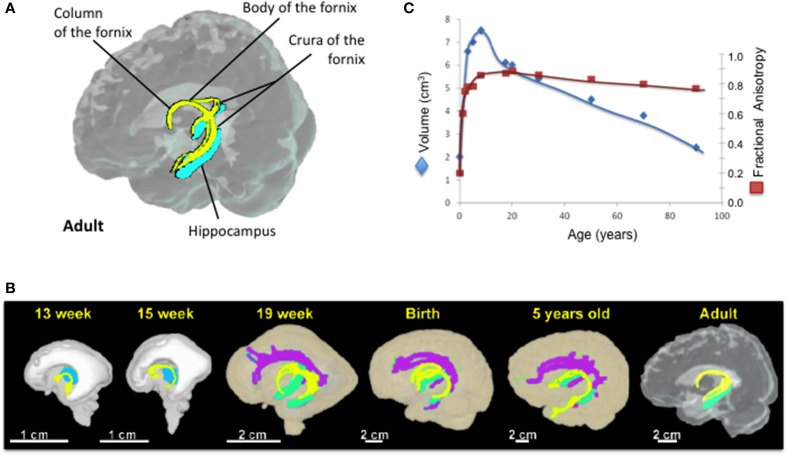
The fornix is a part of the limbic system and constitutes the major efferent and afferent white matter tracts from the hippocampi. The underdevelopment of or injuries to the fornix are strongly associated with memory deficits. Its role in memory impairments was suggested long ago with cases of surgical forniceal transections. However, recent advances in brain imaging techniques, such as diffusion tensor imaging, have revealed that macrostructural and microstructural abnormalities of the fornix correlated highly with declarative and episodic memory performance. This structure appears to provide a robust and early imaging predictor for memory deficits not only in neurodegenerative and neuroinflammatory diseases, such as Alzheimer's disease and multiple sclerosis, but also in schizophrenia and psychiatric disorders, and during neurodevelopment and "typical" aging. The objective of the manuscript is to present a systematic review regarding published brain imaging research on the fornix, including the development of its tracts, its role in various neurological diseases, and its relationship to neurocognitive performance in human studies.
Overview
The claims and statistics of this paper haven’t been checked yet.
Run the check — free, no account needed. It reads the open-access text and checks the sources it cites.
Integrity signals
Registry
Cites
Statistics
Figures
Verification
Claims the paper makes
The claims and statistics of this paper haven’t been checked yet.
Run the check — free, no account needed. It reads the open-access text and checks the sources it cites.
Claims other papers make about this one
Each is checked against this paper’s own text: does it say what the citing paper says it does?
The source backs part of the claim, not all of it — or backs it for a different population, condition or comparison.
In the citing paper
“Compared with other limbic white matter pathways, correlations between white matter microstructure and episodic memory in healthy populations are much more reliably found for the fornix than the cingulum (e.g., Douet and Chang, 2015; Metzler-Baddeley et al., 2011; Rudebeck et al., 2009).”
From The cingulum bundle: Anatomy, function, and dysfunction (2018) · cites this paper as Douet and Chang, 2015
Experimental — accuracy not yet measured
Self-check
Percentages, sums and reported differences re-computed from the paper’s own numbers.
The self-check needs the paper’s full text.
Citations
Cites
Mild cognitive impairment as a diagnostic entity
Petersen, R. C. (2004) · Journal of Internal Medicine · doi:10.1111/j.1365-2796.2004.01388.x · Cited by 7,864
Tract-based spatial statistics: Voxelwise analysis of multi-subject diffusion data
Smith, S. M., Jenkinson, M., Johansen‐Berg, H., Rueckert, D., Nichols, T. E., Mackay, C. E., Watkins, K. E., Ciccarelli, O., Cader, M. Z., Matthews, P. M., et al. (2006) · NeuroImage · doi:10.1016/j.neuroimage.2006.02.024 · Cited by 6,598
Current concepts in mild cognitive impairment
Petersen, R. C., Doody, R., Kurz, A., Mohs, R. C., Morris, J. C., Rabins, P. V., Ritchie, K., Rossor, M., Thal, L., & Winblad, B. (2001) · Archives of Neurology · doi:10.1001/archneur.58.12.1985 · Cited by 4,781
A PROPOSED MECHANISM OF EMOTION
PAPEZ, J. W. (1937) · Archives of Neurology And Psychiatry · doi:10.1001/archneurpsyc.1937.02260220069003 · Cited by 3,563
The Medial Temporal Lobe Memory System
Squire, L. R., & Zola-Morgan, S. (1991) · Science · doi:10.1126/science.1896849 · Cited by 3,282
Diagnostic Criteria for Parkinson Disease
Gelb, D. J., Oliver, E., & Gilman, S. (1999) · Archives of Neurology · doi:10.1001/archneur.56.1.33 · Cited by 2,822
Dysmyelination revealed through MRI as increased radial (but unchanged axial) diffusion of water
Song, S.-K., Sun, S.-W., Ramsbottom, M. J., Chang, C., Russell, J., & Cross, A. H. (2002) · NeuroImage · doi:10.1006/nimg.2002.1267 · Cited by 2,582
Song, S.-K., Sun, S.-W., Ju, W.-K., Lin, S.-J., Cross, A. H., & Neufeld, A. H. (2003) · NeuroImage · doi:10.1016/j.neuroimage.2003.07.005 · Cited by 1,738
Demyelination increases radial diffusivity in corpus callosum of mouse brain
Song, S.-K., Yoshino, J., Le, T. Q., Lin, S.-J., Sun, S.-W., Cross, A. H., & Armstrong, R. C. (2005) · NeuroImage · doi:10.1016/j.neuroimage.2005.01.028 · Cited by 1,658
Diffusion tensor imaging of white matter tract evolution over the lifespan
Lebel, C., Gee, M., Camicioli, R., Wieler, M., Martin, W., & Beaulieu, C. (2012) · NeuroImage · doi:10.1016/j.neuroimage.2011.11.094 · Cited by 1,100
Research works printed but not linked: 3
- Aggleton, 2000, Identifying cortical inputs to the rat hippocampus that subserve allocentric spatial processes: a simple problem with a complex answer, Hippocampus, 10, 466, 10.1002/1098-1063(2000)10:4466::AID-HIPO133.0.CO;2-Y · DOI
- Broca, 1890, Anatomie comparée des circonvolutions cérébrales, 385
- Nolte, 2009, The Human Brain: An Introduction Ot Its Functional Anatomy
Cited by
Sorted by citations
Items 101–122 of 122
Буклина, С. Б., Konovalov, A. N., Pitskhelauri, D. I., Shkarubo, M. A., Poddubskaya, A. A., & Kolycheva, M. V. (2017) · Burdenko s Journal of Neurosurgery · doi:10.17116/neiro201781228-37 · Cited by 2
Li, X., Cedrés, N., Olofsson, J., & Persson, J. (2025) · Human Brain Mapping · doi:10.1002/hbm.70375 · Cited by 1
Cui, A., Choi, Y., Yeganeh, N. M., Hermiston, N., Werker, J. F., & Boyd, L. A. (2025) · Frontiers in Psychology · doi:10.3389/fpsyg.2025.1659705 · Cited by 1
Development of a paediatric model of diffuse traumatic brain injury in ferrets
Krieg, J. L., Hooper, C., Kapuwelle, H., George, R. P., Hood, R. J., O’Brien, W. T., McDonald, S. J., Leonard, A. V., Turner, R. J., & Corrigan, F. (2026) · Scientific Reports · doi:10.1038/s41598-026-37303-6 · Cited by 1
Magnin, É., Béreau, M., Chamard-Witkowski, L., Jary, A., Moreau, T., & Berger, É. (2016) · Revue Neurologique · doi:10.1016/j.neurol.2016.03.008 · Cited by 1
DTI: Functional Anatomy of Key Tracts
Kamali, A., Kumar, V. A., Hasan, K. M., Maheshwari, M., Klein, A. P., Talekar, K. S., Ulmer, J. L., & Faro, S. H. (2023) · Springer eBooks · doi:10.1007/978-3-031-10909-6_45 · Cited by 1
Regional- and cell type-specific changes of the human brain during aging
Chen, Y., Zu, G., Zhang, B. L. F., Bai, Z., Liu, Q., Li, W., & You, L. (2024) · Human Brain · doi:10.37819/hb.3.1780 · Cited by 1
Ghanem, J., Totzek, J. F., Henri-Bellemare, C., Raucher‐Chéné, D., Kiar, G., Patel, R., Chakravarty, M. M., Shah, J. L., Joober, R., MALLA, A. K., et al. (2025) · Progress in Neuro-Psychopharmacology and Biological Psychiatry · doi:10.1016/j.pnpbp.2025.111294 · Cited by 1
Neuropilin-Dependent Signaling and Neuropilin-Independent Signaling of the Guidance Molecule Sema3E
Mann, F., & Chauvet, S. (2017) · Springer eBooks · doi:10.1007/978-3-319-48824-0_5
Disconnectome Associated with Progressive Ischemic Periventricular White Matter LesionsPreprint
Li, Z., Dolui, S., Habes, M., Bassett, D. S., Wolk, D. A., & Detre, J. A. (2019) · bioRxiv (Cold Spring Harbor Laboratory) · doi:10.1101/2019.12.24.888081
Bunyachatakul, S. (2021) · Indian Journal of Public Health Research & Development · doi:10.37506/ijphrd.v12i1.13884
Rim, H. T., Ahn, J. S., Park, J. C., Byun, J., Lee, S., & Park, W. (2022) · Research Square · doi:10.21203/rs.3.rs-1397430/v1
Ferraciolli, S. F. (2021) · Digital Library of Theses and Dissertations (Universidade de São Paulo) · doi:10.11606/t.5.2021.tde-05042022-124523
Zhang, B., Fullmer, N. H., Dunn, S. L., Zheng, Z. S., Caroline Schnakers, A., & Rosario, E. R. (2024) · medRxiv · doi:10.1101/2024.10.24.24315709
The cellular associates of late life changes in white matter microstructurePreprint
Chin, R., Zhang, X.-H., Anderson, K. M., Yendiki, A., & Holmes, A. J. (2025) · bioRxiv (Cold Spring Harbor Laboratory) · doi:10.1101/2025.09.02.673734 · Cited by 1
Popoviciu, A., Rajaram, S., Brackins, T., & Richmond, L. L. (2026) · Neuropsychologia · doi:10.1016/j.neuropsychologia.2026.109394
Stanslaski, S. R., & Netoff, T. I. (2026) · Brain stimulation · doi:10.1016/j.brs.2026.103070
Antonenko, D., Thams, F., Flöel, A., & Hayek, D. (2019) · OSF Preprints (OSF Preprints)
Genetically linked brain imaging markers of memory decline in aging and Alzheimer's disease
Yang, Y., Lorenz, A., Sathe, A., Schilling, K. G., Gaynor, L. S., Choi, S., Lee, M. L., Scollard, P., Trittschuh, E. H., Mukherjee, S., et al. (2026) · Alzheimer s & Dementia · doi:10.1002/alz.71663
LeFevre, J. D., Vyas, Y., Sathe, A., Shashikumar, N., Pechman, K. R., Yang, Y., Durant, A., Kanakaraj, P., Kim, M. E., Gao, C., et al. (2026) · Alzheimer s & Dementia · doi:10.1002/alz.71698
Salbacak, A., & Aygün, T. (2026) · Medical Hypotheses · doi:10.1016/j.mehy.2026.112145
Metadata
- Authors
- Douet, V., & Chang, L.
- Affiliations
- University of Hawaiʻi at Mānoa (US)
- Corresponding author
- Douet, Vanessa
- Chang, Linda
- Venue
- Frontiers in Aging Neuroscience
- Publisher
- Frontiers Media SA
- Published
- 2015-01-14
- Volume
- 6
- Pages
- 343-343
- ISSN
- 1663-4365 (ISSN-L)
- PMC
- PMC4294158
- OpenAlex
- W2070157918
- Cited by
- 122
- References
- 145
- Citations by year
- 2026: 6 · 2025: 8 · 2024: 10 · 2023: 21 · 2022: 11 · 2021: 14 · 2020: 17 · 2019: 12 · 2018: 11 · 2017: 6 · 2016: 5 · 2015: 1
- FWCI
- 1.22
- Type
- journal article
- Language
- en
- Open access
- gold
- doi.org
- Licence
- cc-by
- Locations
- Frontiers in Aging Neuroscience (publishedVersion, cc-by)
- Europe PMC (PubMed Central) (publishedVersion, cc-by)
- PubMed Central (publishedVersion, cc-by)
- PubMed (publishedVersion)
- DOAJ (DOAJ: Directory of Open Access Journals) (submittedVersion, cc-by-sa)
- Funders
- National Institutes of Health (MD007601)
- National Institute on Drug Abuse (K24DA016170)
- National Institute on Minority Health and Health Disparities (G12MD007601)
- Topics
- Health Sciences › Medicine › Radiology, Nuclear Medicine and Imaging › Advanced Neuroimaging Techniques and Applications
- Health Sciences › Medicine › Psychiatry and Mental health › Dementia and Cognitive Impairment Research
- Life Sciences › Neuroscience › Cognitive Neuroscience › Memory and Neural Mechanisms
- Keywords
- fornix, episodic memory, memory impairment, healthy aging, Alzheimer's disease, multiple sclerosis, schizophrenia, neuroinflammation, neurodegeneration, diffusion tensor imaging, white matter microstructure, hippocampal connectivity, limbic system, brain development, imaging biomarkers
About these checks
Registry, retraction, Cites and statistics results are computed, not judged, with one exception: a printed reference that no index lists is read once by a model before it counts as a citation error. Claims are extracted by a model and kept only when their quote appears verbatim in the text; each cited source is then checked, with a model’s help, for whether it says what the claim relies on. Those verdicts can be wrong — read the claim’s quote and the cited source before relying on one.
Statistics and arithmetic are recomputed, not judged.
Confidence bands are shown only for fields calibrated on our annotated test set. None is calibrated yet, so no band is shown.
What counts as a claim ›How we measure confidence ›How we check this ›
Are you an author, or do you see a problem with this page? Request a review
