

The DOI matches its Crossref record; 90 of the 92 references it cites were found in a registry or an index.
Comprehensive evaluation of the 5xFAD mouse model for preclinical testing applications: A MODEL-AD study.
Open accessAbstract
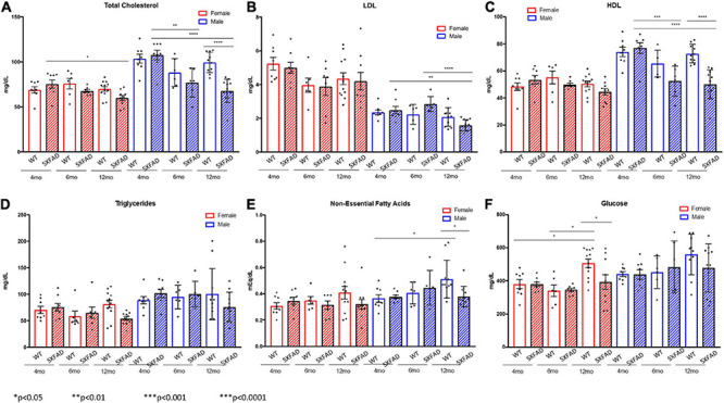
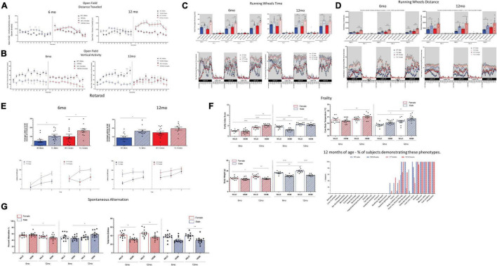
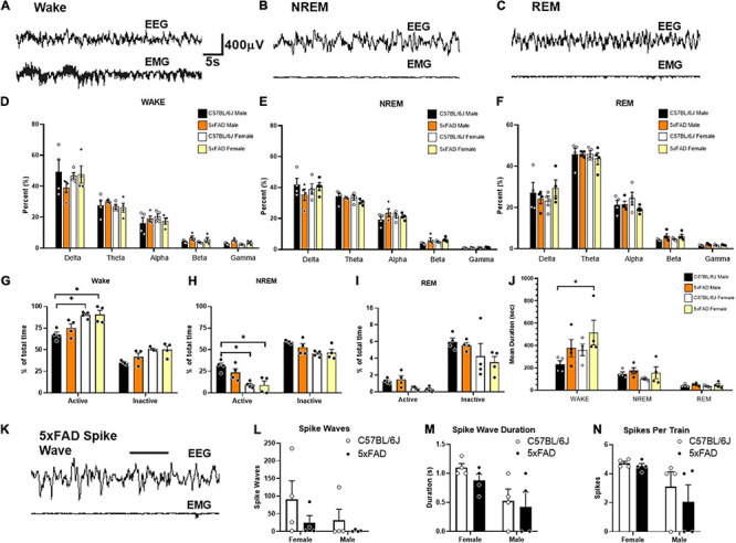
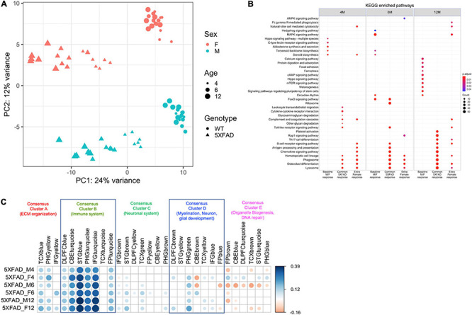
The ability to investigate therapeutic interventions in animal models of neurodegenerative diseases depends on extensive characterization of the model(s) being used. There are numerous models that have been generated to study Alzheimer’s disease (AD) and the underlying pathogenesis of the disease. While transgenic models have been instrumental in understanding AD mechanisms and risk factors, they are limited in the degree of characteristics displayed in comparison with AD in humans, and the full spectrum of AD effects has yet to be recapitulated in a single mouse model. The Model Organism Development and Evaluation for Late-Onset Alzheimer’s Disease (MODEL-AD) consortium was assembled by the National Institute on Aging (NIA) to develop more robust animal models of AD with increased relevance to human disease, standardize the characterization of AD mouse models, improve preclinical testing in animals, and establish clinically relevant AD biomarkers, among other aims toward enhancing the translational value of AD models in clinical drug design and treatment development. Here we have conducted a detailed characterization of the 5XFAD mouse, including transcriptomics, electroencephalogram, in vivo imaging, biochemical characterization, and behavioral assessments. The data from this study is publicly available through the AD Knowledge Portal.
Overview
The claims and statistics of this paper haven’t been checked yet.
Run the check — free, no account needed. It reads the open-access text and checks the sources it cites.
Integrity signals
Registry
Cites
Statistics
Figures
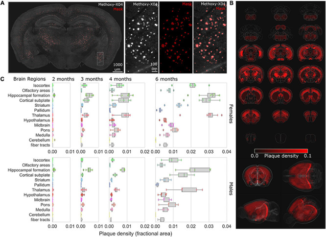
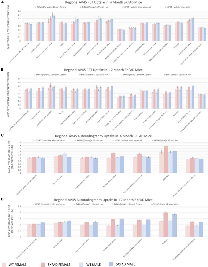
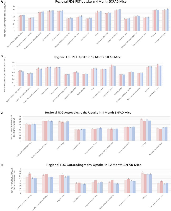
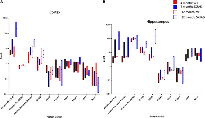
Verification
Claims the paper makes
The claims and statistics of this paper haven’t been checked yet.
Run the check — free, no account needed. It reads the open-access text and checks the sources it cites.
Claims other papers make about this one
Each is checked against this paper’s own text: does it say what the citing paper says it does?
The source backs part of the claim, not all of it — or backs it for a different population, condition or comparison.
In the citing paper
“In 5xFAD mice, amyloid plaques are first observed between two and four months of age in the cortical layer V and in the subiculum of the hippocampal formation (Oblak et al., 2021).”
From Age-dependent microstructure alterations in 5xFAD mice by high-resolution diffusion tensor imaging (2022) · cites this paper as Oblak et al., 2021
Experimental — accuracy not yet measuredThe source backs part of the claim, not all of it — or backs it for a different population, condition or comparison.
In the citing paper
“Given the robust Aβ deposition in the subiculum in 5xFAD animals, 16 we selected this region to analyze the morphology of diffuse (6E10 + , green) and dense-core (X34 + , blue) plaques, along with neuronal abundance using the neuronal nuclear protein (NeuN, red) in matched brain sections (Figure 2A).”
From INPP5D deficiency attenuates amyloid pathology in a mouse model of Alzheimer's disease (2023) · cites this paper as [16]
Experimental — accuracy not yet measuredThe source backs part of the claim, not all of it — or backs it for a different population, condition or comparison.
In the citing paper
“Our positive findings contrast sharply with negative findings of the MODEL-AD consortium16 and other recent studies that have also used T1/T2 MRI and DTI methods7,44 (Supplementary Tables 2 and 3).”
From Magnetic resonance microscopy maps widespread effects of Alzheimer’s disease on brain structures and behavior in mice (2026) · cites this paper as [16]
Experimental — accuracy not yet measuredThe source backs part of the claim, not all of it — or backs it for a different population, condition or comparison.
In the citing paper
“This raises the possibility that rostral-to-caudal effects we have detected are downstream of regional differences in promoter activity. This can probably be ruled out because many teams have shown strong expression of the transgene and high levels of amyloid plaque deposition in thalamus and midbrain81—regions in which we have demonstrated highly significant reductions in volume (Ref20, their”
From Magnetic resonance microscopy maps widespread effects of Alzheimer’s disease on brain structures and behavior in mice (2026) · cites this paper as [16]
Experimental — accuracy not yet measuredThe source backs part of the claim, not all of it — or backs it for a different population, condition or comparison.
In the citing paper
“Despite the aggressive rate of pathological Aβ aggregation and lysosomal disruption in the 5XFAD and other FAD mice, they do not display robust ageor pathology-associated cognitive deficits on hippocampal and non-hippocampal memory tasks (Jankowsky & Zheng, 2017;Oblak et al., 2021)”
From Hippocampal TMEM55B overexpression in the 5XFAD mouse model of Alzheimer's disease (2024) · cites this paper as Oblak et al., 2021
Experimental — accuracy not yet measured
The cited source says what the claim attributes to it.
In the citing paper
“Given the reported sex differences in some disease pathology phenotypes46,47,62, mice from both sexes were used unless otherwise specified. When both male and female mice were used, an analysis for sex differences was performed; unless otherwise noted, no sex differences were observed.”
From Lymphoid gene expression supports neuroprotective microglia function (2025) · cites this paper as [47]
The cited source says what the claim attributes to it.
In the citing paper
“5xFAD mice also develop prominent gliosis, inflammation, neuronal loss, behavioral impairments, and sex-distinct systemic metabolic changes39,38,40.”
From Amyloid-β induces lipid droplet-mediated microglial dysfunction via the enzyme DGAT2 in Alzheimer’s disease (2025) · cites this paper as [39]
The cited source says what the claim attributes to it.
In the citing paper
“Previous deep phenotyping of 5XFAD mice highlights the difficulty of modeling AD-like memory deficits in 5XFAD mice despite recapitulation of other AD-like phenotypes (Forner et al., 2015;Oblak et al., 2021).”
From Hippocampal TMEM55B overexpression in the 5XFAD mouse model of Alzheimer's disease (2024) · cites this paper as Oblak et al., 2021
In the citing paper
“at 2 months of age to intervene in adult mice at the very earliest stages of Aβ plaque development (Forner et al., 2015;Oblak et al., 2021).”
From Hippocampal TMEM55B overexpression in the 5XFAD mouse model of Alzheimer's disease (2024) · cites this paper as Oblak et al., 2021
Self-check
Percentages, sums and reported differences re-computed from the paper’s own numbers.
The self-check needs the paper’s full text.
Citations
Cites
Moderated estimation of fold change and dispersion for RNA-seq data with DESeq2
Love, M. I., Huber, W., & Anders, S. (2014) · Genome Biology · doi:10.1186/s13059-014-0550-8 · Cited by 95,106
STAR: ultrafast universal RNA-seq aligner
Dobin, A., Davis, C. A., Schlesinger, F., Drenkow, J., Zaleski, C., Jha, S., Batut, P., Chaisson, M., & Gingeras, T. R. (2013) · Bioinformatics · doi:10.1093/bioinformatics/bts635 · Cited by 51,709
clusterProfiler: an R Package for Comparing Biological Themes Among Gene Clusters
Yu, G., Wang, L.-G., Han, Y., & He, Q.-Y. (2012) · OMICS: A Journal of Integrative Biology · doi:10.1089/omi.2011.0118 · Cited by 36,259
Frailty Consensus: A Call to Action
Morley, J. E., Vellas, B. J., van Kan, G. A., Anker, S. D., Bauer, J. M., Bernabei, R., Cesari, M., Chumlea, W. C., Doehner, W., Evans, J., et al. (2013) · Journal of the American Medical Directors Association · doi:10.1016/j.jamda.2013.03.022 · Cited by 4,201
Association of apolipoprotein E allele ϵ4 with late‐onset familial and sporadic Alzheimer's disease
Saunders, A. M., Strittmatter, W. J., Schmechel, D., St. George-Hyslop, P. H., Pericak-Vance, M. A., Joo, S. H., Rosi, B. L., Gusella, J. F., Crapper-MacLachlan, D. R., Alberts, M. J., et al. (1993) · Neurology · doi:10.1212/wnl.43.8.1467 · Cited by 3,786
Oakley, H., Cole, S. L., Logan, S., Maus, E., Shao, P., Craft, J., Guillozet-Bongaarts, A., Ohno, M., Disterhoft, J., Van Eldik, L., et al. (2006) · The Journal of Neuroscience · doi:10.1523/jneurosci.1202-06.2006 · Cited by 3,530
Alzheimer disease in the United States (2010–2050) estimated using the 2010 census
Hebert, L. E., Weuve, J., Scherr, P. A., & Evans, D. A. (2013) · Neurology · doi:10.1212/wnl.0b013e31828726f5 · Cited by 2,642
A mesoscale connectome of the mouse brain
Oh, S. W., Harris, J. A., Ng, L., Winslow, B., Cain, N., Mihalas, S., Wang, Q., Lau, C., Kuan, L., Henry, A. M., et al. (2014) · Nature · doi:10.1038/nature13186 · Cited by 2,612
Alzheimer’s disease drug-development pipeline: Few candidates, frequent failures
Cummings, J. L., Morstorf, T., & Zhong, K. (2014) · Alzheimer's Research & Therapy · doi:10.1186/alzrt269 · Cited by 1,744
The Allen Mouse Brain Common Coordinate Framework: A 3D Reference Atlas
Wang, Q., Ding, S., Li, Y., Royall, J. J., Feng, D. H., Lesnar, P., Graddis, N., Naeemi, M., Facer, B. A. C., Ho, A., et al. (2020) · Cell · doi:10.1016/j.cell.2020.04.007 · Cited by 1,464
Cited by
Sorted by citations
Restoring hippocampal glucose metabolism rescues cognition across Alzheimer’s disease pathologies
Minhas, P. S., Jones, J. R., Latif‐Hernandez, A., Sugiura, Y., Durairaj, A. S., Wang, Q., Mhatre, S. D., Uenaka, T., Crapser, J. D., Conley, T. E., et al. (2024) · Science · doi:10.1126/science.abm6131 · Cited by 244
Updates on mouse models of Alzheimer’s disease
Zhong, M. Z., Peng, T., Duarte, M. L., Wang, M., & Cai, D. (2024) · Molecular Neurodegeneration · doi:10.1186/s13024-024-00712-0 · Cited by 189
Spatial proteomics in three-dimensional intact specimens
Bhatia, H. S., Brunner, A.-D., Öztürk, F., Kapoor, S., Rong, Z., Mai, H., Thielert, M., Ali, M., Al-Maskari, R., Paetzold, J. C., et al. (2022) · Cell · doi:10.1016/j.cell.2022.11.021 · Cited by 129
Gamma Oscillations in Alzheimer’s Disease and Their Potential Therapeutic Role
Traikapi, A., & Konstantinou, N. (2021) · Frontiers in Systems Neuroscience · doi:10.3389/fnsys.2021.782399 · Cited by 123
Prakash, P., Manchanda, P., Paouri, E., Bisht, K., Sharma, K., Rajpoot, J., Wendt, V., Hossain, A., Wijewardhane, P. R., Randolph, C. E., et al. (2025) · Immunity · doi:10.1016/j.immuni.2025.04.029 · Cited by 104
Metadata
- Authors
- Oblak, A. L., Lin, P. B., Kotredes, K. P., Pandey, R. S., Garceau, D., Williams, H. M., Uyar, A., O’Rourke, R., O’Rourke, S., Ingraham, C., Bednarczyk, D., Belanger, M., Cope, Z. A., Little, G. J., Williams, S.-P. G., Ash, C., Bleckert, A., Ragan, T., Logsdon, B. A., Mangravite, L. M., Sukoff Rizzo, S. J., Territo, P. R., Carter, G. W., Howell, G. R., Sasner, M., & Lamb, B. T.
- Affiliations
- Indiana University School of Medicine (US)
- Jackson Laboratory (US)
- University of Pittsburgh (US)
- The Jackson Laboratory for Genomic Medicine (US)
- Sage Bionetworks (US)
- Corresponding author
- Oblak, Adrian L.
- ORCID
- Venue
- Frontiers in Aging Neuroscience
- Publisher
- Frontiers Media SA
- Published
- 2021-07-23
- Volume
- 13
- Pages
- 713726-713726
- ISSN
- 1663-4365 (ISSN-L)
- PMC
- PMC8346252
- OpenAlex
- W3184377705
- Cited by
- 325
- References
- 92
- Citations by year
- 2026: 65 · 2025: 73 · 2024: 72 · 2023: 66 · 2022: 45 · 2021: 4
- FWCI
- 29.69
- Type
- journal article
- Language
- en
- Open access
- gold
- doi.org
- Licence
- Locations
- Frontiers in Aging Neuroscience (publishedVersion, cc-by)
- PubMed Central (publishedVersion, cc-by)
- PubMed (publishedVersion)
- DOAJ (DOAJ: Directory of Open Access Journals) (submittedVersion, cc-by-sa)
- The Mouseion at the JAXlibrary (Jackson Laboratory) (submittedVersion, cc-by)
- IUScholarWorks (Indiana University) (submittedVersion, cc-by)
- Purdue University Indianapolis (Indiana University) (submittedVersion, cc-by)
- Funders
- National Institute on Aging (U54AG054345, K01AG054753)
- Topics
- Life Sciences › Neuroscience › Cellular and Molecular Neuroscience › Alzheimer's disease research and treatments
- Health Sciences › Medicine › Psychiatry and Mental health › Dementia and Cognitive Impairment Research
- Life Sciences › Neuroscience › Neurology › Neuroinflammation and Neurodegeneration Mechanisms
- Keywords
- Alzheimer's disease, transgenic mouse model, preclinical drug testing, model validation, transcriptomics, EEG, in vivo imaging, behavioral assessment, biomarkers, translational research
About these checks
Registry, retraction, Cites and statistics results are computed, not judged, with one exception: a printed reference that no index lists is read once by a model before it counts as a citation error. Claims are extracted by a model and kept only when their quote appears verbatim in the text; each cited source is then checked, with a model’s help, for whether it says what the claim relies on. Those verdicts can be wrong — read the claim’s quote and the cited source before relying on one.
Statistics and arithmetic are recomputed, not judged.
Confidence bands are shown only for fields calibrated on our annotated test set. None is calibrated yet, so no band is shown.
What counts as a claim ›How we measure confidence ›How we check this ›
Are you an author, or do you see a problem with this page? Request a review
