

The DOI matches its Crossref record; 30 of the 31 references it cites were found in a registry or an index.
Different scaling of white matter volume, cortical connectivity, and gyrification across rodent and primate brains
Open accessAbstract
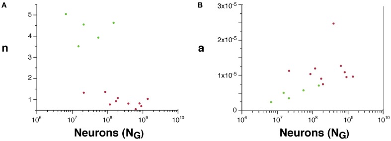
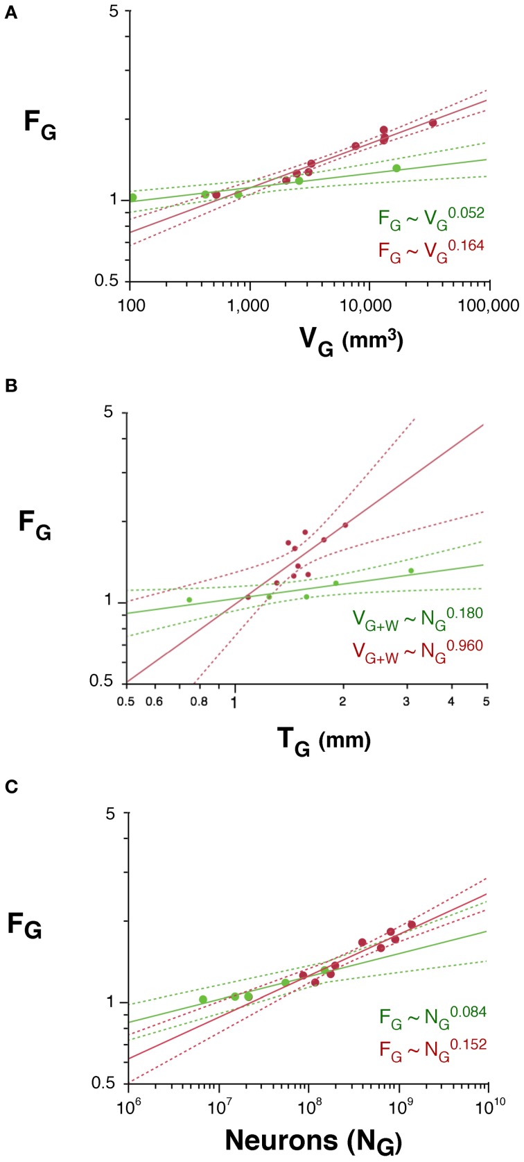
Expansion of the cortical gray matter in evolution has been accompanied by an even faster expansion of the subcortical white matter volume and by folding of the gray matter surface, events traditionally considered to occur homogeneously across mammalian species. Here we investigate how white matter expansion and cortical folding scale across species of rodents and primates as the gray matter gains neurons. We find very different scaling rules of white matter expansion across the two orders, favoring volume conservation and smaller propagation times in primates. For a similar number of cortical neurons, primates have a smaller connectivity fraction and less white matter volume than rodents; moreover, as the cortex gains neurons, there is a much faster increase in white matter volume and in its ratio to gray matter volume in rodents than in primates. Order-specific scaling of the white matter can be attributed to different scaling of average fiber caliber and neuronal connectivity in rodents and primates. Finally, cortical folding increases as different functions of the number of cortical neurons in rodents and primates, scaling faster in the latter than in the former. While the neuronal rules that govern gray and white matter scaling are different across rodents and primates, we find that they can be explained by the same unifying model, with order-specific exponents. The different scaling of the white matter has implications for the scaling of propagation time and computational capacity in evolution, and calls for a reappraisal of developmental models of cortical expansion in evolution.
Overview
The claims and statistics of this paper haven’t been checked yet.
Run the check — free, no account needed. It reads the open-access text and checks the sources it cites.
Integrity signals
Registry
Cites
Statistics
Figures
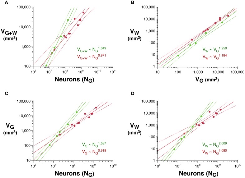
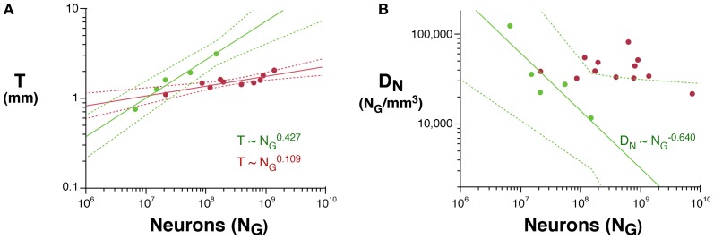
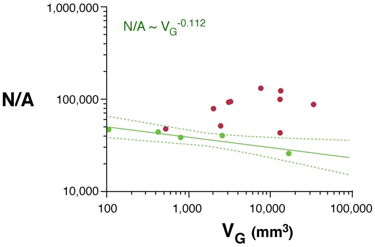
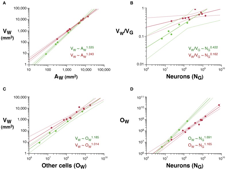
Verification
Claims the paper makes
The claims and statistics of this paper haven’t been checked yet.
Run the check — free, no account needed. It reads the open-access text and checks the sources it cites.
Claims other papers make about this one
Each is checked against this paper’s own text: does it say what the citing paper says it does?
The source backs part of the claim, not all of it — or backs it for a different population, condition or comparison.
In the citing paper
“The field has typically relied on mouse models to gain understanding of the impact of T21, but differences in cortical development between mice and humans — including the complexity of progenitor types or the formation of a lissencephalic rather than gyrencephalic brain153–155 — limit their ability to define mechanisms relevant to the human phenotype.”
From Consequences of trisomy 21 for brain development in Down syndrome (2024) · cites this paper as [155]
Experimental — accuracy not yet measured
Self-check
Percentages, sums and reported differences re-computed from the paper’s own numbers.
The self-check needs the paper’s full text.
Citations
Cites
Collective dynamics of ‘small-world’ networks
Watts, D. J., & Strogatz, S. H. (1998) · Nature · doi:10.1038/30918 · Cited by 43,060
Azevedo, F. A. C., Carvalho, L. R. B., Grinberg, L. T., Farfel, J. M., Ferretti, R. E. L., Leite, R. E. P., Filho, W. J., Lent, R., & Herculano‐Houzel, S. (2009) · Journal of Comparative Neurology · doi:10.1002/cne.21974 · Cited by 2,337
Self-citationThe human brain in numbers: a linearly scaled-up primate brain
Herculano-Houzel, S. (2009) · Frontiers in Human Neuroscience · doi:10.3389/neuro.09.031.2009 · Cited by 1,778 · cited 1×
“…have more folded cerebral cortices primates with similarly sized brains (Pillay and Manger, 2007), they probably have not more but rather far fewer cortical neurons than primates, due to much lower neuronal densities (Tower, 1954; Herculano-Houzel, 2009; and our unpublished observations).”
BackgroundSelf-citationThe Small World of the Cerebral Cortex
Sporns, O., & Zwi, J. D. (2004) · Neuroinformatics · doi:10.1385/ni:2:2:145 · Cited by 1,378
Collective dynamics of 'small-world' networks
Watts, D. J., & Strogatz, S. H. (2011) · Princeton University Press eBooks · doi:10.1515/9781400841356.301 · Cited by 1,087
THE BASIC UNIFORMITY IN STRUCTURE OF THE NEOCORTEX
Röckel, A., Hiorns, R. W., & Powell, T. P. S. (1980) · Brain · doi:10.1093/brain/103.2.221 · Cited by 890
Sporns, O., & Kötter, R. (2004) · PLoS Biology · doi:10.1371/journal.pbio.0020369 · Cited by 812
A universal scaling law between gray matter and white matter of cerebral cortex
Zhang, K., & Sejnowski, T. J. (2000) · Proceedings of the National Academy of Sciences · doi:10.1073/pnas.090504197 · Cited by 723
: Cellular scaling rules for rodent brains.
Herculano-Houzel, S., Mota, B., & Lent, R. (2006) · Proceedings of the National Academy of Sciences · doi:10.1073/pnas.0604911103 · Cited by 579
Self-citationHerculano-Houzel, S., & Lent, R. (2005) · The Journal of Neuroscience · doi:10.1523/jneurosci.4526-04.2005 · Cited by 551
Self-citationCellular scaling rules for primate brains.
Herculano-Houzel, S., Collins, C. E., Wong, P., & Kaas, J. H. (2007) · Proceedings of the National Academy of Sciences · doi:10.1073/pnas.0611396104 · Cited by 422
Self-citationControl of oligodendrocyte number in the developing rat optic nerve
Barres, B. A., & RAFF, M. C. (1994) · Neuron · doi:10.1016/0896-6273(94)90305-0 · Cited by 393
Axonal Control of Oligodendrocyte Development
Barres, B. A., & Raff, M. C. (1999) · The Journal of Cell Biology · doi:10.1083/jcb.147.6.1123 · Cited by 360
Functional Trade-Offs in White Matter Axonal Scaling
Wang, S. S. ‐., Shultz, J. R., Burish, M. J., Harrison, K. H., Hof, P. R., Towns, L. C., WAGERS, M. W., & Wyatt, K. D. (2008) · Journal of Neuroscience · doi:10.1523/jneurosci.5559-05.2008 · Cited by 304
Size and Shape of the Cerebral Cortex in Mammals
Hofman, M. A. (2008) · Brain Behavior and Evolution · doi:10.1159/000116529 · Cited by 257
The basic nonuniformity of the cerebral cortex
Herculano‐Houzel, S., Collins, C. E., Wong, P., Kaas, J. H., & Lent, R. (2008) · Proceedings of the National Academy of Sciences · doi:10.1073/pnas.0805417105 · Cited by 206
Self-citationTower, D. B. (1954) · Journal of Comparative Neurology · doi:10.1002/cne.901010103 · Cited by 203
Not All Brains Are Made the Same: New Views on Brain Scaling in Evolution
Herculano‐Houzel, S. (2011) · Brain Behavior and Evolution · doi:10.1159/000327318 · Cited by 183
Self-citationConnectivity-driven white matter scaling and folding in primate cerebral cortex
Herculano‐Houzel, S., Mota, B., Wong, P., & Kaas, J. H. (2010) · Proceedings of the National Academy of Sciences · doi:10.1073/pnas.1012590107 · Cited by 166
Self-citation(2011) · doi:10.1159/000330825 · Cited by 138
Self-citationPrinciples underlying mammalian neocortical scaling
Changizi, M. A. (2001) · Biological Cybernetics · doi:10.1007/s004220000205 · Cited by 137
Order‐specific quantitative patterns of cortical gyrification
Pillay, P., & Manger, P. R. (2007) · European Journal of Neuroscience · doi:10.1111/j.1460-9568.2007.05524.x · Cited by 130
Cellular Scaling Rules for the Brains of an Extended Number of Primate Species
Gabi, M., Collins, C. E., Wong, P., Torres, L. B., Kaas, J. H., & Herculano-Houzel, S. (2010) · Brain, Behavior and Evolution · doi:10.1159/000319872 · Cited by 102
Self-citationNeuronal scaling rules for primate brains
Herculano‐Houzel, S. (2012) · Progress in brain research · doi:10.1016/b978-0-444-53860-4.00015-5 · Cited by 82
Self-citationMota, B., & Herculano‐Houzel, S. (2012) · Frontiers in Neuroanatomy · doi:10.3389/fnana.2012.00003 · Cited by 80
Self-citationManger, P. R., Prowse, M., Haagensen, M., & Hemingway, J. (2012) · The Journal of Comparative Neurology · doi:10.1002/cne.23046 · Cited by 63
First-order transition in small-world networks
de Menezes, M. A., Moukarzel, C. F., & Penna, T. J. P. (2000) · Europhysics Letters (EPL) · doi:10.1209/epl/i2000-00308-1 · Cited by 53
Karbowski, J. (2003) · Journal of Computational Neuroscience · doi:10.1023/a:1027467911225 · Cited by 48
Scaling of cortical neuron density and white matter volume in mammals.
Prothero, J. W. (1997) · PubMed · Cited by 35
Axonal control of oligodendrocyte development
Barres, B. A., & RAFF, M. C. (1997) · Oxford University Press eBooks · doi:10.1093/acprof:oso/9781872748542.003.0004 · Cited by 13
Research works printed but not linked: 2
- Le Gros Clark W. (1945). Deformation patterns on the cerebral cortex, in Essays on Growth and Form, eds Le Gros Clark W. E. Medawar P. B. (Oxford: Oxford University Press), 1– 23
- Welker W. (1990). Why does cerebral cortex fissure and fold? A review of determinants of gyri and sulci, in Cerebral Cortex, Comparative structure and evolution of cerebral cortex, part II, Vol. 8B, eds Jones E. G. Peters A. (New York, NY: Plenum Press), 3– 136
Cited by
Sorted by citations
Phillips, K. A., Bales, K. L., Capitanio, J. P., Conley, A. J., Czoty, P. W., Hart, B. A. ‘., Hopkins, W. D., Hu, S., Miller, L. A., Nader, M. A., et al. (2014) · American Journal of Primatology · doi:10.1002/ajp.22281 · Cited by 649
Cortical folding scales universally with surface area and thickness, not number of neurons
Mota, B., & Herculano‐Houzel, S. (2015) · Science · doi:10.1126/science.aaa9101 · Cited by 286
Self-citation(2014) · doi:10.3389/fnana.2014.00077 · Cited by 206
Self-citationGămănuț, R., Kennedy, H., Toroczkai, Z., Ercsey-Ravasz, M., Essen, D. C. V., Knoblauch, K., & Burkhalter, A. (2018) · Neuron · doi:10.1016/j.neuron.2017.12.037 · Cited by 215
Herculano-Houzel, S., Avelino-de-Souza, K., Neves, K., PorfÃrio, J., Messeder, D., Mattos Feijó, L., Maldonado, J., & Manger, P. R. (2014) · Frontiers in Neuroanatomy · doi:10.3389/fnana.2014.00046 · Cited by 153
Self-citation
Metadata
- Authors
- Ventura-Antunes, L., Mota, B., & Herculano-Houzel, S.
- Affiliations
- Universidade Federal do Rio de Janeiro (BR)
- ORCID
- Mota, Bruno
- Venue
- Frontiers in Neuroanatomy
- Publisher
- Frontiers Media SA
- Published
- 2013
- Volume
- 7
- Pages
- 3-3
- ISSN
- 1662-5129 (ISSN-L)
- PMC
- PMC3620553
- OpenAlex
- W2044032178
- Cited by
- 129
- References
- 31
- Citations by year
- 2026: 8 · 2025: 12 · 2024: 13 · 2023: 7 · 2022: 13 · 2021: 8 · 2020: 12 · 2019: 7 · 2018: 6 · 2017: 12 · 2016: 15 · 2015: 8 · 2014: 6 · 2013: 2
- FWCI
- 8.02
- Type
- journal article
- Language
- en
- Open access
- gold
- doi.org
- Licence
- cc-by
- Locations
- Frontiers in Neuroanatomy (publishedVersion, cc-by)
- Europe PMC (PubMed Central) (publishedVersion, cc-by)
- PubMed Central (publishedVersion, cc-by)
- PubMed (publishedVersion)
- DOAJ (DOAJ: Directory of Open Access Journals) (submittedVersion, cc-by-sa)
- Funders
- James S. McDonnell Foundation
- Coordenação de Aperfeiçoamento de Pessoal de Nível Superior
- Conselho Nacional de Desenvolvimento Científico e Tecnológico
- Fundação Carlos Chagas Filho de Amparo à Pesquisa do Estado do Rio de Janeiro
- Topics
- Health Sciences › Medicine › Radiology, Nuclear Medicine and Imaging › Advanced Neuroimaging Techniques and Applications
- Life Sciences › Agricultural and Biological Sciences › Ecology, Evolution, Behavior and Systematics › Primate Behavior and Ecology
- Life Sciences › Neuroscience › Developmental Neuroscience › Neurogenesis and neuroplasticity mechanisms
- Keywords
- white matter volume, cortical folding, cortical connectivity, primate brain evolution, propagation time, cortical expansion, scaling exponents, brain evolution
About these checks
Registry, retraction, Cites and statistics results are computed, not judged, with one exception: a printed reference that no index lists is read once by a model before it counts as a citation error. Claims are extracted by a model and kept only when their quote appears verbatim in the text; each cited source is then checked, with a model’s help, for whether it says what the claim relies on. Those verdicts can be wrong — read the claim’s quote and the cited source before relying on one.
Statistics and arithmetic are recomputed, not judged.
Confidence bands are shown only for fields calibrated on our annotated test set. None is calibrated yet, so no band is shown.
What counts as a claim ›How we measure confidence ›How we check this ›
Are you an author, or do you see a problem with this page? Request a review
