Cited by 104Neuroscience and Neuropharmacology ResearchAmino Acid Enzymes and MetabolismNicotinic Acetylcholine Receptors Study

The DOI matches its Crossref record; 56 of the 56 references it cites were found in a registry or an index.

GluN2D-containing NMDA receptors-mediate synaptic currents in hippocampal interneurons and pyramidal cells in juvenile mice

Open access
Engelhardt, Jakob von, Bocklisch, Christina, Tönges, Lars, Herb, Anne, Mishina, Masayoshi, Monyer, Hannah·2015·Frontiers in Cellular Neuroscience

Abstract

The differential regulation of the two major N-methyl-D-aspartate receptor (NMDAR) subunits GluN2A and GluN2B during development in forebrain pyramidal cells has been thoroughly investigated. In contrast, much less is known about the role of GluN2D, which is expressed at low levels and is downregulated following the second postnatal week. However, it appears that few cells, presumably interneurons, continue to express GluN2D also in juvenile mice. To investigate which hippocampal cell types express this subunit, we generated transgenic mice with EGFP-tagged GluN2D receptors. The expression of the transgene was confined to hippocampal interneurons, most of which were parvalbumin- and/or somatostatin-positive. Electrophysiological and morphological analyses showed that GluN2D was present mainly in fast spiking basket and axo-axonic cells. Based on pharmacological evidence and electrophysiological analysis of GluN2D knockout mice, we conclude that GluN2D-containing NMDARs mediate synaptic currents in hippocampal interneurons of young and juvenile mice and in CA1 pyramidal neurons of newborn mice.

Overview

The claims and statistics of this paper haven’t been checked yet.

Run the check — free, no account needed. It reads the open-access text and checks the sources it cites.

A few free checks a day · papers already verified open instantly · results are public

Integrity signals

Registry

Crossref · 10.3389/fncel.2015.00095

Cites

56 of 56 references verified0 citation errors · 0 not checkable

Statistics

Figures

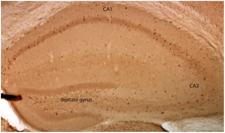
Figure 1 GluN2D-EGFP is expressed in the hippocampus. Visualization of EGFP-GluN2D in the hippocampus with an anti-EGFP antibody and DAB chromogen. EGFP-GluN2D-positive cells are localized in the CA1, CA2, dentate gyrus, and CA3 subregion. Most positive cells are close to or within the principal cell layer.
Figure 1 GluN2D-EGFP is expressed in the hippocampus. Visualization of EGFP-GluN2D in the hippocampus with an anti-EGFP antibody and DAB chromogen. EGFP-GluN2D-positive cells are localized in the CA1, CA2, dentate gyrus, and CA3 subregion. Most positive cells are close to or within the principal cell layer.CC BY · PubMed Central
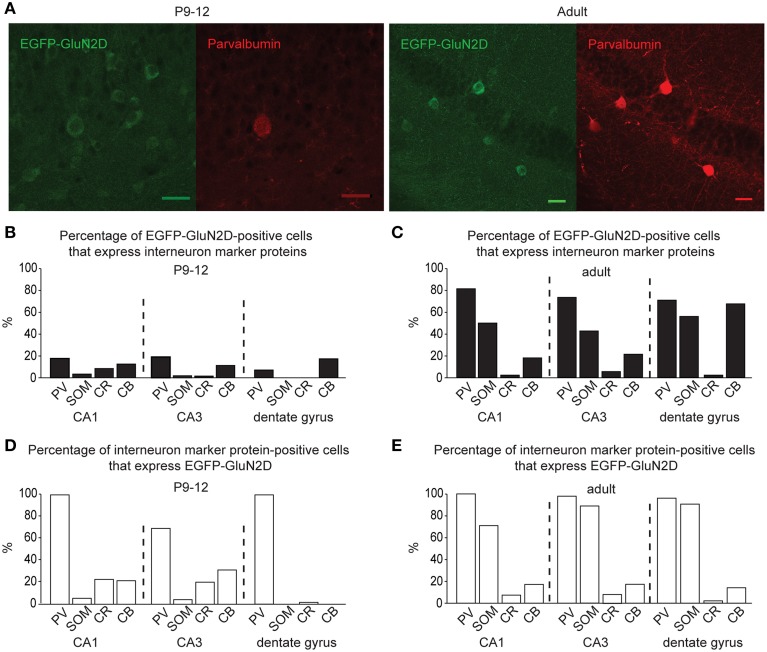
Figure 2 GluN2D-EGFP is expressed in hippocampal interneurons. (A) Immunohistochemical images of hippocampal EGFP-GluN2D-positive cells co-expressing PV in brains of P12 and adult mice. Scale bars = 30μm. (B,C) Quantification of EGFP-GluN2D-positive cells that express with PV, SOM, CR and CB in CA1, CA3 and dentate gyrus in P9-12 (B) and adult mice (C), respectively. (D,E) Quantification of the percentage of interneuron marker-positive cells that are EGFP-GluN2D-positive in CA1, CA3, and dentate gyrus in P9-12 (D) and adult mice (E), respectively.
Figure 2 GluN2D-EGFP is expressed in hippocampal interneurons. (A) Immunohistochemical images of hippocampal EGFP-GluN2D-positive cells co-expressing PV in brains of P12 and adult mice. Scale bars = 30μm. (B,C) Quantification of EGFP-GluN2D-positive cells that express with PV, SOM, CR and CB in CA1, CA3 and dentate gyrus in P9-12 (B) and adult mice (C), respectively. (D,E) Quantification of the percentage of interneuron marker-positive cells that are EGFP-GluN2D-positive in CA1, CA3, and dentate gyrus in P9-12 (D) and adult mice (E), respectively.CC BY · PubMed Central
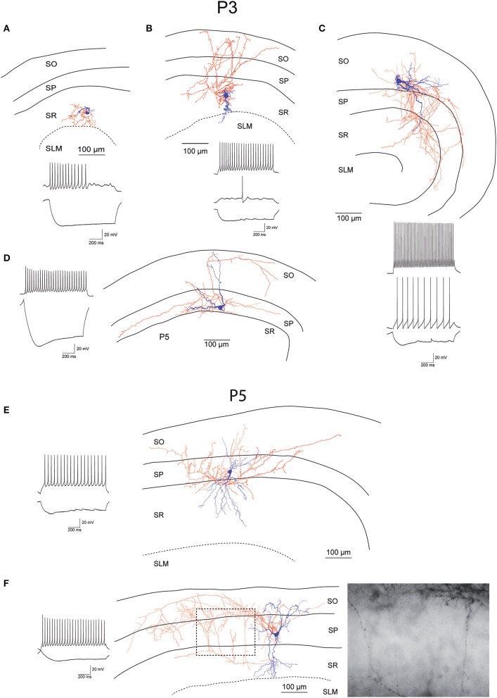
Figure 3 Morphology and firing pattern of GluN2D-EGFP-positive cells in P3-5 transgenic mice. (A–D) Reconstruction of four EGFP-GluN2D-positive cells (P3) with voltage responses to hyperpolarizing and depolarizing current injections. (E,F) Reconstruction of two EGFP-GluN2D-positive cells (P5) with voltage responses to hyperpolarizing and depolarizing current injections. Localization of cell body, dendritic and axonal arbors indicate that the two neurons may be an immature basket (upper) and an immature axo-axonic cell. The image of part of the axon in the stratum pyramidale of the lower cell (see dashed box in reconstruction) shows vertical rows of boutons (parallel to axon initial segments), typical for axo-axonic cells. SO, Stratum oriens; SP, stratum pyramidale; SR, stratum radiatum; SLM, stratum lacunosum-moleculare.
Figure 3 Morphology and firing pattern of GluN2D-EGFP-positive cells in P3-5 transgenic mice. (A–D) Reconstruction of four EGFP-GluN2D-positive cells (P3) with voltage responses to hyperpolarizing and depolarizing current injections. (E,F) Reconstruction of two EGFP-GluN2D-positive cells (P5) with voltage responses to hyperpolarizing and depolarizing current injections. Localization of cell body, dendritic and axonal arbors indicate that the two neurons may be an immature basket (upper) and an immature axo-axonic cell. The image of part of the axon in the stratum pyramidale of the lower cell (see dashed box in reconstruction) shows vertical rows of boutons (parallel to axon initial segments), typical for axo-axonic cells. SO, Stratum oriens; SP, stratum pyramidale; SR, stratum radiatum; SLM, stratum lacunosum-moleculare.CC BY · PubMed Central
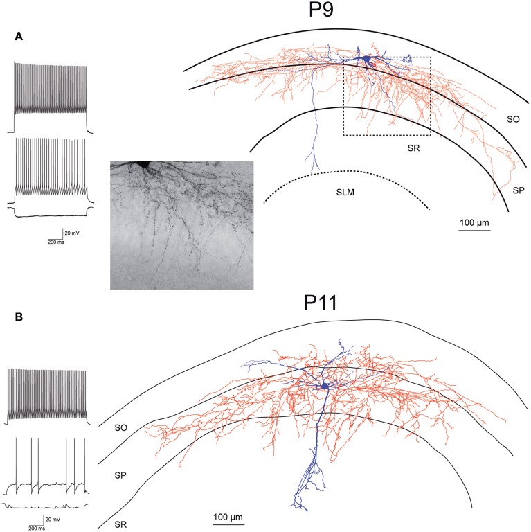
Figure 4 Morphology and firing pattern of GluN2D-EGFP-positive cells in P9-11 transgenic mice. (A) Reconstruction of an EGFP-GluN2D-positive cell (P9) with voltage responses to hyperpolarizing and depolarizing current injections. Firing pattern, localization of cell body, dendritic, and especially the axonal arbor at the border of SO and SP indicate that this is a fast-spiking axo-axonic cell. The image of part of the axon in the stratum pyramidale (see dashed box in reconstruction) shows vertical rows of boutons (parallel to axon initial segments), typical for axo-axonic cells. (B) Reconstruction of an EGFP-GluN2D-positive cell (P11) with voltage responses to hyperpolarizing and depolarizing current injections. Firing pattern and morphology (e.g., axonal arbor in SP) indicate that this is a fast-spiking basket cell. SO, Stratum oriens; SP, stratum pyramidale; SR, stratum radiatum; SLM, stratum lacunosum-moleculare.
Figure 4 Morphology and firing pattern of GluN2D-EGFP-positive cells in P9-11 transgenic mice. (A) Reconstruction of an EGFP-GluN2D-positive cell (P9) with voltage responses to hyperpolarizing and depolarizing current injections. Firing pattern, localization of cell body, dendritic, and especially the axonal arbor at the border of SO and SP indicate that this is a fast-spiking axo-axonic cell. The image of part of the axon in the stratum pyramidale (see dashed box in reconstruction) shows vertical rows of boutons (parallel to axon initial segments), typical for axo-axonic cells. (B) Reconstruction of an EGFP-GluN2D-positive cell (P11) with voltage responses to hyperpolarizing and depolarizing current injections. Firing pattern and morphology (e.g., axonal arbor in SP) indicate that this is a fast-spiking basket cell. SO, Stratum oriens; SP, stratum pyramidale; SR, stratum radiatum; SLM, stratum lacunosum-moleculare.CC BY · PubMed Central
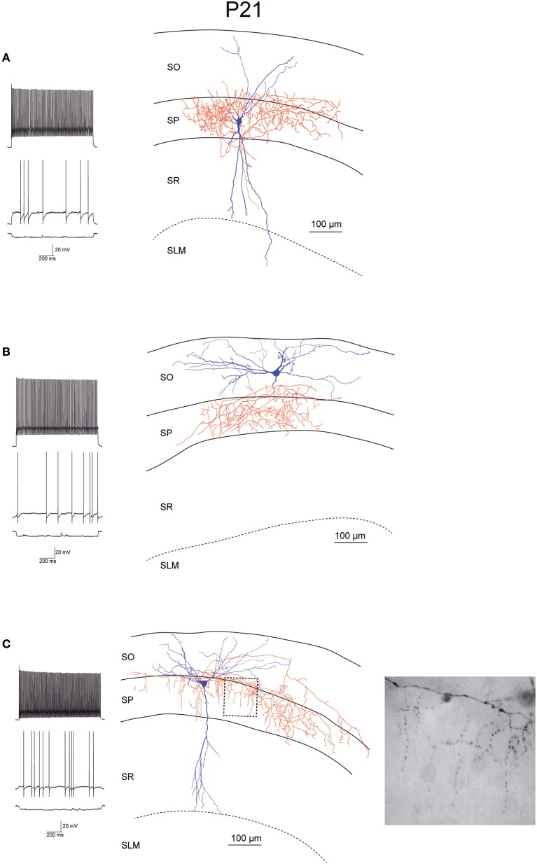
Figure 5 Morphology and firing pattern of GluN2D-EGFP-positive cells in P21 transgenic mice. (A–C) Reconstruction of three EGFP-GluN2D-positive cells (P21) with voltage responses to hyperpolarizing and depolarizing current injections. All three cells display fast spiking firing patterns. The axonal arbors of the upper two cells (A,B) are consistent with basket cells while the lower cell (C) resembles an axo-axonic cell. The image of part of the axon in the stratum pyramidale of the lower cell (see dashed box in reconstruction) shows vertical rows of boutons (parallel to axon initial segments), typical for axo-axonic cells. SO, Stratum oriens; SP, stratum pyramidale; SR, stratum radiatum; SLM, stratum lacunosum-moleculare.
Figure 5 Morphology and firing pattern of GluN2D-EGFP-positive cells in P21 transgenic mice. (A–C) Reconstruction of three EGFP-GluN2D-positive cells (P21) with voltage responses to hyperpolarizing and depolarizing current injections. All three cells display fast spiking firing patterns. The axonal arbors of the upper two cells (A,B) are consistent with basket cells while the lower cell (C) resembles an axo-axonic cell. The image of part of the axon in the stratum pyramidale of the lower cell (see dashed box in reconstruction) shows vertical rows of boutons (parallel to axon initial segments), typical for axo-axonic cells. SO, Stratum oriens; SP, stratum pyramidale; SR, stratum radiatum; SLM, stratum lacunosum-moleculare.CC BY · PubMed Central
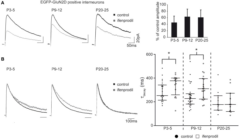
Figure 6 Effects of ifenprodil on NMDAR-mediated EPSCs in GluN2D-EGFP-positive cells. (A) Example traces of NMDAR-mediated EPSCs before and after application of the GluN2B specific antagonist ifenprodil. EPSCs were recorded at a holding potential of +40 mV in CA1 GluN2D-EGFP-positive interneurons in P3-5, P9-12, and P20-25 mice. The quantification of the remaining current after ifenprodil application shows that NMDARs containing other subunits than GluN2B contribute to synaptic currents at all three ages. (B) Example traces of NMDAR-mediated EPSCs before and after ifenprodil application normalized to the peak. The quantification of EPSC τdecay shows that NMDARs with slower decay kinetics (i.e., GluN2D) than that of diheteromeric GluN2B-containing contributed to synaptic currents in GluN2D-EGFP-positive cells in P3-5 and P9-12 mice. The age-dependent decrease of τdecay is consistent with an up-regulation of the “fast” GluN2A subunit. *p < 0.05.
Figure 6 Effects of ifenprodil on NMDAR-mediated EPSCs in GluN2D-EGFP-positive cells. (A) Example traces of NMDAR-mediated EPSCs before and after application of the GluN2B specific antagonist ifenprodil. EPSCs were recorded at a holding potential of +40 mV in CA1 GluN2D-EGFP-positive interneurons in P3-5, P9-12, and P20-25 mice. The quantification of the remaining current after ifenprodil application shows that NMDARs containing other subunits than GluN2B contribute to synaptic currents at all three ages. (B) Example traces of NMDAR-mediated EPSCs before and after ifenprodil application normalized to the peak. The quantification of EPSC τdecay shows that NMDARs with slower decay kinetics (i.e., GluN2D) than that of diheteromeric GluN2B-containing contributed to synaptic currents in GluN2D-EGFP-positive cells in P3-5 and P9-12 mice. The age-dependent decrease of τdecay is consistent with an up-regulation of the “fast” GluN2A subunit. *p < 0.05.CC BY · PubMed Central
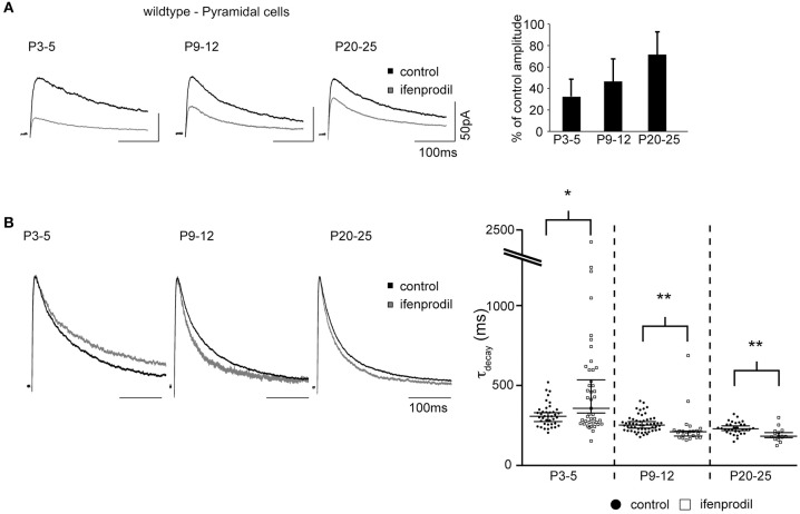
Figure 7 Effects of ifenprodil on NMDAR-mediated EPSCs in pyramidal cells of wildtype mice. (A) Example traces of NMDAR-mediated EPSCs before and after application of ifenprodil. EPSCs were recorded in CA1 pyramidal cells in P3-5, P9-12, and P20-25 wildtype mice. The quantification of the remaining current after ifenprodil application shows that NMDARs containing other subunits than GluN2B contribute to synaptic currents at all three ages. The decrease of blocking efficiency speaks for a developmental reduction in the proportional contribution of diheteromeric GluN2B-containing NMDARs. (B) Example traces of NMDAR-mediated EPSCs before and after ifenprodil application normalized to the peak. The quantification of EPSC τdecay shows that NMDARs with slower decay kinetics than that of diheteromeric GluN2B-containing NMDARs contribute to synaptic currents in pyramidal cells in P3-5 mice. In contrast, ifenprodil decreases τdecay in P9-12 and P20-25 mice, indicating that GluN2A-containing NMDARs contribute to synaptic currents. *p < 0.05, **p < 0.01.
Figure 7 Effects of ifenprodil on NMDAR-mediated EPSCs in pyramidal cells of wildtype mice. (A) Example traces of NMDAR-mediated EPSCs before and after application of ifenprodil. EPSCs were recorded in CA1 pyramidal cells in P3-5, P9-12, and P20-25 wildtype mice. The quantification of the remaining current after ifenprodil application shows that NMDARs containing other subunits than GluN2B contribute to synaptic currents at all three ages. The decrease of blocking efficiency speaks for a developmental reduction in the proportional contribution of diheteromeric GluN2B-containing NMDARs. (B) Example traces of NMDAR-mediated EPSCs before and after ifenprodil application normalized to the peak. The quantification of EPSC τdecay shows that NMDARs with slower decay kinetics than that of diheteromeric GluN2B-containing NMDARs contribute to synaptic currents in pyramidal cells in P3-5 mice. In contrast, ifenprodil decreases τdecay in P9-12 and P20-25 mice, indicating that GluN2A-containing NMDARs contribute to synaptic currents. *p < 0.05, **p < 0.01.CC BY · PubMed Central
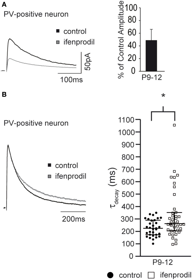
Figure 8 Effects of ifenprodil on NMDAR-mediated EPSCs in P9-12 PV-EGFP-positive cells. (A) Example traces of NMDAR-mediated EPSCs before and after application of ifenprodil. EPSCs were recorded in CA1 PV-EGFP interneurons of P9-12 mice. The quantification of the remaining current after ifenprodil application shows that NMDARs containing other subunits than GluN2B contribute to synaptic currents. (B) Example traces of NMDAR-mediated EPSCs before and after ifenprodil application normalized to the peak. The quantification of EPSC τdecay shows that NMDARs with slower decay kinetics than that of diheteromeric GluN2B-containing NMDARs contribute to synaptic currents in PV-EGFP interneurons in P9-12 mice. *p < 0.05.
Figure 8 Effects of ifenprodil on NMDAR-mediated EPSCs in P9-12 PV-EGFP-positive cells. (A) Example traces of NMDAR-mediated EPSCs before and after application of ifenprodil. EPSCs were recorded in CA1 PV-EGFP interneurons of P9-12 mice. The quantification of the remaining current after ifenprodil application shows that NMDARs containing other subunits than GluN2B contribute to synaptic currents. (B) Example traces of NMDAR-mediated EPSCs before and after ifenprodil application normalized to the peak. The quantification of EPSC τdecay shows that NMDARs with slower decay kinetics than that of diheteromeric GluN2B-containing NMDARs contribute to synaptic currents in PV-EGFP interneurons in P9-12 mice. *p < 0.05.CC BY · PubMed Central
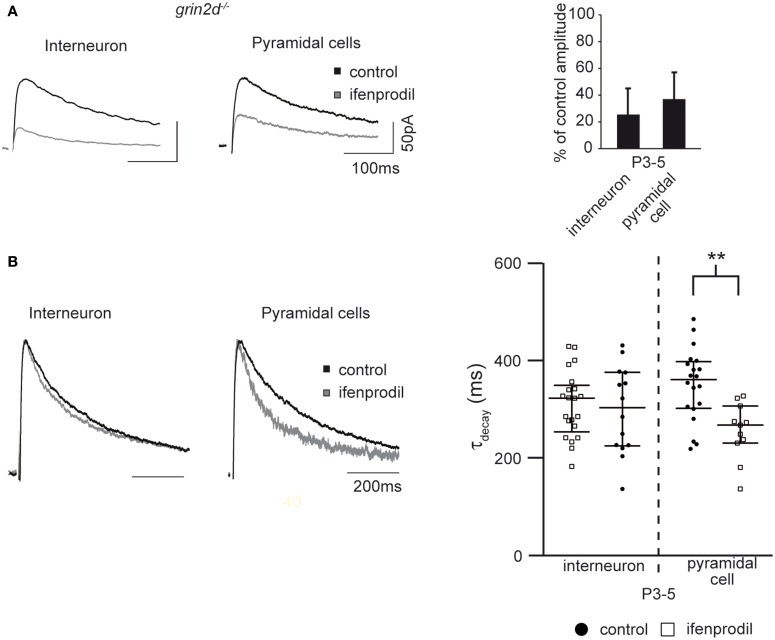
Figure 9 Effects of ifenprodil on NMDAR-mediated EPSCs in pyramidal cells and stratum oriens interneurons in P3-5 grin2d−/− mice. (A) Example traces of NMDAR-mediated EPSCs before and after application of the GluN2B specific antagonist ifenprodil. EPSCs were recorded in CA1 stratum oriens interneurons and CA1 pyramidal cells of P3-5 grin2d−/− mice. The quantification of the remaining current after ifenprodil application is shown on the right. (B) Example traces of NMDAR-mediated EPSCs before and after ifenprodil application normalized to the peak. The quantification shows that ifenprodil does not alter τdecay of EPSC in interneurons and decreases τdecay of EPSC in pyramidal cells, suggesting that slow GluN2D subunits do not contribute to synaptic NMDARs in P3-5 grin2d−/− mice, in contrast to their contribution in EGFP-GluN2D, PV-EGFP, and wildtype mice. **p < 0.01.
Figure 9 Effects of ifenprodil on NMDAR-mediated EPSCs in pyramidal cells and stratum oriens interneurons in P3-5 grin2d−/− mice. (A) Example traces of NMDAR-mediated EPSCs before and after application of the GluN2B specific antagonist ifenprodil. EPSCs were recorded in CA1 stratum oriens interneurons and CA1 pyramidal cells of P3-5 grin2d−/− mice. The quantification of the remaining current after ifenprodil application is shown on the right. (B) Example traces of NMDAR-mediated EPSCs before and after ifenprodil application normalized to the peak. The quantification shows that ifenprodil does not alter τdecay of EPSC in interneurons and decreases τdecay of EPSC in pyramidal cells, suggesting that slow GluN2D subunits do not contribute to synaptic NMDARs in P3-5 grin2d−/− mice, in contrast to their contribution in EGFP-GluN2D, PV-EGFP, and wildtype mice. **p < 0.01.CC BY · PubMed Central

Verification

Claims the paper makes

The claims and statistics of this paper haven’t been checked yet.

Run the check — free, no account needed. It reads the open-access text and checks the sources it cites.

A few free checks a day · papers already verified open instantly · results are public

Claims other papers make about this one

Each is checked against this paper’s own text: does it say what the citing paper says it does?

Self-check

Percentages, sums and reported differences re-computed from the paper’s own numbers.

The self-check needs the paper’s full text.

Citations

Cites

Show all 56 references

Research works printed but not linked: 2

  1. Dunah, 1996, NMDA receptor subunit NR2D identified using a polyclonal antiserum, FASEB J, 10, 808
  2. Wisden, 1994, In situ hybridization with synthetic oligonucleotide probes, 9

Cited by

Sorted by citations

See all 104

Metadata

Authors
Engelhardt, J. V., Bocklisch, C., Tönges, L., Herb, A., Mishina, M., & Monyer, H.
Affiliations
  • German Cancer Research Center (DE)
  • Ritsumeikan University (JP)
Corresponding author
Monyer, Hannah
Venue
Frontiers in Cellular Neuroscience
Published
2015-03-25
Volume
9
Pages
95-95
ISSN
1662-5102 (ISSN-L)
OpenAlex
W1983068587
Cited by
103
References
56
Citations by year
2026: 6 · 2025: 10 · 2024: 9 · 2023: 8 · 2022: 9 · 2021: 11 · 2020: 11 · 2019: 11 · 2018: 7 · 2017: 10 · 2016: 6 · 2015: 5
FWCI
4.33
Type
journal article
Language
en
Open access
Licence
cc-by
Locations
  • Frontiers in Cellular Neuroscience (publishedVersion, cc-by)
  • Europe PMC (PubMed Central) (publishedVersion, cc-by)
  • PubMed Central (publishedVersion, cc-by)
  • PubMed (publishedVersion)
  • DOAJ (DOAJ: Directory of Open Access Journals) (submittedVersion, cc-by-sa)
  • DZNE Pub (submittedVersion, cc-by)
Funders
Japan Society for the Promotion of Science (24249014, 22123008)
Topics
  • Life Sciences › Neuroscience › Cellular and Molecular Neuroscience › Neuroscience and Neuropharmacology Research
  • Life Sciences › Biochemistry, Genetics and Molecular Biology › Biochemistry › Amino Acid Enzymes and Metabolism
  • Life Sciences › Biochemistry, Genetics and Molecular Biology › Molecular Biology › Nicotinic Acetylcholine Receptors Study
Keywords
GluN2D, NMDA receptor subunits, hippocampal interneurons, pyramidal neurons, basket cells, axo-axonic cells, parvalbumin, somatostatin, synaptic currents, juvenile development, CA1, fast spiking

About these checks

Registry, retraction, Cites and statistics results are computed, not judged, with one exception: a printed reference that no index lists is read once by a model before it counts as a citation error. Claims are extracted by a model and kept only when their quote appears verbatim in the text; each cited source is then checked, with a model’s help, for whether it says what the claim relies on. Those verdicts can be wrong — read the claim’s quote and the cited source before relying on one.

Statistics and arithmetic are recomputed, not judged.

Confidence bands are shown only for fields calibrated on our annotated test set. None is calibrated yet, so no band is shown.

What counts as a claim ›How we measure confidence ›How we check this ›

Are you an author, or do you see a problem with this page? Request a review