

The DOI matches its Crossref record; 191 of the 191 references it cites were found in a registry or an index.
Neuropathobiology of COVID-19: The Role for Glia
Open accessAbstract
SARS-CoV-2, which causes the Coronavirus Disease 2019 (COVID-19) pandemic, has a brain neurotropism through binding to the receptor angiotensin-converting enzyme 2 expressed by neurones and glial cells, including astrocytes and microglia. Systemic infection which accompanies severe cases of COVID-19 also triggers substantial increase in circulating levels of chemokines and interleukins that compromise the blood-brain barrier, enter the brain parenchyma and affect its defensive systems, astrocytes and microglia. Brain areas devoid of a blood-brain barrier such as the circumventricular organs are particularly vulnerable to circulating inflammatory mediators. The performance of astrocytes and microglia, as well as of immune cells required for brain health, is considered critical in defining the neurological damage and neurological outcome of COVID-19. In this review, we discuss the neurotropism of SARS-CoV-2, the implication of neuroinflammation, adaptive and innate immunity, autoimmunity, as well as astrocytic and microglial immune and homeostatic functions in the neurological and psychiatric aspects of COVID-19. The consequences of SARS-CoV-2 infection during ageing, in the presence of systemic comorbidities, and for the exposed pregnant mother and foetus are also covered.
Overview
The claims and statistics of this paper haven’t been checked yet.
Run the check — free, no account needed. It reads the open-access text and checks the sources it cites.
Integrity signals
Registry
Cites
Statistics
Figures
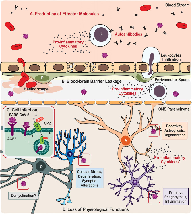
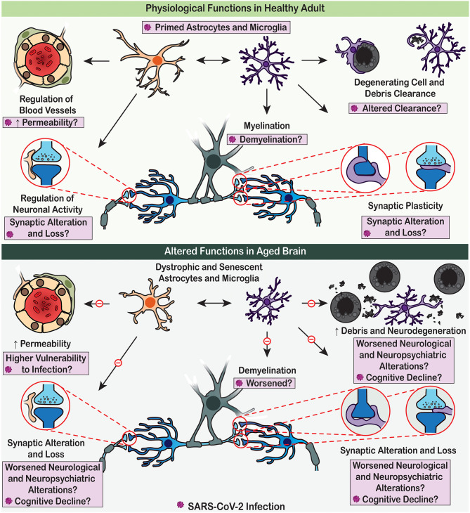
Verification
Claims the paper makes
The claims and statistics of this paper haven’t been checked yet.
Run the check — free, no account needed. It reads the open-access text and checks the sources it cites.
Claims other papers make about this one
Each is checked against this paper’s own text: does it say what the citing paper says it does?
In the citing paper
“Once the virus enters the CNS, it affects neurons, microglia, oligodendrocytes, and especially astrocytes, undermining neurons viability [17, 179, 180].”
From Cognitive decline following acute viral infections: literature review and projections for post-COVID-19 (2022) · cites this paper as [179]
In the citing paper
“In SARS-CoV-2, trans-nasal and trans-synaptic mechanisms might allow the virus to invade the olfactory bulb and brainstem, with both being the possible initial site for CNS invasion [179].”
From Cognitive decline following acute viral infections: literature review and projections for post-COVID-19 (2022) · cites this paper as [179]
Self-check
Percentages, sums and reported differences re-computed from the paper’s own numbers.
The self-check needs the paper’s full text.
Citations
Cites
Clinical features of patients infected with 2019 novel coronavirus in Wuhan, China
Huang, C., Wang, Y., Li, X., Ren, L., Zhao, J., Hu, Y., Zhang, L., Fan, G., Xu, J., Gu, X., et al. (2020) · The Lancet · doi:10.1016/s0140-6736(20)30183-5 · Cited by 48,884
Wu, Z., & McGoogan, J. M. (2020) · JAMA · doi:10.1001/jama.2020.2648 · Cited by 17,016 · cited 2×
“…it, can spread into the other organs including the central nervous system (CNS) and cause severe neurological outcomes during the disease development and progression, especially for patients with comorbidities and/or elderly people (Huang et al., 2020; Wolfel et al., 2020; Wu and McGoogan, 2020).”
BackgroundPolazzi, E., Mengoni, I., Caprini, M., Peña‐Altamira, E., Kurtys, E., & Monti, B. (2012) · Neurosignals · doi:10.1159/000337115 · Cited by 9,023
Neurologic manifestations of hospitalized patients with coronavirus disease 2019 in Wuhan, China
Mao, L., Jin, H., Wang, M., Hu, Y., Chen, S., He, Q., Chang, J., Hong, C., Zhou, Y., Wang, D., et al. (2020) · JAMA Neurology · doi:10.1001/jamaneurol.2020.1127 · Cited by 7,166
Virological assessment of hospitalized patients with COVID-2019
Wölfel, R., Corman, V. M., Guggemos, W., Seilmaier, M., Zange, S., Müller, M. A., Niemeyer, D., Jones, T. C., Vollmar, P., Rothe, C., et al. (2020) · Nature · doi:10.1038/s41586-020-2196-x · Cited by 6,661
Effects of stress throughout the lifespan on the brain, behaviour and cognition
Lupien, S. J., McEwen, B. S., Gunnar, M. R., & Heim, C. (2009) · Nature Reviews Neuroscience · doi:10.1038/nrn2639 · Cited by 6,256 · cited 1×
“Indeed, many epidemiological studies suggest that early-life adversity can be associated with an increased occurrence of neurodevelopmental, neurological or neuropsychiatric disorders later in life (Halligan et al., 2007; Lupien et al., 2009; Knuesel et al., 2014).”
BackgroundResting microglial cells are highly dynamic surveillants of brain parenchyma in vivo
Nimmerjahn, A., Kirchhoff, F., & Helmchen, F. (2005) · Science · doi:10.1126/science.1110647 · Cited by 5,827 · cited 1×
“These microglia have an amoeboid instead of a surveilling (derived from to “surveil,” which means to keep a place or a person under surveillance) morphology (Davalos et al., 2005; Nimmerjahn et al., 2005) and express markers that suggest they are ready to cope with invading agents using phagocytosis and the release of…”
BackgroundIliff, J. J., Wang, M., Liao, Y., Plogg, B. A., Peng, W., Gundersen, G. A., Benveniste, H., Vates, G. E., Deane, R., Goldman, S. A., et al. (2012) · Science Translational Medicine · doi:10.1126/scitranslmed.3003748 · Cited by 5,824 · cited 1×
“Furthermore, the astroglia-based glymphatic waste collection system (Iliff et al., 2012) declines with age, which affects clearance of numerous by-products and toxic substances (Kress et al., 2014).”
BackgroundAstrocyte-endothelial interactions at the blood-brain barrier
Abbott, N. J., Rönnbäck, L., & Hansson, E. (2006) · Nature Reviews Neuroscience · doi:10.1038/nrn1824 · Cited by 5,595 · cited 1×
“All these structures are highly vascularised and they lack the blood-brain-barrier (BBB) (Duvernoy and Risold, 2007), which is formed by the endothelial cells lining brain vessels, together with pericytes, astrocytes, microglia, and extracellular components such as the basement membrane that together form the neurovas…”
BackgroundCryan, J. F., O'Riordan, K. J., Cowan, C. S. M., Sandhu, K. V., Bastiaanssen, T. F. S., Boehme, M., Codagnone, M. G., Cussotto, S., Fulling, C., Golubeva, A. V., et al. (2019) · Physiological Reviews · doi:10.1152/physrev.00018.2018 · Cited by 5,286
Cited by
Sorted by citations
Microglia states and nomenclature: A field at its crossroads
Paolicelli, R. C., Sierra, A., Stevens, B., Tremblay, M.-E., Aguzzi, A., Ajami, B., Amit, I., Audinat, E., Bechmann, I., Bennett, M., et al. (2022) · Neuron · doi:10.1016/j.neuron.2022.10.020 · Cited by 1,973
Self-citationAstrocytes in human central nervous system diseases: a frontier for new therapies
Verkhratsky, A., Butt, A., Li, B., Illes, P., Zorec, R., Semyanov, A., Tang, Y., & Sofroniew, M. V. (2023) · Signal Transduction and Targeted Therapy · doi:10.1038/s41392-023-01628-9 · Cited by 428
Self-citationPost-COVID-19 Depressive Symptoms: Epidemiology, Pathophysiology, and Pharmacological Treatment
Mazza, M. G., Palladini, M., Poletti, S., & Benedetti, F. (2022) · CNS Drugs · doi:10.1007/s40263-022-00931-3 · Cited by 248
“Accordingly, increased microglial activation in association with high levels of IL-6 and TNF-α have been observed in MDD episodes [82], whereas microglia activation has been implicated in both acute and chronic neurological complications of COVID-19 [83], and increased microglial activation has been demonstrated in po…”
BackgroundS1-Leitlinie Post-COVID/Long-COVID
Koczulla, A. R., Ankermann, T., Behrends, U., Berlit, P., Böing, S., Brinkmann, F., Franke, C., Glöckl, R., Gogoll, C., Hummel, T. R., et al. (2021) · Pneumologie · doi:10.1055/a-1551-9734 · Cited by 164
“[150] Tremblay ME, Madore C, Bordeleau M et al.”
BackgroundCould SARS-CoV-2 Spike Protein Be Responsible for Long-COVID Syndrome?
Theoharides, T. C. (2022) · Molecular Neurobiology · doi:10.1007/s12035-021-02696-0 · Cited by 156
“Increasing evidence indicates the involvement of neuroinflammation [71, 88, 89] that may damage brain blood vessels [90, 91], as well as brain cells [88, 92, 93], possibly via activation of microglia [94, 95] and mast cells [96].”
BackgroundNeurological complications and infection mechanism of SARS-CoV-2
Wan, D., Du, T., Hong, W., Chen, L., Que, H., Lu, S., & Peng, X. (2021) · Signal Transduction and Targeted Therapy · doi:10.1038/s41392-021-00818-7 · Cited by 140
COVID-19 and cognitive impairment: neuroinvasive and blood‒brain barrier dysfunction
Chen, Y., Yang, W., Chen, F., & Cui, L. (2022) · Journal of Neuroinflammation · doi:10.1186/s12974-022-02579-8 · Cited by 134
Neuroinvasion and Neurotropism by SARS-CoV-2 Variants in the K18-hACE2 Mouse
Seehusen, F., Clark, J. J., Sharma, P., Bentley, E. G., Kirby, A., Subramaniam, K. S., Wunderlin-Giuliani, S., Hughes, G. L., Patterson, E. I., Michael, B. D., et al. (2022) · Viruses · doi:10.3390/v14051020 · Cited by 116
Koczulla, A. R., Ankermann, T., Behrends, U., Berlit, P., Berner, R., Böing, S., Brinkmann, F., Frank, U., Franke, C., Glöckl, R., et al. (2022) · Pneumologie · doi:10.1055/a-1946-3230 · Cited by 118
“[281] Tremblay ME, Madore C, Bordeleau M et al.”
BackgroundLong COVID and neuropsychiatric manifestations (Review)
Efstathiou, V., Stefanou, M., Demetriou, M., Siafakas, N., Makris, M. P., Tsivgoulis, G. K., Zoumpourlis, V., Kympouropoulos, S. P., Tsoporis, J. N., Spandidos, D. A., et al. (2022) · Experimental and Therapeutic Medicine · doi:10.3892/etm.2022.11290 · Cited by 118
“Patients affected by ‘long COVID’ include also those who initially had mild or asymptomatic disease, pointing to residual effects that involve multiple organ systems, including the peripheral and central nervous system (CNS) (23,31,32).”
InfluentialDamiano, R. F., Guedes, B. F., de Rocca, C. C., de Pádua Serafim, A., Castro, L. H. M., Munhoz, C. D., Nitrini, R., Filho, G. B., Miguel, E. C., Lucchetti, G., et al. (2022) · European Archives of Psychiatry and Clinical Neuroscience · doi:10.1007/s00406-021-01286-4 · Cited by 98
“Once the virus enters the CNS, it affects neurons, microglia, oligodendrocytes, and especially astrocytes, undermining neurons viability [17, 179, 180].”
BackgroundBoroujeni, M. E., Simani, L., Bluyssen, H. A. R., Samadikhah, H. R., Benisi, S. Z., Hassani, S., Dilmaghani, N. A., Fathi, M., Vakili, K. G., Mahmoudiasl, G., et al. (2021) · ACS Chemical Neuroscience · doi:10.1021/acschemneuro.1c00111 · Cited by 81
Petersen, M., Nägele, F. L., Mayer, C., Schell, M., Petersen, E. L., Kühn, S., Gallinat, J., Fiehler, J., Pasternak, O., Matschke, J., et al. (2023) · Proceedings of the National Academy of Sciences · doi:10.1073/pnas.2217232120 · Cited by 83
“Evidence of neuroinflammation was reported in histopathological and clinical studies of COVID-19 patients: virus invasion (24, 25), activation of glial cells (9, 26, 27), and a cytokine response in the cerebrospinal fluid accompanying neurological and psychiatric COVID-19 symptoms (10).”
Can COVID-19 pandemic boost the epidemic of neurodegenerative diseases?
Verkhratsky, A., Li, Q., Melino, S., Melino, G., & Shi, Y. (2020) · Biology Direct · doi:10.1186/s13062-020-00282-3 · Cited by 81
Self-citationTheoharides, T. C., & Kempuraj, D. (2023) · Cells · doi:10.3390/cells12050688 · Cited by 82
“We advance the premise that brain perivascular inflammation is a critical pathogenetic factor in long COVID mostly due to SARS-CoV-2 activating brain mast cells and microglia resulting in the release of inflammatory, neurotoxic, and vasoactive mediators [60,61].”
BackgroundElucidating the Neuropathologic Mechanisms of SARS-CoV-2 Infection
Pacheco‐Herrero, M., Soto-Rojas, L. O., Harrington, C. R., Flores-Martinez, Y. M., Villegas-Rojas, M. M., León-Aguilar, A. M., Martínez-Gómez, P. A., Campa-Córdoba, B. B., Apátiga-Pérez, R., Corniel-Taveras, C. N., et al. (2021) · Frontiers in Neurology · doi:10.3389/fneur.2021.660087 · Cited by 71
Neurotropic Viruses, Astrocytes, and COVID-19
Verdev, P. T., Potokar, M., Kolenc, M., Korva, M., Avšič‐Županc, T., Zorec, R., & Jorgačevski, J. (2021) · Frontiers in Cellular Neuroscience · doi:10.3389/fncel.2021.662578 · Cited by 72
“This phenomenon is typical for severe COVID-19 pathogenesis, which eventually leads to BBB alterations and neuroinflammation (Najjar et al., 2020; Tremblay et al., 2020).”
BackgroundAwogbindin, I. O., Ben‐Azu, B., Olusola, B. A., Akinluyi, E. T., Adeniyi, P. A., Paolo, T. D., & Tremblay, M. (2021) · Frontiers in Cellular Neuroscience · doi:10.3389/fncel.2021.670298 · Cited by 61
“Despite their CNS homeostasis and neuronal cell functions, microglia and astrocytes are mainly involved in the protective immune responses during neuroinflammation (Facci et al., 2018; Jha et al., 2019; Tremblay et al., 2020).”
BackgroundSelf-citationDid the COVID-19 Pandemic Increase the Incidence of Acute Macular Neuroretinopathy?
Azar, G., Bonnin, S., Vasseur, V., Faure, C., Salviat, F., Vignal‐Clermont, C., Titah, C., Farès, S., Boulanger-Scemama, É., Derrien, S., et al. (2021) · Journal of Clinical Medicine · doi:10.3390/jcm10215038 · Cited by 58
Proust, A. M., Queval, C. J., Harvey, R., Adams, L., Bennett, M., & Wilkinson, R. J. (2023) · Journal of Neuroinflammation · doi:10.1186/s12974-023-02861-3 · Cited by 55
SARS-CoV-2 involvement in central nervous system tissue damage
Sabra, M., Kobeissy, F. H., Bizri, M., Haidar, M. A., Shakkour, Z., Reslan, M. A., Al-Haj, N., Chamoun, P., Habashy, K. J., Kaafarani, H., et al. (2021) · Neural Regeneration Research · doi:10.4103/1673-5374.327323 · Cited by 58
COVID-19, Oxidative Stress, and Neuroinflammation in the Depression Route
Mingoti, M. E. D., Bertollo, A. G., Simões, J. L. B., Francisco, G. R., Bagatini, M. D., & Ignácio, Z. M. (2022) · Journal of Molecular Neuroscience · doi:10.1007/s12031-022-02004-y · Cited by 60
“Given the presence of ACE2 receptors in microglia and astrocytes, it may be possible to relate SARS-CoV-2 with glial activation (Tremblay et al. 2020).”
BackgroundBrain microstructural changes and fatigue after COVID-19
Bispo, D. D. D. C., Brandão, P. R. D. P., Pereira, D. A., Maluf, F. B., Dias, B. A., Paranhos, H. R., von Glehn, F., de Oliveira, A. C. P., Regattieri, N. A. T., Silva, L. S., et al. (2022) · Frontiers in Neurology · doi:10.3389/fneur.2022.1029302 · Cited by 56
“Neurons, oligodendrocytes, and other glial cells may have impaired physiological functions during SARS-CoV-2 inflammatory insult, leading to a disturbance of brain homeostasis (62).”
BackgroundNeurological complications associated with Covid‐19; molecular mechanisms and therapeutic approaches
Mehrabani, M. M., Karvandi, M. S., Maafi, P., & Doroudian, M. (2022) · Reviews in Medical Virology · doi:10.1002/rmv.2334 · Cited by 53
Abdi, A., AlOtaiby, S., Badarin, F. A., Khraibi, A., Hamdan, H., & Nader, M. (2022) · Biomedicine & Pharmacotherapy · doi:10.1016/j.biopha.2021.112518 · Cited by 47
Depressive and Neurocognitive Disorders in the Context of the Inflammatory Background of COVID-19
Dąbrowska, E., Galińska-Skok, B., & Waszkiewicz, N. (2021) · Life · doi:10.3390/life11101056 · Cited by 47
Stoian, A., Stoian, M., Bajkó, Z., Maier, S., Andone, S. R., Cioflinc, R. A., Moțățăianu, A., Bărcuţean, L., & Bălașa, R. (2022) · Biomedicines · doi:10.3390/biomedicines10040774 · Cited by 42
“The brain’s defensive system and systemic inflammation can influence the CNS through both innate and adaptive immune systems through their cellular elements [64,97].”
BackgroundInfluentialCOVID-19 in People With Schizophrenia: Potential Mechanisms Linking Schizophrenia to Poor Prognosis
Mohan, M., Perry, B. I., Saravanan, P., & Singh, S. P. (2021) · Frontiers in Psychiatry · doi:10.3389/fpsyt.2021.666067 · Cited by 40
de Andrade, E. G., Šimončičová, E., Carrier, M., Vecchiarelli, H. A., Robert, M.-È., & Tremblay, M. (2021) · Frontiers in Cellular Neuroscience · doi:10.3389/fncel.2021.647378 · Cited by 39
“Besides, reactive microglia generally adopt different morphologies, for example, shifting to amoeboid shapes with bigger somas and less ramified processes, as opposed to the steady-state, highly branched “surveilling” phenotype (Figure 1; Savage et al., 2019; Tremblay et al., 2020).”
BackgroundSelf-citationVarghese, S. M., Patel, S., Nandan, A., Jose, A., Ghosh, S., Sah, R. K., Menon, B., Athira, K. V., & Chakravarty, S. (2024) · Molecular Neurobiology · doi:10.1007/s12035-024-04205-5 · Cited by 39
Naidu, S. A. G., Wallace, T. C., Davies, K. J. A., & Naidu, A. S. (2021) · Journal of Dietary Supplements · doi:10.1080/19390211.2021.1922567 · Cited by 35
“Furthermore, microglia modulate the CNS micro-milieu, thereby reduce the severity of demyelination, and influence neuronal repair in cerebral cortex (Lavi and Cong 2020; Tremblay et al. 2020).”
BackgroundNeurological sequela and disruption of neuron-glia homeostasis in SARS-CoV-2 infection
Savelieff, M. G., Feldman, E. L., & Stino, A. M. (2022) · Neurobiology of Disease · doi:10.1016/j.nbd.2022.105715 · Cited by 37
“The virus also triggers a pro-inflammatory response, which can render the BBB susceptible to damage, leading to immune cell infiltration into the brain along with viral penetration (Tremblay et al., 2020; Solomon, 2021).”
Szabo, M. P., Iba, M., Nath, A., Masliah, E., & Kim, C. (2022) · Experimental & Molecular Medicine · doi:10.1038/s12276-022-00755-7 · Cited by 37
Chiappalupi, S., Salvadori, L., Donato, R., Riuzzi, F., & Sorci, G. (2021) · Biomolecules · doi:10.3390/biom11060876 · Cited by 35
“The increase in circulating levels of chemokines and interleukins subsequent to SARS-COV-2 pulmonary infection compromises the blood–brain barrier (BBB), favoring the entry of the virus into the brain parenchyma and leading to inflammation, with the subsequent risk of neurological damage [107].”
BackgroundTREM2, microglial and ischemic stroke
Wang, H., Li, X., Wang, Q., Ma, J., Gao, X., & Wang, M. (2023) · Journal of Neuroimmunology · doi:10.1016/j.jneuroim.2023.578108 · Cited by 32
Defining Microglial States and Nomenclature: A Roadmap to 2030Preprint
Paolicelli, R. C., Sierra, A., Stevens, B., Tremblay, M., Aguzzi, A., Ajami, B., Amit, I., Audinat, É., Bechmann, I., Bennett, M. L., et al. (2022) · SSRN Electronic Journal · doi:10.2139/ssrn.4065080 · Cited by 33
Self-citationWhat can neuroimmunology teach us about the symptoms of long-COVID?
Mondelli, V., & Pariante, C. M. (2021) · Oxford Open Immunology · doi:10.1093/oxfimm/iqab004 · Cited by 32
Müller, K., Poppele, I., Ottiger, M., Zwingmann, K., Berger, I., Thomas, A., Wastlhuber, A., Ortwein, F., Schultz, A.-L., Weghofer, A., et al. (2023) · International Journal of Environmental Research and Public Health · doi:10.3390/ijerph20021468 · Cited by 31
Role of Demyelination in the Persistence of Neurological and Mental Impairments after COVID-19
Khodanovich, M. Y., Kamaeva, D. A., & Naumova, A. V. (2022) · International Journal of Molecular Sciences · doi:10.3390/ijms231911291 · Cited by 32
Astrocytes and the Psychiatric Sequelae of COVID-19: What We Learned from the Pandemic
Steardo, L., Steardo, L., & Scuderi, C. (2022) · Neurochemical Research · doi:10.1007/s11064-022-03709-7 · Cited by 34
COVID-19: a new emerging respiratory disease from the neurological perspective
El-Sayed, A., Aleya, L., & Kamel, M. S. (2021) · Environmental Science and Pollution Research · doi:10.1007/s11356-021-12969-9 · Cited by 30
“All these routes were proposed based on current evidences (Satarker and Nampoothiri 2020; Tremblay et al. 2020; Lima et al. 2020; Jakhmola et al. 2020).”
BackgroundTheoharides, T. C. (2021) · Journal of Personalized Medicine · doi:10.3390/jpm11090860 · Cited by 29
Disrupted Sleep During a Pandemic
Kumar, N., & Gupta, R. M. (2022) · Sleep Medicine Clinics · doi:10.1016/j.jsmc.2021.10.006 · Cited by 33
“Once inside the brain, SARS-CoV2 invades tissues with ACE-2 receptors including the frontotemporal cortex, circumventricular organs, thalamus, and brainstem involving the rostral ventrolateral medulla among others.(28,29) ACE-2 expression has also been reported in the hypothalamus and pituitary gland.”
BackgroundZhang, Y., Archie, S. R., Ghanwatkar, Y., Sharma, S., Nozohouri, S., Burks, E., Mdzinarishvili, A., Liu, Z., & Abbruscato, T. J. (2022) · Fluids and Barriers of the CNS · doi:10.1186/s12987-022-00339-7 · Cited by 29
“Given that the large number of functions performed by glial cells in the neuroinflammatory response, it could be expected that activation of astrocytes has a major impact on the CNS during SARSCoV-2 infection [23, 24].”
BackgroundVestibular disorders in patients after COVID-19 infection
Pazdro-Zastawny, K., Dorobisz, K., Misiak, P., Kruk-Krzemień, A., & Zatoński, T. (2022) · Frontiers in Neurology · doi:10.3389/fneur.2022.956515 · Cited by 33
Plasmalogens and platelet‐activating factor roles in chronic inflammatory diseases
Tremblay, M., Almsherqi, Z. A., & Deng, Y. (2022) · BioFactors · doi:10.1002/biof.1916 · Cited by 30
“Genetic variants and environmental risk factors altering these microglial functions are further associated with most if not all CNS disorders, including neurodevelopmental, neuropsychiatric, and neurodegenerative.(99,100) These findings highlight the importance of evaluating the outcomes of decreased, but also restore…”
BackgroundSelf-citationTsilioni, I., & Theoharides, T. C. (2023) · Molecular Neurobiology · doi:10.1007/s12035-023-03493-7 · Cited by 29
“Increasing evidence indicates the involvement of neuro-inflammation [64–66] that may damage brain blood vessels [67, 68] and brain cells [64, 69, 70] possibly via activation of microglia [71, 72].”
BackgroundCOVID ‐19 Induces Neuroinflammation and Suppresses Peroxisomes in the Brain
Roczkowsky, A., Limonta, D., Fernandes, J. P., Branton, W. G., Clarke, M., Hlavay, B. A., Noyce, R. S., Joseph, J. T., Ogando, N. S., Das, S., et al. (2023) · Annals of Neurology · doi:10.1002/ana.26679 · Cited by 29
Principles of Astrogliopathology
Verkhratsky, A., Li, B., Scuderi, C., & Parpura, V. (2021) · Advances in Neurobiology · doi:10.1007/978-3-030-77375-5_3 · Cited by 21
“These conditions include neurotrauma (Burda et al. 2016; Faulkner et al. 2004), systemic inflammation and sepsis (Shulyatnikova and Verkhratsky 2019; Tremblay et al. 2020), microbial or viral neuroinfection (Soung and Klein 2018; Zorec et al. 2019), toxic encephalopathies (Li et al. 2021;…”
BackgroundSelf-citationFu, Y., Guo, Z., Wang, Y., Zhang, H., Zhang, F., Xu, Z., Shen, X., Roppongi, R. T., Mo, S., Gu, W., et al. (2022) · Frontiers in Immunology · doi:10.3389/fimmu.2022.967356 · Cited by 22
Sideratou, C.-M., & Papaneophytou, C. P. (2023) · Infectious Disease Reports · doi:10.3390/idr15060072 · Cited by 23
“This cascade of events can dysregulate neurogenesis and disrupt the normal functioning of neurons, oligodendrocytes, and neuroglial cells [100].”
BackgroundGiorgio, A. D., Mirijello, A., Gennaro, C. D., Fontana, A., Alboini, P. E., Florio, L., Inchingolo, V., Zarrelli, M. M., Miscio, G., Raggi, P., et al. (2022) · Diagnostics · doi:10.3390/diagnostics12020544 · Cited by 19
Acute and long-term effects of COVID-19 on brain and mental health: A narrative review
Bremner, J. D., Russo, S. J., Gallagher, R., & Simon, N. M. (2024) · Brain Behavior and Immunity · doi:10.1016/j.bbi.2024.11.007 · Cited by 19
Yan, L., Jayaram, M., Chithanathan, K., Zharkovsky, A., & Tian, L. (2021) · Frontiers in Cellular Neuroscience · doi:10.3389/fncel.2021.750373 · Cited by 19
“Glia-induced neuroinflammation is a common pathological observation in neurological cases of COVID-19 (Tremblay et al., 2020; Vargas et al., 2020), as microglia and astrocytes are key pathogen-combating players in the brain during SARS-CoV-2 infection (McMahon et al.”
BackgroundSelf-citationCăliman-Sturdza, O. A., Gheorghita, R. E., & Lobiuc, A. (2025) · Life · doi:10.3390/life15030439 · Cited by 32
Newhouse, A. L., Kritzer, M. D., Eryilmaz, H., Praschan, N. C., Camprodon, J. A., Fricchione, G. L., & Chemali, Z. N. (2022) · Journal of the Academy of Consultation-Liaison Psychiatry · doi:10.1016/j.jaclp.2022.08.007 · Cited by 17
“24 In addition, studies of single-nucleus transcriptomes of frontal cortex and the choroid plexus in 30 SARS-CoV-2 patients reveal cellular changes in COVID-19-associated microglia and astrocytes that share features with pathological cell states in neurodegenerative disorders, such as Alzheimer…”
Fetal brain vulnerability to SARS-CoV-2 infection
McMahon, C. L., Castro, J., Silvas, J. A., Perez, A. M., Estrada, M., Carrion, R., & Hsieh, J. (2023) · Brain Behavior and Immunity · doi:10.1016/j.bbi.2023.06.015 · Cited by 17
Alterations in microbiota of patients with COVID‐19: implications for therapeutic interventions
Qiu, Y., Mo, C., Chen, L., Ye, W., Chen, G., & Zhu, T. (2024) · MedComm · doi:10.1002/mco2.513 · Cited by 18
“119 Glial activation and neuroinflammation may play an important role in cognitive impairment caused by COVID-19.”
BackgroundHayden, M. R. (2021) · Neuroglia · doi:10.3390/neuroglia2010004 · Cited by 19
“Furthermore, the major histocompatibility complex (MHC)positive antigen presenting cells will present these viral antigens to the acquired immune system’s auto-reactive T-cells that will form the antibody which will then develop the antigen–antibody complex [94,99].”
BackgroundImmunological Interfaces: The COVID-19 Pandemic and Depression
Perlmutter, A. (2021) · Frontiers in Neurology · doi:10.3389/fneur.2021.657004 · Cited by 18
Genetics of COVID ‐19 and myalgic encephalomyelitis/chronic fatigue syndrome: a systematic review
Tziastoudi, M., Cholevas, C., Stefanidis, I., & Theoharides, T. C. (2022) · Annals of Clinical and Translational Neurology · doi:10.1002/acn3.51631 · Cited by 19
“Microglia have been implicated in COVID-19(228) and were also associated with neuroinflammation.(229) Microglia have important functions both in health and disease of the central nervous system (CNS), especially with respect to neuroinflammation 232 and neurodegenerative diseases.”
BackgroundAstrocytes in the pathophysiology of neuroinfection
Zorec, R., & Verkhratsky, A. (2022) · Essays in Biochemistry · doi:10.1042/ebc20220082 · Cited by 16
Self-citationSARS-CoV-2 Infection: Paving Way for Sleep Disorders in Long Term!
Gupta, R. M., & Pandi-Perumal, S. R. (2021) · Sleep and Vigilance · doi:10.1007/s41782-021-00145-5 · Cited by 16
“, brainstem, circumventricular organ, subfornical area, paraventricular nucleus, and rostroventrolateral medulla [10].”
BackgroundPost‐COVID‐19 neuropsychiatric syndrome: Is maladaptive glial recovery to blame?
Steardo, L., Steardo, L., Verkhratsky, A., & Scuderi, C. (2021) · Acta Physiologica · doi:10.1111/apha.13717 · Cited by 15
Self-citationVolk, P., Manesh, M. R., Warren, M. E., Besko, K., de Andrade, E. G., Wicki‐Stordeur, L. E., & Swayne, L. A. (2023) · Journal of Neurochemistry · doi:10.1111/jnc.16016 · Cited by 14
Microglia as potential key regulators in viral-induced neuroinflammation
Ismail, F. S., Faustmann, T. J., Faustmann, P. M., & Corvace, F. (2024) · Frontiers in Cellular Neuroscience · doi:10.3389/fncel.2024.1426079 · Cited by 13
Nelson, B. K., Farah, L. N., Grier, A., Su, W., Chen, J., Sossi, V., Sekhon, M. S., Stoessl, A. J., Wellington, C. L., Honer, W. G., et al. (2024) · NeuroImage · doi:10.1016/j.neuroimage.2024.120859 · Cited by 16
“We found lower MD in the long COVID group, but no differences in other DTI metrics or cognitive performance.”
Backgroundda Cunha, L. N. O. L., Tizziani, T., Souza, G. B., Moreira, M. A., Neto, J. S. S., Santos, C. V. D. D., de Carvalho, M. G., Dalmarco, E. M., Turqueti, L. B., Scotti, L., et al. (2021) · Current Medicinal Chemistry · doi:10.2174/0929867328666210726094955 · Cited by 12
Neuroinvasion and neurotropism by SARS-CoV-2 variants in the K18-hACE2 mousePreprint
Seehusen, F., Clark, J. J., Sharma, P., Bentley, E. G., Kirby, A., Subramaniam, K. S., Wunderlin, S., Hughes, G. L., Patterson, E. I., Michael, B. D., et al. (2021) · bioRxiv (Cold Spring Harbor Laboratory) · doi:10.1101/2021.04.16.440173 · Cited by 12
DiNicolantonio, J. J., McCarty, M. F., Barroso-Aranda, J., Assanga, S. B. I., Luján, L. M. L., & O’Keefe, J. H. (2021) · Open Heart · doi:10.1136/openhrt-2021-001663 · Cited by 11
Valenza, M., Steardo, L., Steardo, L., Verkhratsky, A., & Scuderi, C. (2021) · Frontiers in Cellular Neuroscience · doi:10.3389/fncel.2021.748136 · Cited by 12
Self-citationGonçalves, C., Sesterheim, P., Wartchow, K. M., Bobermin, L. D., Leipnitz, G., & Quincozes‐Santos, A. (2022) · Frontiers in Cellular Neuroscience · doi:10.3389/fncel.2022.905218 · Cited by 11
“Another very important aspect of COVID-19 is the greater vulnerability of elderly patients; possibly the aging of astrocytes makes these individuals more susceptible to Sars-CoV-2-induced dysfunction (Tremblay et al., 2020).”
BackgroundKhullar, S., & Wang, D. (2023) · Human Molecular Genetics · doi:10.1093/hmg/ddad009 · Cited by 12
Autopsy findings of pediatric COVID-19: a systematic review
Octavius, G. S., Wijaya, J. H., Tan, A. O., Muljono, M. P., Chandra, S., & Juliansen, A. (2022) · Egyptian Journal of Forensic Sciences · doi:10.1186/s41935-022-00288-0 · Cited by 11
SARS-CoV-2 infection is associated with increased odds of insomnia, RLS and dream enactment behavior
Kumar, N., Goyal, A., Hussain, A., Saini, L. K., Chawla, O., Arora, P. K., Daga, R., Tikka, S. K., Goyal, S., Kanchan, S., et al. (2022) · Indian Journal of Psychiatry · doi:10.4103/indianjpsychiatry.indianjpsychiatry_586_21 · Cited by 10
“[17] Thus, the present study was planned to study the effect of SARS-CoV-2 infection to the occurrence of insomnia, RLS and DEB.”
Peripheral nervous system involvement associated with COVID-19. A systematic review of literature
Hanganu, A. R., Constantin, A. M., Moise, E.-S., Niculae, C., Olaru, I. D., Băicuş, C., & Hristea, A. (2023) · PLoS ONE · doi:10.1371/journal.pone.0283827 · Cited by 10
SARS-CoV-2 hampers dopamine production in iPSC-derived dopaminergic neurons
Cappelletti, G., Carsana, E. V., Lunghi, G., Breviario, S., Vanetti, C., Fonzo, A. D., Frattini, E., Magni, M., Zecchini, S., Clerici, M., et al. (2023) · Experimental and Molecular Pathology · doi:10.1016/j.yexmp.2023.104874 · Cited by 10
Dialogue between Neuroinflammation and Neurodegenerative Diseases in COVID-19
Chowdhury, B., Sharma, A., Satarker, S., Mudgal, J., & Nampoothiri, M. (2021) · Journal of Environmental Pathology Toxicology and Oncology · doi:10.1615/jenvironpatholtoxicoloncol.2021038365 · Cited by 10
SARS-CoV-2-Induced Amyloidgenesis: Not One, but Three Hypotheses for Cerebral COVID-19 Outcomes
Gonçalves, C., Bobermin, L. D., Sesterheim, P., & Netto, C. A. (2022) · Metabolites · doi:10.3390/metabo12111099 · Cited by 10
“Note that the brain endothelium is closely surrounded by the feet of astrocytes, forming the BBB [19].”
BackgroundInfluentialEvidence of Microglial Immune Response Following Coronavirus PHEV Infection of CNS
Zhang, J., Li, Z., Lu, H., Shi, J., Gao, R., Ma, Y., Lan, Y., Guan, J., Zhao, K., Gao, F., et al. (2022) · Frontiers in Immunology · doi:10.3389/fimmu.2021.804625 · Cited by 10
COVID‐19 and cognitive impairment: From evidence to SARS‐CoV‐2 mechanism
Pan, H., Niu, J., Feng, L., Yin, Y. L., Dang, C., Lu, Y., Li, L., Ji, J., Yang, K., Wang, L., et al. (2024) · Brain‐X · doi:10.1002/brx2.58 · Cited by 9
Crosstalk between SARS-CoV-2 Infection and Neurological Disorders: A Review
Azhar, A., Wali, M. A., Rashid, Q., Khan, W. H., Alhosaini, K. A., Owais, M., & Kamal, M. A. (2022) · CNS & Neurological Disorders - Drug Targets · doi:10.2174/1871527321666220418114009 · Cited by 8
Post-Acute Sequelae of SARS-CoV-2 Infection: A Descriptive Clinical Study
Gutiérrez-Martínez, L., Karten, J., Kritzer, M. D., Josephy‐Hernandez, S. E., Kim, D. D., Newhouse, A. L., Pasinski, M., Praschan, N. C., Razafsha, M., Rubin, D. B., et al. (2022) · Journal of Neuropsychiatry · doi:10.1176/appi.neuropsych.21070193 · Cited by 8
Human Brain In Vitro Model for Pathogen Infection-Related Neurodegeneration Study
Yan, Y., & Cho, A. (2024) · International Journal of Molecular Sciences · doi:10.3390/ijms25126522 · Cited by 7
“During severe COVID-19, the increase in circulating chemokines and interleukins can compromise the BBB, allowing these inflammatory mediators to affect glial cells [174].”
BackgroundRole Of Retroelements In The Development Of COVID-19 Neurological Consequences
Mustafin, R. N., Kazantseva, A. V., Kovas, Y. V., & Хуснутдинова, Э. К. (2022) · Russian Open Medical Journal · doi:10.15275/rusomj.2022.0313 · Cited by 7
“The key factor in CNS inflammation caused by SARS-CoV-2 is the activation of astrocytes and microglia [95], which are the main sources of cytokines in the inflamed CNS, including interferon and interleukins involved in the development of a cytokine storm in the brain [96].”
BackgroundChoi, S. S. A., Chan, H. H., Chan, C. M., Wang, X., Webb, S., Leung, K. W., Tsim, K. W. K., & Miller, A. L. (2022) · Journal of Virology · doi:10.1128/jvi.01418-22 · Cited by 7
“It has been proposed that such entry pathways might contribute to the long-term effects of COVID-19 (long-COVID) infection (75, 115, 116, 150), which at present are not well understood and might have the potential to be targeted by antiviral agents (53, 114, 151).”
BackgroundDeveloping brain under renewed attack: viral infection during pregnancy
Recaioglu, H., & Kolk, S. M. (2023) · Frontiers in Neuroscience · doi:10.3389/fnins.2023.1119943 · Cited by 6
“Exceptionally, yolk sac-derived microglial cells appearing and migrating to the developing brain (GW4-24; Menassa and Gomez-Nicola, 2018) not only participate in brain development in critical stages but also are the resident macrophages of the brain acting as first-line defenders against pathogens (Tremblay et al., 20…”
BackgroundOneyear longitudinal study on biomarkers of blood–brain barrier permeability in COVID-19 patients
Wallensten, J., Havervall, S., Power, Y., Åsberg, M., Borg, K., Nager, A., Thålin, C., & Mobarrez, F. (2024) · Scientific Reports · doi:10.1038/s41598-024-73321-y · Cited by 7
“One hypothesis is that SARS-CoV-2 activates microglia and astrocytes, inducing an inflammatory response and increasing cytokine production, which results in a feedback loop that affects glia cells in the brain 9 and subsequently the BBB integrity 3 .”
BackgroundGoldstein, M. R., & Cheslock, M. (2021) · Medical Hypotheses · doi:10.1016/j.mehy.2021.110645 · Cited by 8
Neurotropic SARS-CoV-2: Causalities and Realities
Pardasani, M., & Abraham, N. M. (2022) · IntechOpen eBooks · doi:10.5772/intechopen.108573 · Cited by 5
Impact of COVID-19 on Anxiety and Depression - Biopsychosocial Factors
Bertollo, A. G., Galvan, A. C. L., Mingoti, M. E. D., Dallagnol, C., & Ignácio, Z. M. (2023) · CNS & Neurological Disorders - Drug Targets · doi:10.2174/1871527322666230210100048 · Cited by 6
Groppa, S. A., Ciolac, D., Duarte, C. L., Garcia, C., Gasnaș, D., Leahu, P., Efremova, D., Alexandru, G., Bălănuță, T., Mîrzac, D., et al. (2021) · Advances in experimental medicine and biology · doi:10.1007/5584_2021_675 · Cited by 6
SARS-CoV-2 Attacks in the Brain: Focus on the Sialome
Wielgat, P., Narejko, K., & Car, H. (2022) · Cells · doi:10.3390/cells11091458 · Cited by 6
Mora, V. P., Kalergis, A. M., & Böhmwald, K. (2024) · Microorganisms · doi:10.3390/microorganisms12081713 · Cited by 6
“The potential disruption of the BBB, a protective barrier against foreign molecules, facilitates the virus’s ability to infect the brain [22,69].”
BackgroundInfluentialCappelletti, G., Brambilla, L., Strizzi, S., Limanaqi, F., Melzi, V., Rizzuti, M., Nizzardo, M., Saulle, I., Trabattoni, D., Corti, S. P., et al. (2025) · The FASEB Journal · doi:10.1096/fj.202401604rrr · Cited by 5
“59,116,117 The initiation of an inflammatory reaction in SARS- CoV- 2-infected HCOs was further substantiated by the increased expression of genes associated with the inflammasome pathway, particularly NLRP3 and IL-18.”
Lipid metabolism, microglia, and stroke
Chen, L., Zhang, M., Wei, W., Li, Q., Wang, L., Zhao, M., He, L., Xu, H., Yang, P., & Zhang, P. (2025) · Neural Regeneration Research · doi:10.4103/nrr.nrr-d-24-01523 · Cited by 7
“Microglia are extremely dynamic and constantly responding to episodes of health or disease by adapting diverse states and fulfilling different functions (Vay et al., 2018; Tremblay et al., 2020).”
SARS-CoV-2 and Brain Health: New Challenges in the Era of the Pandemic
Ahmed, W., Jia, F., Zhang, Y., & Chen, L. (2023) · Microorganisms · doi:10.3390/microorganisms11102511 · Cited by 5
Kato, Y., Tago, K., Fukatsu, S., Okabe, M., Shirai, R., Oizumi, H., Ohbuchi, K., Yamamoto, M., Mizoguchi, K., Miyamoto, Y., et al. (2022) · Non-Coding RNA · doi:10.3390/ncrna8030042 · Cited by 5
“As a result, the cell composition in the CNS collapses and neuronal cells can also be seriously damaged [11,13,14].”
BackgroundMild chronic hypoxia and the brain: an ambiguous relationship
Wszędybył-Winklewska, M., Czuba, E., Malinowski, K. S., Waśkow, M., Michalak, K., & Winklewski, P. J. (2026) · Journal of Neuroinflammation · doi:10.1186/s12974-025-03652-8 · Cited by 5
“Hypoxia serves as both a direct trigger of CNS inflammation through HIF-1α-mediated immune cell activation and an indirect facilitator through BBB compromise and peripheral immune cell infiltration [112, 133].”
Microglia and putative macrophages of the subfornical organ: structural and functional features
Гусельникова, В. В., Razenkova, V. A., Суфиева, Д. А., & Korzhevsky, D. E. (2022) · Bulletin of Russian State Medical University · doi:10.24075/brsmu.2022.020 · Cited by 4
Metadata
- Authors
- Tremblay, M.-E., Madore, C., Bordeleau, M., Tian, L., & Verkhratsky, A.
- Affiliations
- Institut National de Recherche pour l'Agriculture, l'Alimentation et l'Environnement (FR)
- Université Laval (CA)
- Peking University (CN)
- Ikerbasque (ES)
- Venue
- Frontiers in Cellular Neuroscience
- Publisher
- Frontiers Media SA
- Published
- 2020-11-11
- Volume
- 14
- Pages
- 592214-592214
- ISSN
- 1662-5102 (ISSN-L)
- PMC
- PMC7693550
- OpenAlex
- W3101561456
- Cited by
- 159
- References
- 188
- Citations by year
- 2026: 7 · 2025: 10 · 2024: 20 · 2023: 25 · 2022: 60 · 2021: 36 · 2020: 1
- FWCI
- 3.42
- Type
- journal article
- Language
- en
- Open access
- gold
- doi.org
- Licence
- Locations
- Frontiers in Cellular Neuroscience (publishedVersion, cc-by)
- PubMed Central (publishedVersion, cc-by)
- PubMed (publishedVersion)
- HAL (submittedVersion, cc-by)
- HAL (submittedVersion)
- cIRcle (University of British Columbia) (submittedVersion, cc-by-nc-nd)
- DOAJ (DOAJ: Directory of Open Access Journals) (submittedVersion, cc-by-sa)
- eScholarship@McGill (McGill) (submittedVersion)
- Oskar-Bordeaux (Universite de Bordeaux) (submittedVersion, cc-by)
- Research Explorer (The University of Manchester) (submittedVersion, other-oa)
- Open Collections
- Funders
- European Commission
- Eesti Teadusagentuur (MOBTT77)
- Fondation pour la Recherche Médicale
- Canadian Institutes of Health Research (Tier 2)
- Topics
- Health Sciences › Medicine › Clinical Neurology › Long-Term Effects of COVID-19
- Life Sciences › Neuroscience › Neurology › Neuroinflammation and Neurodegeneration Mechanisms
- Health Sciences › Medicine › Infectious Diseases › SARS-CoV-2 and COVID-19 Research
- Keywords
- COVID-19 pandemic, SARS-CoV-2, glial cells, astrocytes, microglia, neurotropism, ACE2 receptor, blood-brain barrier, neuroinflammation, chemokines, interleukins, circumventricular organs, autoimmunity, innate immunity, adaptive immunity, neurological complications, psychiatric aspects, aging, comorbidities, pregnancy, fetus
About these checks
Registry, retraction, Cites and statistics results are computed, not judged, with one exception: a printed reference that no index lists is read once by a model before it counts as a citation error. Claims are extracted by a model and kept only when their quote appears verbatim in the text; each cited source is then checked, with a model’s help, for whether it says what the claim relies on. Those verdicts can be wrong — read the claim’s quote and the cited source before relying on one.
Statistics and arithmetic are recomputed, not judged.
Confidence bands are shown only for fields calibrated on our annotated test set. None is calibrated yet, so no band is shown.
What counts as a claim ›How we measure confidence ›How we check this ›
Are you an author, or do you see a problem with this page? Request a review
