

The DOI matches its Crossref record; 32 of the 35 references it cites were found in a registry or an index; none of the 27 sources its claims cite is checked for support yet.
The Ascending Reticular Activating System from Pontine Reticular Formation to the Thalamus in the Human Brain
Open accessAbstract
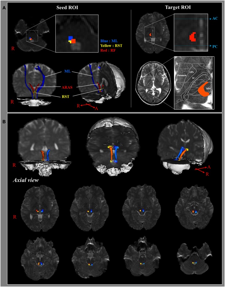
INTRODUCTION: Action of the ascending reticular activating system (ARAS) on the cerebral cortex is responsible for achievement of consciousness. In this study, we attempted to reconstruct the lower single component of the ARAS from the reticular formation (RF) to the thalamus in the normal human brain using diffusion tensor imaging (DTI). METHODS: Twenty six normal healthy subjects were recruited for this study. A 1.5-T scanner was used for scanning of diffusion tensor images, and the lower single component of the ARAS was reconstructed using FMRIB software. We utilized two ROIs for reconstruction of the lower single component of the ARAS: the seed ROI - the RF of the pons at the level of the trigeminal nerve entry zone, the target ROI - the intralaminar nuclei of the thalamus at the level of the commissural plane. RESULTS: The reconstructed ARAS originated from the pontine RF, ascended through the mesencephalic tegmentum just posterior to the red nucleus, and then terminated on the intralaminar nuclei of the thalamus. No significant differences in fractional anisotropy, mean diffusivity, and tract number were observed between hemispheres (p > 0.05). CONCLUSION: We reconstructed the lower single component of the ARAS from the RF to the thalamus in the human brain using DTI. The results of this study might be of value for the diagnosis and prognosis of patients with impaired consciousness.
Overview
AI snapshot
Written from the paper’s full text.
TL;DR
This study used diffusion tensor imaging (DTI) to reconstruct the lower single component of the ascending reticular activating system (ARAS) from the pontine reticular formation to the intralaminar nuclei of the thalamus in 26 healthy adults. The reconstructed pathway was identified in all subjects, with no significant right-left hemispheric differences in FA, MD, or tract number.
Key findings
- The reconstructed ARAS originated from the pontine reticular formation, ascended through the mesencephalic tegmentum just posterior to the red nucleus, and terminated on the intralaminar nuclei of the thalamus at the level of the commissural plane in all subjects.
- Mean DTI values were: right ARAS FA 0.43 ± 0.05, MD 0.96 ± 0.13, tract number 156.96 ± 112.06; left ARAS FA 0.43 ± 0.04, MD 0.94 ± 0.13, tract number 159.73 ± 72.88. No significant differences in FA, MD, or tract number were observed between hemispheres (p > 0.05).
- Pearson correlation showed no significant age-related changes in FA, MD, or tract number (p > 0.05).
Methods
- Design: DTI tractography study in 26 normal healthy subjects (14 males, 12 females; mean age 31.85 ± 9.80 years; range 20–50) with no history of neurologic disease.
- Imaging and analysis: 1.5-T scanner, 32 non-collinear diffusion sensitizing gradients, b = 1000 s/mm², 2.3 mm isotropic voxels; FSL probabilistic tractography (5000 streamline samples, 0.5 mm step lengths, curvature threshold = 0.2) using a seed ROI in the pontine reticular formation at the trigeminal nerve entry zone and a target ROI in the intralaminar nuclei of the thalamus at the commissural plane; FA, MD, and tract number were measured.
- Statistics: paired t-test for right-left hemispheric differences and Pearson correlation for age associations; significance set at p < 0.05.
Limitations
- Stated: the authors could not fully elucidate the entire ARAS because other thalamic and brainstem nuclei involved in the ARAS were not included; the reconstructed tract is described as the main portion, not the entire lower single component of the ARAS.
- Stated: further studies are needed on the clinical usefulness of these findings and on ARAS projections from the thalamus to the cerebral cortex.
- Evident from the text: the sample consisted only of healthy adults, so the diagnostic or prognostic value in patients with impaired consciousness was not directly tested; the study is cross-sectional and uses 1.5-T DTI.
Model-written summary. Check it against the paper before relying on it.
Integrity signals
Registry
Cites
Statistics
Figures
Verification
Claims the paper makes
6 Check pending · 6 No full text of the sources · 4 Own result · 2 Own interpretation · 1 Not checked yet · 1 Own method
A result the paper reports from its own data — it cites no other source for it, so there is none to check. The quote shows it is in the paper.
In the paper
“No significant differences in fractional anisotropy, mean diffusivity, and tract number were observed between hemispheres (p > 0.05).”
- Also stated in: § Results · plain
“No significant differences in FA, MD, and tract number were observed between the right and left hemispheres (p > 0.05) (Table 1).”
- Also stated in: § Discussion · plain
“In addition, values for the FA, MD, and tract numbers of the reconstructed lower single component of the ARAS did not differ significantly between the right and left hemispheres.”
§ The Ascending Reticular Activating System from Pontine Reticular Formation to the Thalamus in the Human Brain > Abstract
plainn = 20- Asserted: passes — The authors put it forward as true.
- Checkable: passes — It can be checked against evidence.
- Load-bearing: unclear — It is unclear whether the paper’s conclusions depend on it.
- Based: passes — The paper gives a reason why it holds.
- Rests on an assumption: In normal white matter, water molecules have relative freedom of movement parallel to nerve fiber tracts, but their movements are restricted across tracts, giving rise to diffusion anisotropy of white matter.
- Also stated in: § Results · plain
In the paper
“In contrast, diffusion tensor imaging (DTI) allows for evaluation of white matter because of its ability to image water diffusion characteristics (Mori et al., 1999).”
§ The Ascending Reticular Activating System from Pontine Reticular Formation to the Thalamus in the Human Brain > Introduction
What the sources say
- Not checked — no full text of the sourcecites Mori et al., 1999 source
plaindirect- Asserted: passes — The authors put it forward as true.
- Checkable: passes — It can be checked against evidence.
- Load-bearing: passes — The paper’s conclusions depend on it.
- Based: passes — The paper gives a reason why it holds.
In the paper
“However, little is known about the whole reconstruction and estimation of the ARAS in the human brain (Edlow et al., 2012).”
§ The Ascending Reticular Activating System from Pontine Reticular Formation to the Thalamus in the Human Brain > Introduction
What the sources say
- Check pendingcites Edlow et al., 2012 source
plain- Asserted: passes — The authors put it forward as true.
- Checkable: passes — It can be checked against evidence.
- Load-bearing: passes — The paper’s conclusions depend on it.
- Based: passes — The paper gives a reason why it holds.
A claim is checked only against the cited paper’s full text, never its abstract, and none is available for its sources.
In the paper
“These neuronal connections originate mainly in the reticular formation (RF) of the brainstem and project through synaptic relays in the intralaminar nucleus of thalamus to the cerebral cortex (Daube, 1986; Paus, 2000; Zeman, 2001; Afifi and Bergman, 2005; Gosseries et al., 2011).”
§ The Ascending Reticular Activating System from Pontine Reticular Formation to the Thalamus in the Human Brain > Introduction
What the sources say
plaindirect- Asserted: passes — The authors put it forward as true.
- Checkable: passes — It can be checked against evidence.
- Load-bearing: passes — The paper’s conclusions depend on it.
- Based: passes — The paper gives a reason why it holds.
In the paper
“Several recent studies have attempted to demonstrate the usefulness of DTI for evaluation of lesions in patients with impaired consciousness and connectivity of specific ARAS nuclei in the human brain (Voss et al., 2006; Perlbarg et al., 2009; Tollard et al., 2009; Tshibanda et al., 2009; Fernandez-Espejo et al., 2010, 2011; Newcombe et al., 2010; Edlow et al., 2012).”
§ The Ascending Reticular Activating System from Pontine Reticular Formation to the Thalamus in the Human Brain > Introduction
What the sources say
plain- Asserted: passes — The authors put it forward as true.
- Checkable: passes — It can be checked against evidence.
- Load-bearing: passes — The paper’s conclusions depend on it.
- Based: passes — The paper gives a reason why it holds.
A conclusion the paper draws from its own results — it cites no other source for it, so there is none to check. The quote shows it is in the paper.
In the paper
“we reconstructed one of the main pathways of the ARAS, the lower single component of the ARAS from the RF to the thalamus in normal subjects”
§ The Ascending Reticular Activating System from Pontine Reticular Formation to the Thalamus in the Human Brain > Discussion
plain- Asserted: passes — The authors put it forward as true.
- Checkable: passes — It can be checked against evidence.
- Load-bearing: passes — The paper’s conclusions depend on it.
- Based: passes — The paper gives a reason why it holds.
What the paper did — it cites no source for this, so there is none to check. The quote shows it is in the paper.
In the paper
“However, one of the limitations of this study is that we were not able to fully elucidate the entire ARAS system because we did not include other thalamic and brainstem nuclei in our analysis which are also involved in the ARAS.”
§ The Ascending Reticular Activating System from Pontine Reticular Formation to the Thalamus in the Human Brain > Discussion
plain- Asserted: passes — The authors put it forward as true.
- Checkable: passes — It can be checked against evidence.
- Load-bearing: unclear — It is unclear whether the paper’s conclusions depend on it.
- Based: passes — The paper gives a reason why it holds.
- Rests on an assumption: The ARAS is composed of several neuronal circuits connecting the brainstem to the cortex.
None of the sources cited for this claim have been checked yet.
In the paper
“Conventional brain MRI, functional neuroimaging techniques, electrophysiological methods, and MR spectroscopy have been used in studies of the ARAS in the human brain (Parvizi and Damasio, 2003; Schiff, 2006; Tshibanda et al., 2009, 2010; Gawryluk et al., 2010).”
§ The Ascending Reticular Activating System from Pontine Reticular Formation to the Thalamus in the Human Brain > Introduction
What the sources say
plain- Asserted: passes — The authors put it forward as true.
- Checkable: passes — It can be checked against evidence.
- Load-bearing: unclear — It is unclear whether the paper’s conclusions depend on it.
- Based: passes — The paper gives a reason why it holds.
A check of a cited source is scheduled, or we don’t hold its full text yet; the claim is checked once it runs.
In the paper
“In addition, several brainstem nuclei (locus coeruleus, dorsal raphe, median raphe, pedunculopontine nucleus, parabrachial nucleus), non-specific thalamic nuclei, hypothalamus, and basal forebrain are also included in the ARAS system (Aston-Jones et al., 2001; Parvizi and Damasio, 2003; Fuller et al., 2011).”
§ The Ascending Reticular Activating System from Pontine Reticular Formation to the Thalamus in the Human Brain > Introduction
What the sources say
plain- Asserted: passes — The authors put it forward as true.
- Checkable: passes — It can be checked against evidence.
- Load-bearing: unclear — It is unclear whether the paper’s conclusions depend on it.
- Based: passes — The paper gives a reason why it holds.
In the paper
“Several studies have demonstrated the clinical usefulness of DTI by estimating some areas of the lower single component of the ARAS from the RF to the thalamus in patients with impaired consciousness (Perlbarg et al., 2009; Tollard et al., 2009; Newcombe et al., 2010; Fernandez-Espejo et al., 2011).”
§ The Ascending Reticular Activating System from Pontine Reticular Formation to the Thalamus in the Human Brain > Discussion
What the sources say
boostedcorrelational- Asserted: passes — The authors put it forward as true.
- Checkable: passes — It can be checked against evidence.
- Load-bearing: unclear — It is unclear whether the paper’s conclusions depend on it.
- Based: passes — The paper gives a reason why it holds.
- It may state two separate claims as one. 6
- Probably not a substantial claim. 7
A result the paper reports from its own data — it cites no other source for it, so there is none to check. The quote shows it is in the paper.
In the paper
“and those of the left ARAS were 0.43 ± 0.04, 0.94 ± 0.13, and 159.73 ± 72.88, respectively.”
§ The Ascending Reticular Activating System from Pontine Reticular Formation to the Thalamus in the Human Brain > Results
plain- Asserted: passes — The authors put it forward as true.
- Checkable: passes — It can be checked against evidence.
- Load-bearing: unclear — It is unclear whether the paper’s conclusions depend on it.
- Based: passes — The paper gives a reason why it holds.
It may state two separate claims as one.
- Rests on an assumption: In normal white matter, water molecules have relative freedom of movement parallel to nerve fiber tracts, but their movements are restricted across tracts, giving rise to diffusion anisotropy of white matter.
A result the paper reports from its own data — it cites no other source for it, so there is none to check. The quote shows it is in the paper.
In the paper
“The reconstructed component of the ARAS originated from the pontine RF, ascended through the mesencephalic tegmentum just posterior to the red nucleus, and then terminated on the intralaminar nuclei of the thalamus at the level of the commissural plane in all subjects (Figure 1B).”
- Also stated in: § Abstract · plain
“The reconstructed ARAS originated from the pontine RF, ascended through the mesencephalic tegmentum just posterior to the red nucleus, and then terminated on the intralaminar nuclei of the thalamus.”
- Also stated in: § Discussion · plain
“Consequently, the lower single component of the ARAS originated from the pontine RF, ascended through the mesencephalic tegmentum posterior to the red nucleus, and then terminated on the intralaminar nuclei of the thalamus.”
§ The Ascending Reticular Activating System from Pontine Reticular Formation to the Thalamus in the Human Brain > Results
plainn = 20- Asserted: passes — The authors put it forward as true.
- Checkable: passes — It can be checked against evidence.
- Load-bearing: passes — The paper’s conclusions depend on it.
- Based: passes — The paper gives a reason why it holds.
It may state two separate claims as one.
- Rests on an assumption: The intralaminar nuclei are the main nuclei of the ARAS among the non-specific thalamic nuclei.
- Rests on an assumption: In normal white matter, water molecules have relative freedom of movement parallel to nerve fiber tracts, but their movements are restricted across tracts, giving rise to diffusion anisotropy of white matter.
- Also stated in: § Abstract · plain
A result the paper reports from its own data — it cites no other source for it, so there is none to check. The quote shows it is in the paper.
In the paper
“Mean values for FA, MD, and fiber volume of the right ARAS were 0.43 ± 0.05, 0.96 ± 0.13, and 156.96 ± 112.06, respectively”
§ The Ascending Reticular Activating System from Pontine Reticular Formation to the Thalamus in the Human Brain > Results
plain- Asserted: passes — The authors put it forward as true.
- Checkable: passes — It can be checked against evidence.
- Load-bearing: unclear — It is unclear whether the paper’s conclusions depend on it.
- Based: passes — The paper gives a reason why it holds.
It may state two separate claims as one.
- Rests on an assumption: In normal white matter, water molecules have relative freedom of movement parallel to nerve fiber tracts, but their movements are restricted across tracts, giving rise to diffusion anisotropy of white matter.
In the paper
“Accordingly, diffusion anisotropy has been used for evaluation of the extent of fiber change in white matter (Chang et al., 2010; Puig et al., 2010).”
§ The Ascending Reticular Activating System from Pontine Reticular Formation to the Thalamus in the Human Brain > Introduction
What the sources say
plaincorrelational- Asserted: passes — The authors put it forward as true.
- Checkable: passes — It can be checked against evidence.
- Load-bearing: passes — The paper’s conclusions depend on it.
- Based: passes — The paper gives a reason why it holds.
Probably not a substantial claim.
In the paper
“The rostral portion of the RF of the brainstem above the trigeminal nerve entry zone is known as the ARAS; in contrast, the caudal portion of the RF is involved in motor function and autonomic function related to cardiac and respiratory function (Daube, 1986).”
§ The Ascending Reticular Activating System from Pontine Reticular Formation to the Thalamus in the Human Brain > Discussion
What the sources say
- Not checked — no full text of the sourcecites Daube, 1986
plain- Asserted: passes — The authors put it forward as true.
- Checkable: passes — It can be checked against evidence.
- Load-bearing: passes — The paper’s conclusions depend on it.
- Based: passes — The paper gives a reason why it holds.
Probably not a substantial claim. It may state two separate claims as one.
A conclusion the paper draws from its own results — it cites no other source for it, so there is none to check. The quote shows it is in the paper.
In the paper
“The results of this study might be of value for the diagnosis and prognosis of patients with impaired consciousness.”
§ The Ascending Reticular Activating System from Pontine Reticular Formation to the Thalamus in the Human Brain > Abstract
hedgedn = 20- Asserted: passes — The authors put it forward as true.
- Checkable: fails — It may be too vague to check against evidence.
- Load-bearing: passes — The paper’s conclusions depend on it.
- Based: fails — The paper may give no reason why it holds.
Probably not a substantial claim. It may be too vague to check against evidence. The paper may give no reason why it holds.
In the paper
“The tract number is determined by the number of voxels contained within a neural tract (Kwak et al., 2010).”
§ The Ascending Reticular Activating System from Pontine Reticular Formation to the Thalamus in the Human Brain > Discussion
What the sources say
- Not checked — no full text of the sourcecites Kwak et al., 2010 source
plaindirect- Asserted: passes — The authors put it forward as true.
- Checkable: passes — It can be checked against evidence.
- Load-bearing: fails — The paper’s conclusions may not depend on it.
- Based: passes — The paper gives a reason why it holds.
Probably not a substantial claim. The paper’s conclusions may not depend on it.
In the paper
“Advantages of probabilistic tractography, which was used in this study, include greater robustness to noise, as well as the ability to detect pathways with sharper angles and to distinguish crossing fibers (Behrens et al., 2007; Winston et al., 2011).”
§ The Ascending Reticular Activating System from Pontine Reticular Formation to the Thalamus in the Human Brain > Materials and Methods > Probabilistic fiber tracking
What the sources say
plain- Asserted: passes — The authors put it forward as true.
- Checkable: passes — It can be checked against evidence.
- Load-bearing: passes — The paper’s conclusions depend on it.
- Based: passes — The paper gives a reason why it holds.
Probably not a substantial claim. It may state two separate claims as one.
A claim is checked only against the cited paper’s full text, never its abstract, and none is available for its sources.
In the paper
“Consciousness is an arousal and awareness of environment and self, which is achieved through action of the ascending reticular activating system (ARAS) on the brain stem and cerebral cortex (Daube, 1986; Paus, 2000; Zeman, 2001; Gosseries et al., 2011).”
§ The Ascending Reticular Activating System from Pontine Reticular Formation to the Thalamus in the Human Brain > Introduction
What the sources say
plaindirect- Asserted: passes — The authors put it forward as true.
- Checkable: passes — It can be checked against evidence.
- Load-bearing: passes — The paper’s conclusions depend on it.
- Based: passes — The paper gives a reason why it holds.
Probably not a substantial claim.
In the paper
“The FA value indicates the degree of directionality and integrity of white matter microstructures such as axons, myelin, and microtubules, and the ADC value indicates the magnitude of water diffusion (Assaf and Pasternak, 2008).”
§ The Ascending Reticular Activating System from Pontine Reticular Formation to the Thalamus in the Human Brain > Discussion
What the sources say
- Not checked — no full text of the sourcecites Assaf and Pasternak, 2008 source
plain- Asserted: passes — The authors put it forward as true.
- Checkable: passes — It can be checked against evidence.
- Load-bearing: fails — The paper’s conclusions may not depend on it.
- Based: passes — The paper gives a reason why it holds.
Probably not a substantial claim. The paper’s conclusions may not depend on it. It may state two separate claims as one.
The paper relies on this without support.
The ARAS cannot be clearly discriminated from adjacent neural structures using conventional brain MRI, functional neuroimaging techniques, electrophysiological methods, and MR spectroscopy, making accurate identification and estimation of the ARAS in the human brain problematic.
“However, because the ARAS cannot be clearly discriminated from adjacent neural structures, accurate identification and estimation of the ARAS in the human brain can be problematic when using these methods.”
The paper relies on this without support.
The intralaminar nuclei are the main nuclei of the ARAS among the non-specific thalamic nuclei.
“We placed the target ROI in the intralaminar nuclei, which are the main nuclei of the ARAS among the non-specific thalamic nuclei.”
The paper relies on this without support.
In normal white matter, water molecules have relative freedom of movement parallel to nerve fiber tracts, but their movements are restricted across tracts, giving rise to diffusion anisotropy of white matter.
“In normal white matter, water molecules have relative freedom of movement parallel to nerve fiber tracts. However, their movements are restricted across tracts, giving rise to diffusion anisotropy of white matter.”
The paper relies on this without support.
The ARAS is composed of several neuronal circuits connecting the brainstem to the cortex.
“The ARAS is composed of several neuronal circuits connecting the brainstem to the cortex.”
Citations
Cites
Yaşlı Kadınlarda Üreme Sağlığı
Kokanalı, D., & Üstün, Y. E. (2019) · DergiPark (Istanbul University) · Cited by 169,621
Advances in functional and structural MR image analysis and implementation as FSL
Smith, S. M., Jenkinson, M., Woolrich, M. W., Beckmann, C. F., Behrens, T. E. J., Johansen-Berg, H., Bannister, P. R., De Luca, M., Drobnjak, I., Flitney, D. E., et al. (2004) · NeuroImage · doi:10.1016/j.neuroimage.2004.07.051 · Cited by 13,630
Probabilistic diffusion tractography with multiple fibre orientations: What can we gain?
Behrens, T. E. J., Berg, H. J., Jbabdi, S., Rushworth, M. F. S., & Woolrich, M. W. (2007) · NeuroImage · doi:10.1016/j.neuroimage.2006.09.018 · Cited by 3,471
Diffusion tensor imaging (DTI)-based white matter mapping in brain research: a review
Assaf, Y., & Pasternak, O. (2008) · Journal of Molecular Neuroscience · doi:10.1007/s12031-007-0029-0 · Cited by 1,537
A neural circuit for circadian regulation of arousal
Aston-Jones, G., Chen, S., Zhu, Y., & Oshinsky, M. L. (2001) · Nature Neuroscience · doi:10.1038/89522 · Cited by 632
Edlow, B. L., Takahashi, E., Wu, O., Benner, T., Dai, G., Bu, L., Grant, P. E., Greer, D. M., Greenberg, S. M., Kinney, H. C., et al. (2012) · Journal of Neuropathology & Experimental Neurology · doi:10.1097/nen.0b013e3182588293 · Cited by 497
Reassessment of the structural basis of the ascending arousal system
Fuller, P., Sherman, D., Pedersen, N. P., Saper, C. B., & Lu, J. (2011) · Journal of Comparative Neurology · doi:10.1002/cne.22559 · Cited by 465
Neuroanatomical correlates of brainstem coma
Parvizi, J. (2003) · Brain · doi:10.1093/brain/awg166 · Cited by 400 · cited 2×
“Conventional brain MRI, functional neuroimaging techniques, electrophysiological methods, and MR spectroscopy have been used in studies of the ARAS in the human brain (Parvizi and Damasio, 2003; Schiff, 2006; Tshibanda et al., 2009, 2010; Gawryluk et al., 2010).”
BackgroundMethodZeman, A. (2001) · Brain · doi:10.1093/brain/124.7.1263 · Cited by 367
Possible axonal regrowth in late recovery from the minimally conscious state
Voss, H. U. (2006) · Journal of Clinical Investigation · doi:10.1172/jci27021 · Cited by 355
Cited by
Sorted by citations
Conlon, B. J., Langguth, B., Hamilton, C., Hughes, S., Meade, E., Connor, C. O., Schecklmann, M., Hall, D. A., Vanneste, S., Leong, S. L., et al. (2020) · Science Translational Medicine · doi:10.1126/scitranslmed.abb2830 · Cited by 118
Sleep disorder-related headaches
Ferini‐Strambi, L., Galbiati, A., & Combi, R. (2019) · Neurological Sciences · doi:10.1007/s10072-019-03837-z · Cited by 103
Structural and functional organization of the midline and intralaminar nuclei of the thalamus
Vertes, R. P., Linley, S. B., & Rojas, A. K. P. (2022) · Frontiers in Behavioral Neuroscience · doi:10.3389/fnbeh.2022.964644 · Cited by 95
Affective Neuroscience Theory and Personality: An Update
Montag, C., & Davis, K. L. (2018) · Personality Neuroscience · doi:10.1017/pen.2018.10 · Cited by 89
The Neurologic Manifestations of Coronavirus Disease 2019 Pandemic: A Systemic Review
Tsai, S.-T., Lu, M., San, S., & Tsai, C. (2020) · Frontiers in Neurology · doi:10.3389/fneur.2020.00498 · Cited by 87
Metadata
- Authors
- Yeo, S. S., Chang, P. H., & Jang, S. H.
- Affiliations
- Yeungnam University (KR)
- Daegu Gyeongbuk Institute of Science and Technology (KR)
- Corresponding author
- Jang, Sung Ho
- Venue
- Frontiers in Human Neuroscience
- Publisher
- Frontiers Media SA
- Published
- 2013
- Volume
- 7
- Pages
- 416-416
- ISSN
- 1662-5161 (ISSN-L)
- PMC
- PMC3722571
- OpenAlex
- W2124163668
- Semantic Scholar
- e3f484fd0bc0f37bcd4849251c32a5f1bd12e3b6
- Cited by
- 162
- References
- 35
- Citations by year
- 2026: 9 · 2025: 7 · 2024: 5 · 2023: 13 · 2022: 13 · 2021: 18 · 2020: 20 · 2019: 15 · 2018: 17 · 2017: 13 · 2016: 14 · 2015: 14 · 2014: 4
- FWCI
- 7.00
- Type
- journal article
- Language
- en
- Open access
- gold
- doi.org
- Licence
- cc-by
- Locations
- Frontiers in Human Neuroscience (publishedVersion, cc-by)
- Europe PMC (PubMed Central) (publishedVersion, cc-by)
- PubMed (publishedVersion)
- PubMed Central (publishedVersion, cc-by)
- DOAJ (DOAJ: Directory of Open Access Journals) (submittedVersion, cc-by-sa)
- Funders
- Daegu Gyeongbuk Institute of Science and Technology
- Topics
- Health Sciences › Medicine › Radiology, Nuclear Medicine and Imaging › Advanced Neuroimaging Techniques and Applications
- Life Sciences › Neuroscience › Cognitive Neuroscience › Functional Brain Connectivity Studies
- Health Sciences › Medicine › Clinical Neurology › Traumatic Brain Injury Research
- Keywords
- ascending reticular activating system, pontine reticular formation, thalamus, intralaminar nuclei, mesencephalic tegmentum, consciousness, diffusion tensor imaging, impaired consciousness, hemispheric lateralization
About these checks
Registry, retraction, Cites and statistics results are computed, not judged, with one exception: a printed reference that no index lists is read once by a model before it counts as a citation error. Claims are extracted by a model and kept only when their quote appears verbatim in the text; each cited source is then checked, with a model’s help, for whether it says what the claim relies on. Those verdicts can be wrong — read the claim’s quote and the cited source before relying on one.
A language model drew the claims from the paper on 2026-11-11, under version 3 of the claim definition (model deepseek-v4.1-flash, prompt extract-v3).
A second model labelled each claim on 2026-11-11 (model jev-1.13-20260917, definition revision gold-guideline-v0.2).
Statistics and arithmetic are recomputed, not judged.
Confidence bands are shown only for fields calibrated on our annotated test set. None is calibrated yet, so no band is shown.
What counts as a claim ›How we measure confidence ›How we check this ›
Are you an author, or do you see a problem with this page? Request a review
