

The DOI matches its Crossref record; 95 of the 95 references it cites were found in a registry or an index.
FAK Structure and Regulation by Membrane Interactions and Force in Focal Adhesions
Open accessAbstract
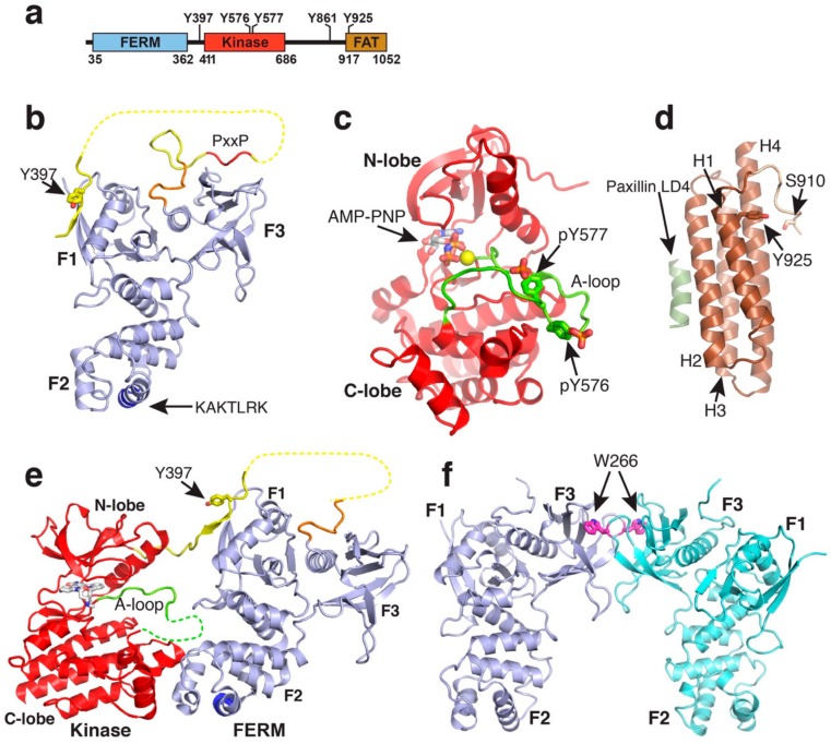
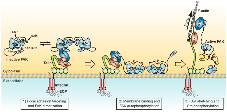
Focal adhesion kinase (FAK) is a non-receptor tyrosine kinase with key roles in the regulation of cell adhesion migration, proliferation and survival. In cancer FAK is a major driver of invasion and metastasis and its upregulation is associated with poor patient prognosis. FAK is autoinhibited in the cytosol, but activated upon localisation into a protein complex, known as focal adhesion complex. This complex forms upon cell adhesion to the extracellular matrix (ECM) at the cytoplasmic side of the plasma membrane at sites of ECM attachment. FAK is anchored to the complex via multiple sites, including direct interactions with specific membrane lipids and connector proteins that attach focal adhesions to the actin cytoskeleton. In migrating cells, the contraction of actomyosin stress fibres attached to the focal adhesion complex apply a force to the complex, which is likely transmitted to the FAK protein, causing stretching of the FAK molecule. In this review we discuss the current knowledge of the FAK structure and how specific structural features are involved in the regulation of FAK signalling. We focus on two major regulatory mechanisms known to contribute to FAK activation, namely interactions with membrane lipids and stretching forces applied to FAK, and discuss how they might induce structural changes that facilitate FAK activation.
Overview
The claims and statistics of this paper haven’t been checked yet.
Run the check — free, no account needed. It reads the open-access text and checks the sources it cites.
Integrity signals
Registry
Cites
Statistics
Figures
Verification
Claims the paper makes
The claims and statistics of this paper haven’t been checked yet.
Run the check — free, no account needed. It reads the open-access text and checks the sources it cites.
Claims other papers make about this one
Each is checked against this paper’s own text: does it say what the citing paper says it does?
The source backs part of the claim, not all of it — or backs it for a different population, condition or comparison.
In the citing paper
“The osteoclast cytoskeleton is made of filamentous structures that belong to one of four categories: polarized actin filaments; polarized microtubules; non-polarized intermediate filaments; or non-polarized septin filaments. The cytoskeleton fulfills essential functions including cell adhesion, migration, contractility, division, vesicular transport, and bone resorption [2,3,4,5,6].”
From Osteoclasts and Microgravity (2020) · cites this paper as [5]
Experimental — accuracy not yet measured
Self-check
Percentages, sums and reported differences re-computed from the paper’s own numbers.
The self-check needs the paper’s full text.
Citations
Cites
Molecular Mechanisms of Stress-Responsive Changes in Collagen and Elastin Networks in Skin
Aziz, J., Shezali, H., Radzi, Z., Yahya, N. A., Abu Kassim, N. H., Czernuszka, J., & Rahman, M. T. (2016) · Skin Pharmacology and Physiology · doi:10.1159/000447017 · Cited by 7,323
Matrix Crosslinking Forces Tumor Progression by Enhancing Integrin Signaling
Levental, K. R., Yu, H., Kass, L., Lakins, J. N., Egeblad, M., Erler, J. T., Fong, S. F. T., Csiszar, K., Giaccia, A., Weninger, W., et al. (2009) · Cell · doi:10.1016/j.cell.2009.10.027 · Cited by 4,101 · cited 1×
“Levental and colleagues [85] used a breast cancer mouse model to show that stiffening of the tumour stroma, induced by increased ECM crosslinking, results in activation of FAK which in turn causes a highly aggressive and invasive behaviour of tumour cells.”
BackgroundReduced cell motility and enhanced focal adhesion contact formation in cells from FAK-deficient mice
llić, D., Furuta, Y., Kanazawa, S., Takeda, N., Sobue, K., Nakatsuji, N., Nomura, S., Fujimoto, J., Okada, M., Yamamoto, T. S., et al. (1995) · Nature · doi:10.1038/377539a0 · Cited by 1,853
Integrin-mediated signal transduction linked to Ras pathway by GRB2 binding to focal adhesion kinase
Schlaepfer, D. D., Hanks, S. K., Hunter, T., & Geer, P. V. D. (1994) · Nature · doi:10.1038/372786a0 · Cited by 1,595 · cited 2×
“In this scenario, force could be a trigger to release the N-terminal FAT helix resulting in exposure of Y925 for phosphorylation by Src. Phosphorylated Y925 is a docking site for the adaptor protein Grb2 [90], which on the one hand links FAK signalling to the Ras/MAPK signalling pathway but was also suggested to recru…”
BackgroundNanoscale architecture of integrin-based cell adhesions
Kanchanawong, P., Shtengel, G. E., Pasapera, A. M., Ramko, E. B., Davidson, M. W., Hess, H. F., & Waterman, C. M. (2010) · Nature · doi:10.1038/nature09621 · Cited by 1,572 · cited 3×
“Super-resolution optical microscopy has defined a layered focal adhesion architecture with a signalling layer closest to the plasma membrane, a central force transduction layer and an actin regulatory layer, the site of actin attachment to the focal adhesion complex [24].”
BackgroundInfluentialMeasuring mechanical tension across vinculin reveals regulation of focal adhesion dynamics
Grashoff, C., Hoffman, B. D., Brenner, M. D., Zhou, R., Parsons, M., Yang, M. T., McLean, M. A., Sligar, S. G., Chen, C. S., Ha, T., et al. (2010) · Nature · doi:10.1038/nature09198 · Cited by 1,549
Stretching Single Talin Rod Molecules Activates Vinculin Binding
Hernández, A. E. D. R., Pérez‐Jiménez, R., Liu, R., Roca‐Cusachs, P., Fernández, J. M., & Sheetz, M. P. (2009) · Science · doi:10.1126/science.1162912 · Cited by 1,492
pp125FAK a structurally distinctive protein-tyrosine kinase associated with focal adhesions.
Schaller, M. D., Borgman, C. A., Cobb, B. S., Vines, R. R., Reynolds, A. B., & Parsons, J. T. (1992) · Proceedings of the National Academy of Sciences · doi:10.1073/pnas.89.11.5192 · Cited by 1,447
FAK in cancer: mechanistic findings and clinical applications
Sulzmaier, F. J., Jean, C., & Schlaepfer, D. D. (2014) · Nature Reviews Cancer · doi:10.1038/nrc3792 · Cited by 1,381 · cited 2×
“The importance of FAK in cell motility is also reflected in disease where FAK plays major roles in tumour invasion and metastasis [11].”
BackgroundFAK integrates growth-factor and integrin signals to promote cell migration
Sieg, D. J., Hauck, C. R., Ilić, D., Klingbeil, C. K., SCHAEFER, E. M., Damsky, C. H., & Schlaepfer, D. D. (2000) · Nature Cell Biology · doi:10.1038/35010517 · Cited by 1,240
Cited by
Sorted by citations
Targeting PI3K/AKT signaling for treatment of idiopathic pulmonary fibrosis
Wang, J., Hu, K., Cai, X., Yang, B., He, Q., Wang, J., & Weng, Q. (2021) · Acta Pharmaceutica Sinica B · doi:10.1016/j.apsb.2021.07.023 · Cited by 476
Dual Role of Reactive Oxygen Species and their Application in Cancer Therapy
Huang, R., Chen, H., Liang, J., Li, Y., Yang, J., Luo, C., Tang, Y., Ding, Y., Liu, X., Yuan, Q., et al. (2021) · Journal of Cancer · doi:10.7150/jca.54699 · Cited by 228
“On the one hand, FAK can lead to focal adhesion, and actin stress fibers located on the inside of the cell membrane adhere to the focal adhesion spot [82].”
BackgroundRoles of focal adhesion proteins in skeleton and diseases
Chen, S., He, T., Zhong, Y., Chen, M., Yao, Q., Chen, D., Shao, Z., & Xiao, G. (2022) · Acta Pharmaceutica Sinica B · doi:10.1016/j.apsb.2022.09.020 · Cited by 157
PROTAC’ing oncoproteins: targeted protein degradation for cancer therapy
Kelm, J. M., Pandey, D. S., Malin, E., Kansou, H. W., Arora, S., Kumar, R., & Gavande, N. S. (2023) · Molecular Cancer · doi:10.1186/s12943-022-01707-5 · Cited by 122
Progress in the Development of Small Molecular Inhibitors of Focal Adhesion Kinase (FAK)
Lu, Y., & Sun, H. (2020) · Journal of Medicinal Chemistry · doi:10.1021/acs.jmedchem.0c01248 · Cited by 104
Metadata
- Authors
- Tapial Martínez, P., López Navajas, P., & Lietha, D.
- Affiliations
- Centro de Investigaciones Biológicas (CIB), Spanish National Research Council (CSIC), Cell Signalling and Adhesion Group, 28040 Madrid, Spain
- Consejo Superior de Investigaciones Científicas (ES)
- Corresponding author
- Lietha, Daniel
- ORCID
- Lietha, Daniel
- Venue
- Biomolecules
- Publisher
- MDPI AG
- Published
- 2020-01-24
- Volume
- 10
- Issue
- 2
- Pages
- 179
- ISSN
- 2218-273X (ISSN-L)
- PMC
- PMC7072507
- OpenAlex
- W3002011373
- Semantic Scholar
- 3830912f29816d1795a68aeaa4e040c80c112f67
- Cited by
- 206
- References
- 95
- Citations by year
- 2026: 16 · 2025: 21 · 2024: 45 · 2023: 44 · 2022: 41 · 2021: 24 · 2020: 15
- FWCI
- 4.32
- Type
- journal article
- Language
- en
- Open access
- gold
- dx.doi.org
- Licence
- Locations
- Biomolecules (publishedVersion, cc-by)
- PubMed Central (publishedVersion, cc-by)
- PubMed (publishedVersion)
- DIGITAL.CSIC (Spanish National Research Council (CSIC)) (submittedVersion, cc-by)
- DOAJ (DOAJ: Directory of Open Access Journals) (submittedVersion, cc-by-sa)
- Funders
- Ministerio de Ciencia, Innovación y Universidades (RTI2018-099318-B-I00, RTI2018)
- Ministerio de Asuntos Económicos y Transformación Digital, Gobierno de España (BFU2016-77665-R)
- European Regional Development Fund (BFU2016, RTI2018, BFU2016-77665-R)
- Agencia Estatal de Investigación
- Topics
- Health Sciences › Medicine › Immunology and Allergy › Cell Adhesion Molecules Research
- Life Sciences › Biochemistry, Genetics and Molecular Biology › Cell Biology › Cellular Mechanics and Interactions
- Life Sciences › Biochemistry, Genetics and Molecular Biology › Molecular Biology › Protein Kinase Regulation and GTPase Signaling
- Keywords
- focal adhesion kinase, focal adhesions, membrane lipid interactions, mechanical forces, FAK activation, non-receptor tyrosine kinase, cell adhesion, extracellular matrix, metastasis, actin cytoskeleton
- MeSH terms
- Cell Adhesion, Cell Movement, Humans, Membranes, Actin Cytoskeleton, Phosphorylation, Signal Transduction, Focal Adhesions, Mechanotransduction, Cellular, Focal Adhesion Kinase 1
About these checks
Registry, retraction, Cites and statistics results are computed, not judged, with one exception: a printed reference that no index lists is read once by a model before it counts as a citation error. Claims are extracted by a model and kept only when their quote appears verbatim in the text; each cited source is then checked, with a model’s help, for whether it says what the claim relies on. Those verdicts can be wrong — read the claim’s quote and the cited source before relying on one.
Statistics and arithmetic are recomputed, not judged.
Confidence bands are shown only for fields calibrated on our annotated test set. None is calibrated yet, so no band is shown.
What counts as a claim ›How we measure confidence ›How we check this ›
Are you an author, or do you see a problem with this page? Request a review
