

The DOI matches its Crossref record; 95 of the 95 references it cites were found in a registry or an index.
FAK Structure and Regulation by Membrane Interactions and Force in Focal Adhesions
Open accessAbstract
Focal adhesion kinase (FAK) is a non-receptor tyrosine kinase with key roles in the regulation of cell adhesion migration, proliferation and survival. In cancer FAK is a major driver of invasion and metastasis and its upregulation is associated with poor patient prognosis. FAK is autoinhibited in the cytosol, but activated upon localisation into a protein complex, known as focal adhesion complex. This complex forms upon cell adhesion to the extracellular matrix (ECM) at the cytoplasmic side of the plasma membrane at sites of ECM attachment. FAK is anchored to the complex via multiple sites, including direct interactions with specific membrane lipids and connector proteins that attach focal adhesions to the actin cytoskeleton. In migrating cells, the contraction of actomyosin stress fibres attached to the focal adhesion complex apply a force to the complex, which is likely transmitted to the FAK protein, causing stretching of the FAK molecule. In this review we discuss the current knowledge of the FAK structure and how specific structural features are involved in the regulation of FAK signalling. We focus on two major regulatory mechanisms known to contribute to FAK activation, namely interactions with membrane lipids and stretching forces applied to FAK, and discuss how they might induce structural changes that facilitate FAK activation.
Overview
The claims and statistics of this paper haven’t been checked yet.
Run the check — free, no account needed. It reads the open-access text and checks the sources it cites.
Integrity signals
Registry
Cites
Statistics
Figures
Verification
Claims the paper makes
The claims and statistics of this paper haven’t been checked yet.
Run the check — free, no account needed. It reads the open-access text and checks the sources it cites.
Claims other papers make about this one
Each is checked against this paper’s own text: does it say what the citing paper says it does?
The source backs part of the claim, not all of it — or backs it for a different population, condition or comparison.
In the citing paper
“The osteoclast cytoskeleton is made of filamentous structures that belong to one of four categories: polarized actin filaments; polarized microtubules; non-polarized intermediate filaments; or non-polarized septin filaments. The cytoskeleton fulfills essential functions including cell adhesion, migration, contractility, division, vesicular transport, and bone resorption [2,3,4,5,6].”
From Osteoclasts and Microgravity (2020) · cites this paper as [5]
Experimental — accuracy not yet measured
Self-check
Percentages, sums and reported differences re-computed from the paper’s own numbers.
The self-check needs the paper’s full text.
Citations
Cites
Molecular Mechanisms of Stress-Responsive Changes in Collagen and Elastin Networks in Skin
Aziz, J., Shezali, H., Radzi, Z., Yahya, N. A., Abu Kassim, N. H., Czernuszka, J., & Rahman, M. T. (2016) · Skin Pharmacology and Physiology · doi:10.1159/000447017 · Cited by 7,323
Matrix Crosslinking Forces Tumor Progression by Enhancing Integrin Signaling
Levental, K. R., Yu, H., Kass, L., Lakins, J. N., Egeblad, M., Erler, J. T., Fong, S. F. T., Csiszar, K., Giaccia, A., Weninger, W., et al. (2009) · Cell · doi:10.1016/j.cell.2009.10.027 · Cited by 4,101 · cited 1×
“Levental and colleagues [85] used a breast cancer mouse model to show that stiffening of the tumour stroma, induced by increased ECM crosslinking, results in activation of FAK which in turn causes a highly aggressive and invasive behaviour of tumour cells.”
BackgroundReduced cell motility and enhanced focal adhesion contact formation in cells from FAK-deficient mice
llić, D., Furuta, Y., Kanazawa, S., Takeda, N., Sobue, K., Nakatsuji, N., Nomura, S., Fujimoto, J., Okada, M., Yamamoto, T. S., et al. (1995) · Nature · doi:10.1038/377539a0 · Cited by 1,853
Integrin-mediated signal transduction linked to Ras pathway by GRB2 binding to focal adhesion kinase
Schlaepfer, D. D., Hanks, S. K., Hunter, T., & Geer, P. V. D. (1994) · Nature · doi:10.1038/372786a0 · Cited by 1,595 · cited 2×
“In this scenario, force could be a trigger to release the N-terminal FAT helix resulting in exposure of Y925 for phosphorylation by Src. Phosphorylated Y925 is a docking site for the adaptor protein Grb2 [90], which on the one hand links FAK signalling to the Ras/MAPK signalling pathway but was also suggested to recru…”
BackgroundNanoscale architecture of integrin-based cell adhesions
Kanchanawong, P., Shtengel, G. E., Pasapera, A. M., Ramko, E. B., Davidson, M. W., Hess, H. F., & Waterman, C. M. (2010) · Nature · doi:10.1038/nature09621 · Cited by 1,572 · cited 3×
“Super-resolution optical microscopy has defined a layered focal adhesion architecture with a signalling layer closest to the plasma membrane, a central force transduction layer and an actin regulatory layer, the site of actin attachment to the focal adhesion complex [24].”
BackgroundInfluentialMeasuring mechanical tension across vinculin reveals regulation of focal adhesion dynamics
Grashoff, C., Hoffman, B. D., Brenner, M. D., Zhou, R., Parsons, M., Yang, M. T., McLean, M. A., Sligar, S. G., Chen, C. S., Ha, T., et al. (2010) · Nature · doi:10.1038/nature09198 · Cited by 1,549
Stretching Single Talin Rod Molecules Activates Vinculin Binding
Hernández, A. E. D. R., Pérez‐Jiménez, R., Liu, R., Roca‐Cusachs, P., Fernández, J. M., & Sheetz, M. P. (2009) · Science · doi:10.1126/science.1162912 · Cited by 1,492
pp125FAK a structurally distinctive protein-tyrosine kinase associated with focal adhesions.
Schaller, M. D., Borgman, C. A., Cobb, B. S., Vines, R. R., Reynolds, A. B., & Parsons, J. T. (1992) · Proceedings of the National Academy of Sciences · doi:10.1073/pnas.89.11.5192 · Cited by 1,447
FAK in cancer: mechanistic findings and clinical applications
Sulzmaier, F. J., Jean, C., & Schlaepfer, D. D. (2014) · Nature Reviews Cancer · doi:10.1038/nrc3792 · Cited by 1,381 · cited 2×
“The importance of FAK in cell motility is also reflected in disease where FAK plays major roles in tumour invasion and metastasis [11].”
BackgroundFAK integrates growth-factor and integrin signals to promote cell migration
Sieg, D. J., Hauck, C. R., Ilić, D., Klingbeil, C. K., SCHAEFER, E. M., Damsky, C. H., & Schlaepfer, D. D. (2000) · Nature Cell Biology · doi:10.1038/35010517 · Cited by 1,240
Functional atlas of the integrin adhesome
Zaidel‐Bar, R., Itzkovitz, S., Ma’ayan, A., Iyengar, R., & Geiger, B. (2007) · Nature Cell Biology · doi:10.1038/ncb0807-858 · Cited by 1,182 · cited 1×
“Reconstitution studies will likely require a reductionist approach, focusing on the core focal adhesion components, rather than the full adhesome of the order of ~150 proteins [20,21].”
BackgroundTargeting focal adhesion kinase renders pancreatic cancers responsive to checkpoint immunotherapy
Jiang, H., Hegde, S., Knolhoff, B. L., Zhu, Y., Herndon, J. M., Meyer, M. A., Nywening, T. M., Hawkins, W. G., Shapiro, I. M., Weaver, D. T., et al. (2016) · Nature Medicine · doi:10.1038/nm.4123 · Cited by 1,060
How cryo-EM is revolutionizing structural biology
Bai, X., McMullan, G., & Scheres, S. H. W. (2014) · Trends in Biochemical Sciences · doi:10.1016/j.tibs.2014.10.005 · Cited by 967 · cited 1×
“Due to the challenges described above, progress will likely come stepwise in the form of substructures and will perhaps lag behind the explosion of high resolution cellular mega-complexes being reported in the post genomic area as a result of the recent revolutionary advances in cryo-electron microscopy [93].”
BackgroundTRPV1 structures in nanodiscs reveal mechanisms of ligand and lipid action
Gao, Y., Cao, E., Julius, D. J., & Cheng, Y. (2016) · Nature · doi:10.1038/nature17964 · Cited by 830 · cited 1×
“Regarding membrane systems, nanodiscs have proven a suitable system for high resolution cryo-EM studies [91].”
Focal adhesion protein-tyrosine kinase phosphorylated in response to cell attachment to fibronectin.
Hanks, S. K., Calalb, M. B., Harper, M. C., & Patel, S. K. (1992) · Proceedings of the National Academy of Sciences · doi:10.1073/pnas.89.18.8487 · Cited by 820 · cited 1×
“Focal adhesion kinase (FAK) was discovered almost three decades ago as a kinase that is highly phosphorylated in response to cell adhesion and localisation into the focal adhesion complex [1,2].”
BackgroundThe Tail of Integrins, Talin, and Kindlins
Moser, M., Legate, K. R., Zent, R., & Fässler, R. (2009) · Science · doi:10.1126/science.1163865 · Cited by 782 · cited 1×
“On the other hand, a high-affinity integrin state initiating ECM binding can be triggered by the binding of intracellular components, such as talin, kindlin, and RIAM [22,23].”
BackgroundStructural Basis of Integrin Activation by Talin
Wegener, K. L., Partridge, A. W., Han, J., Pickford, A. R., Liddington, R., Ginsberg, M. H., & Campbell, I. D. (2007) · Cell · doi:10.1016/j.cell.2006.10.048 · Cited by 677 · cited 1×
“Talin is via its N-terminal FERM domain attached to cytoplasmic tails of β-integrin receptors and the membrane [40,71,72].”
BackgroundPearson, M. A., Reczek, D., Bretscher, A. P., & Karplus, P. A. (2000) · Cell · doi:10.1016/s0092-8674(00)80836-3 · Cited by 614 · cited 1×
“The clover-leaf assembly of the FAK FERM domain is overall very similar to the one seen in the ERM family members ezrin [36], radixin [37], and moesin [38], but different to the FERM domain of talin, which surprisingly is found to arrange its lobes in a linear arrangement [39].”
BackgroundHirata, E., Girotti, M. R., Viros, A., Hooper, S., Spencer-Dene, B., Matsuda, M., Larkin, J., Marais, R., & Sahai, E. (2015) · Cancer Cell · doi:10.1016/j.ccell.2015.03.008 · Cited by 593
Defining Single Molecular Forces Required to Activate Integrin and Notch Signaling
Wang, X., & Ha, T. (2013) · Science · doi:10.1126/science.1231041 · Cited by 572 · cited 1×
“5–10 pN [80,81] with peak forces reaching ~40 pN for molecules engaged with integrin receptors [82].”
BackgroundHorton, E. R., Byron, A., Askari, J. A., Ng, D., Millon‐Frémillon, A., Robertson, J. M., Koper, E., Paul, N. R., Warwood, S., Knight, D., et al. (2015) · Nature Cell Biology · doi:10.1038/ncb3257 · Cited by 557
Focal adhesion kinase links mechanical force to skin fibrosis via inflammatory signaling
Wong, V. W., Rustad, K. C., Akaishi, S., Sorkin, M., Glotzbach, J. P., Januszyk, M., Nelson, E. R., Levi, K., Paterno, J., Vial, I. N., et al. (2012) · Nature Medicine · doi:10.1038/nm.2574 · Cited by 515 · cited 1×
“Cellular strain, applied to cells by stretching of an elastic substrate the cells are growing on, directly increases the force experienced in focal adhesions and is shown to cause increased FAK phosphorylation levels in different cellular systems [28,29].”
BackgroundFocal adhesion kinase is involved in mechanosensing during fibroblast migration
Wang, H.-B., Dembo, M., Hanks, S. K., & Wang, Y. (2001) · Proceedings of the National Academy of Sciences · doi:10.1073/pnas.201201198 · Cited by 514 · cited 1×
“This property is lost upon deletion of FAK [86].”
BackgroundStructural Determinants of Integrin Recognition by Talin
García‐Álvarez, B., de Pereda, J. M., Calderwood, D., Ulmer, T. S., Critchley, D. R., Campbell, I. D., Ginsberg, M. H., & Liddington, R. (2003) · Molecular Cell · doi:10.1016/s1097-2765(02)00823-7 · Cited by 514 · cited 2×
“Talin is via its N-terminal FERM domain attached to cytoplasmic tails of β-integrin receptors and the membrane [40,71,72].”
BackgroundStructural Basis for the Autoinhibition of Focal Adhesion Kinase
Lietha, D., Cai, X., Ceccarelli, D. F. J., Li, Y., Schaller, M. D., & Eck, M. J. (2007) · Cell · doi:10.1016/j.cell.2007.05.041 · Cited by 510 · cited 7×
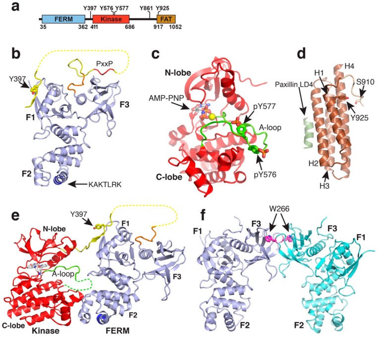
“The phosphorylated Y577 residue interacts with several conserved basic residues in the kinase C-lobe resulting in structuring the activation loop into a typical β-hairpin structure as seen in several other phosphorylated kinase structures [35].”
BackgroundInfluentialSelf-citationType Iγ phosphatidylinositol phosphate kinase targets and regulates focal adhesions
Ling, K., Doughman, R. L., Firestone, A. J., Bunce, M. W., & Anderson, R. A. (2002) · Nature · doi:10.1038/nature01082 · Cited by 443
Di Paolo, G., Pellegrini, L., Letinic, K., Cestra, G., Zoncu, R., Voronov, S., Chang, S., Guo, J., Wenk, M. R., & De Camilli, P. (2002) · Nature · doi:10.1038/nature01147 · Cited by 441 · cited 1×
“The membrane at focal adhesion sites contains increased levels of PIP2 due to the enzyme phosphatidylinositol-4-phosphate-5-kianse type Iγ (PIP5KIγ), which is recruited to focal adhesions by talin to locally generate PIP2 [66,67].”
BackgroundInteraction between focal adhesion kinase and Crk-associated tyrosine kinase substrate p130Cas.
Polte, T. R., & Hanks, S. K. (1995) · Proceedings of the National Academy of Sciences · doi:10.1073/pnas.92.23.10678 · Cited by 420 · cited 1×
“It contains two PxxP motives, site I (P712PKP) interacts with the SH3 domain of p130Cas [47] and site II (P874PKKPP) with the SH3 domains of the small GTPase activating proteins GRAF and ASAP [48,49].”
BackgroundInteraction of Focal Adhesion Kinase with Cytoskeletal Protein Talin
Chen, H.-C., Appeddu, P. A., Parsons, J. T., Hildebrand, J. D., Schaller, M. D., & Guan, J. (1995) · Journal of Biological Chemistry · doi:10.1074/jbc.270.28.16995 · Cited by 392 · cited 1×
“The FAT domain is reported to interact with paxillin [50] and talin [51,52], but only for the paxillin interaction there is structural information available that shows that the FAT domain interacts via two faces with helical LD2 and LD4 motifs of paxillin [53,54].”
BackgroundStructural basis of the membrane‐targeting and unmasking mechanisms of the radixin FERM domain
Hamada, K., Shimizu, T., Matsui, T., Tsukita, S., Tsukita, S., & Hakoshima, T. (2000) · The EMBO Journal · doi:10.1093/emboj/19.17.4449 · Cited by 388 · cited 2×
“The clover-leaf assembly of the FAK FERM domain is overall very similar to the one seen in the ERM family members ezrin [36], radixin [37], and moesin [38], but different to the FERM domain of talin, which surprisingly is found to arrange its lobes in a linear arrangement [39].”
BackgroundBrown, M. C., Perrotta, J. A., & Turner, C. E. (1996) · The Journal of Cell Biology · doi:10.1083/jcb.135.4.1109 · Cited by 368
Schiller, H. B., Friedel, C. C., Boulègue, C., & Fässler, R. (2011) · EMBO Reports · doi:10.1038/embor.2011.5 · Cited by 350 · cited 1×
“Reconstitution studies will likely require a reductionist approach, focusing on the core focal adhesion components, rather than the full adhesome of the order of ~150 proteins [20,21].”
BackgroundExtracellular rigidity sensing by talin isoform-specific mechanical linkages
Austen, K., Ringer, P., Mehlich, A., Chrostek‐Grashoff, A., Kluger, C., Klingner, C., Sabass, B., Zent, R., Rief, M., & Grashoff, C. (2015) · Nature Cell Biology · doi:10.1038/ncb3268 · Cited by 348 · cited 1×
“5–10 pN [80,81] with peak forces reaching ~40 pN for molecules engaged with integrin receptors [82].”
BackgroundHildebrand, J. D., Taylor, J. M., & Parsons, J. T. (1996) · Molecular and Cellular Biology · doi:10.1128/mcb.16.6.3169 · Cited by 342 · cited 1×
“It contains two PxxP motives, site I (P712PKP) interacts with the SH3 domain of p130Cas [47] and site II (P874PKKPP) with the SH3 domains of the small GTPase activating proteins GRAF and ASAP [48,49].”
BackgroundMolecular mechanism of vinculin activation and nanoscale spatial organization in focal adhesions
Case, L. B., Baird, M. A., Shtengel, G. E., Campbell, S. L., Hess, H. F., Davidson, M. W., & Waterman, C. M. (2015) · Nature Cell Biology · doi:10.1038/ncb3180 · Cited by 305 · cited 1×
“In agreement with such a scenario, vinculin is shown to move away from the membrane towards the actin regulatory layer during myosin II mediated focal adhesion maturation [87].”
BackgroundStructural Basis for the Autoinhibition of Talin in Regulating Integrin Activation
Goksoy, E., Ma, Y., Wang, X., Kong, X., Perera, D. H., Plow, E. F., & Qin, J. (2008) · Molecular Cell · doi:10.1016/j.molcel.2008.06.011 · Cited by 265 · cited 1×
“Talin is via its N-terminal FERM domain attached to cytoplasmic tails of β-integrin receptors and the membrane [40,71,72].”
BackgroundA conserved protonation-dependent switch controls drug binding in the Abl kinase
Shan, Y., Seeliger, M. A., Eastwood, M. P., Frank, F., Xu, H., Jensen, M. Ø., Dror, R. O., Kuriyan, J., & Shaw, D. E. (2008) · Proceedings of the National Academy of Sciences · doi:10.1073/pnas.0811223106 · Cited by 264 · cited 1×
“Although a crystal structure of FAK in the “DFG-out” conformation exists bound to a type II inhibitor [45], it is not clear whether FAK adopts this conformation merely as a result of specific ligand interactions, or whether it might be part of a native regulation or catalytic cycle as proposed for other kinases includ…”
BackgroundTalin contains three actin-binding sites each of which is adjacent to a vinculin-binding site
Hemmings, L., Rees, D. J. G., Ohanian, V., Bolton, S. J., Gilmore, A. P., PATEL, B., Priddle, H., Trevithick, J. E., Hynes, R. O., & Critchley, D. R. (1996) · Journal of Cell Science · doi:10.1242/jcs.109.11.2715 · Cited by 260
Direct association of pp125FAK with paxillin, the focal adhesion-targeting mechanism of pp125FAK.
Tachibana, K., Sato, T., D'Avirro, N., & Morimoto, C. (1995) · The Journal of Experimental Medicine · doi:10.1084/jem.182.4.1089 · Cited by 253 · cited 1×
“The FAT domain is reported to interact with paxillin [50] and talin [51,52], but only for the paxillin interaction there is structural information available that shows that the FAT domain interacts via two faces with helical LD2 and LD4 motifs of paxillin [53,54].”
BackgroundFAK promotes recruitment of talin to nascent adhesions to control cell motility
Lawson, C., Lim, S., Uryu, S. A., Chen, X. L., Calderwood, D., & Schlaepfer, D. D. (2012) · The Journal of Cell Biology · doi:10.1083/jcb.201108078 · Cited by 237
Jockusch, B. M., & Isenberg, G. (1981) · Proceedings of the National Academy of Sciences · doi:10.1073/pnas.78.5.3005 · Cited by 234 · cited 1×
“Vinculin acts as an enforcer of the linkage to actin by connecting the talin rod via several vinculin binding sites to actin [75,76].”
BackgroundHayashi, I., Vuori, K., & Liddington, R. (2002) · Nature Structural Biology · doi:10.1038/nsb755 · Cited by 227
Nowakowski, J., Cronin, C. N., McRee, D. E., Knuth, M. W., Nelson, C. G., Pavletich, N. P., Rogers, J., Sang, B., Scheibe, D. N., Swanson, R. V., et al. (2002) · Structure · doi:10.1016/s0969-2126(02)00907-3 · Cited by 227
Dissecting the molecular architecture of integrin adhesion sites by cryo-electron tomography
Patla, I., Volberg, T., Elad, N., Hirschfeld‐Warneken, V. C., Grashoff, C., Fässler, R., Spatz, J. P., Geiger, B., & Medalia, O. (2010) · Nature Cell Biology · doi:10.1038/ncb2095 · Cited by 223 · cited 1×
“A past study has identified donut shaped structures in focal adhesions [92].”
BackgroundMcLean, G. W., Komiyama, N. H., Serrels, B., Asano, H., Reynolds, L. E., Conti, F. J. A., Hodivala‐Dilke, K., METZGER, D., Chambon, P., Grant, S. G. N., et al. (2004) · Genes & Development · doi:10.1101/gad.316304 · Cited by 223 · cited 1×
“In mouse models, FAK is shown to be responsible for skin and breast cancer invasion as well as breast to lung metastasis [12,13].”
BackgroundFocal adhesion kinase signaling in unexpected places
Kleinschmidt, E. G., & Schlaepfer, D. D. (2017) · Current Opinion in Cell Biology · doi:10.1016/j.ceb.2017.01.003 · Cited by 219 · cited 1×
“Although first identified as a focal adhesion protein, FAK is today also known to play important roles in the nucleus, endosomes, and adherens junctions (reviewed in [30]).”
BackgroundWeng, Z., Taylor, J. A., Turner, C. E., Brugge, J. S., & Seidel‐Dugan, C. (1993) · Journal of Biological Chemistry · doi:10.1016/s0021-9258(18)82425-5 · Cited by 214
Gingras, A. R., Ziegler, W. H., Frank, R., Barsukov, I., Roberts, G. C. K., Critchley, D. R., & Emsley, J. (2005) · Journal of Biological Chemistry · doi:10.1074/jbc.m508060200 · Cited by 211 · cited 1×
“Vinculin acts as an enforcer of the linkage to actin by connecting the talin rod via several vinculin binding sites to actin [75,76].”
BackgroundThe structure of the C‐terminal actin‐binding domain of talin
Gingras, A. R., Bate, N., Goult, B. T., Hazelwood, L., Canestrelli, I. L., Grossmann, J. G., Liu, H., Putz, N., Roberts, G. C. K., Volkmann, N., et al. (2007) · The EMBO Journal · doi:10.1038/sj.emboj.7601965 · Cited by 198 · cited 1×
“The long C-terminal rod domain of talin reaches across the force transduction layer and interacts via its most C-terminal helix with actin [73,74].”
BackgroundMammary epithelial-specific disruption of the focal adhesion kinase blocks mammary tumor progression
Lahlou, H., Sanguin‐Gendreau, V., Zuo, D., Cardiff, R. D., McLean, G. W., Frame, M. C., & Muller, W. J. (2007) · Proceedings of the National Academy of Sciences · doi:10.1073/pnas.0710091104 · Cited by 196 · cited 1×
“In mouse models, FAK is shown to be responsible for skin and breast cancer invasion as well as breast to lung metastasis [12,13].”
BackgroundTorsoni, A. S., Constancio, S. S., Nadruz, W., Hanks, S. K., & Franchini, K. G. (2003) · Circulation Research · doi:10.1161/01.res.0000081595.25297.1b · Cited by 195 · cited 1×
“Cellular strain, applied to cells by stretching of an elastic substrate the cells are growing on, directly increases the force experienced in focal adhesions and is shown to cause increased FAK phosphorylation levels in different cellular systems [28,29].”
BackgroundAshton, G. H. S., Morton, J. P., Myant, K., Phesse, T. J., Ridgway, R. A., Marsh, V. L., Wilkins, J. A., Athineos, D., Muncan, V., Kemp, R., et al. (2010) · Developmental Cell · doi:10.1016/j.devcel.2010.07.015 · Cited by 195
Seong, J., Tajik, A., Sun, J., Guan, J., Humphries, M. J., Craig, S. E., Shekaran, A., Garcı́a, A. J., Lu, S., Lin, M. Z., et al. (2013) · Proceedings of the National Academy of Sciences · doi:10.1073/pnas.1307405110 · Cited by 194 · cited 1×
“Further, plating of cells on substrates with different stiffness, where stiffer substrates allow the generation of higher forces in focal adhesions, shows that FAK activity increases on stiffer substrates [27].”
BackgroundSpatial and Temporal Regulation of Focal Adhesion Kinase Activity in Living Cells
Cai, X., Lietha, D., Ceccarelli, D. F. J., Karginov, A. V., Rajfur, Z., Jacobson, K., Hahn, K. M., Eck, M. J., & Schaller, M. D. (2007) · Molecular and Cellular Biology · doi:10.1128/mcb.01324-07 · Cited by 183 · cited 3×
“FAK is a key component of the focal adhesion signalling layer and is shown to directly interact with membrane lipids [25,26].”
BackgroundSelf-citationMechanoresponsive stem cells acquire neural crest fate in jaw regeneration
Ransom, R. C., Carter, A. C., Salhotra, A., Leavitt, T., Marecic, O., Murphy, M. P., Lopez, M. L., Wei, Y., Marshall, C. D., Shen, E. Z., et al. (2018) · Nature · doi:10.1038/s41586-018-0650-9 · Cited by 180
Liu, Y.-H., Loijens, J. C., Martin, K. H., Karginov, A. V., & Parsons, J. (2002) · Molecular Biology of the Cell · doi:10.1091/mbc.e02-01-0018 · Cited by 176 · cited 1×
“It contains two PxxP motives, site I (P712PKP) interacts with the SH3 domain of p130Cas [47] and site II (P874PKKPP) with the SH3 domains of the small GTPase activating proteins GRAF and ASAP [48,49].”
BackgroundFAK promotes recruitment of talin to nascent adhesions to control cell motility
Lawson, C., Lim, S., Uryu, S. A., Chen, X. L., Calderwood, D., & Schlaepfer, D. D. (2012) · The Journal of Cell Biology · doi:10.1083/jcb.2011080781963c · Cited by 170 · cited 2×
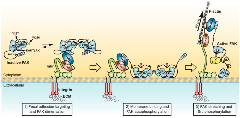
“To this end, in mouse embryo fibroblasts it was observed that FAK is efficiently autophosphorylated in nascent focal adhesions independent of force application [52].”
BackgroundEndothelial-cell FAK targeting sensitizes tumours to DNA-damaging therapy
Tavora, B., Reynolds, L. E., Batista, S., Demircioglu, F., Fernandez, I., Lechertier, T., Lees, D. M., Wong, P.-P., Alexopoulou, A., Elia, G., et al. (2014) · Nature · doi:10.1038/nature13541 · Cited by 163
The Structure of the Talin Head Reveals a Novel Extended Conformation of the FERM Domain
Elliott, P. R., Goult, B. T., Kopp, P., Bate, N., Grossmann, J. G., Roberts, G. C. K., Critchley, D. R., & Barsukov, I. (2010) · Structure · doi:10.1016/j.str.2010.07.011 · Cited by 159
The Structural Basis of Localization and Signaling by the Focal Adhesion Targeting Domain
Arold, S. T., Hoellerer, M. K., & Noble, M. E. M. (2002) · Structure · doi:10.1016/s0969-2126(02)00717-7 · Cited by 158 · cited 1×
“To this end, in one crystal structure the first helix in FAT is swapped between two neighbouring FAT molecules in the crystal [33].”
BackgroundThe Rap1-RIAM-talin axis of integrin activation and blood cell function
Lagarrigue, F., Kim, C., & Ginsberg, M. H. (2016) · Blood · doi:10.1182/blood-2015-12-638700 · Cited by 154 · cited 1×
“On the other hand, a high-affinity integrin state initiating ECM binding can be triggered by the binding of intracellular components, such as talin, kindlin, and RIAM [22,23].”
BackgroundWOOD, C. K., Turner, C. E., Jackson, P., & Critchley, D. R. (1994) · Journal of Cell Science · doi:10.1242/jcs.107.2.709 · Cited by 151
Goñi, G., Epifano, C., Boskovic, J., Camacho‐Artacho, M., Zhou, J., Bronowska, A. K., Martín, M. T., Eck, M. J., Kremer, L., Gräter, F., et al. (2014) · Proceedings of the National Academy of Sciences · doi:10.1073/pnas.1317022111 · Cited by 148 · cited 10×
“mediated FAK activation is shown to be promoted by the plasma membrane lipid phosphatidylinositol 4,5-bisphosphate (PIP2) [26].”
BackgroundInfluentialSelf-citationFAK dimerization controls its kinase‐dependent functions at focal adhesions
Brami‐Cherrier, K., Gervasi, N., Arsenieva, D., Walkiewicz, K. W., Boutterin, M., Ortega, Á., Leonard, P. G., Seantier, B., Gasmi, L., Bouceba, T., et al. (2014) · The EMBO Journal · doi:10.1002/embj.201386399 · Cited by 145 · cited 4×
“These structures have defined the mechanism of FAK autoinhibition in the cytosol [35] and identified a number of interactions, including a mode of recruitment into focal adhesions via paxillin [53,54] and FERM mediated dimerization important for activation [44].”
BackgroundInfluentialFAK activity sustains intrinsic and acquired ovarian cancer resistance to platinum chemotherapy
Diaz Osterman, C. J., Ozmadenci, D., Kleinschmidt, E. G., Taylor, K. N., Barrie, A. M., Jiang, S., Bean, L. M., Sulzmaier, F. J., Jean, C., Tancioni, I., et al. (2019) · eLife · doi:10.7554/elife.47327 · Cited by 145
Ciobănaşu, C., Faivre, B., & Clainche, C. L. (2014) · Nature Communications · doi:10.1038/ncomms4095 · Cited by 141 · cited 1×
“The stretching of talin is shown to reveal cryptic binding sites for vinculin, resulting in stabilization of the focal adhesion complex and a stronger attachment to actin [83,84].”
BackgroundCrystal Structure of the FERM Domain of Focal Adhesion Kinase
Ceccarelli, D. F. J., Song, H. K., Poy, F., Schaller, M. D., & Eck, M. J. (2005) · Journal of Biological Chemistry · doi:10.1074/jbc.m509188200 · Cited by 139 · cited 4×
“Crystallised FERM domains contain most of the linker residues following the FERM domain (residues 362–399 or 362–405, [31]).”
BackgroundMethodInfluentialStructural and mechanistic insights into mechanoactivation of focal adhesion kinase
Bauer, M. S., Baumann, F., Daday, C., Redondo, P. N., Durner, E., Jobst, M. A., Milles, L. F., Mercadante, D., Pippig, D. A., Gaub, H. E., et al. (2019) · Proceedings of the National Academy of Sciences · doi:10.1073/pnas.1820567116 · Cited by 139 · cited 2×
“Further, using atomic force microscopy (AFM) based single molecule force spectroscopy, it was shown that forces applied to FAK molecules via the KAKTLRK lipid binding site in the FERM domain and the C-terminus of the kinase domain (the FAT domain was not included in these studies) results in rupture of autoinhibitory…”
MethodSelf-citationChen, S.-Y., & Chen, H. (2006) · Molecular and Cellular Biology · doi:10.1128/mcb.02186-05 · Cited by 139 · cited 1×
“This sequence is implicated in a number of interactions, including in binding to phosphorylated tails of the Met receptor [43], phosphoinositide lipids [25,26] (see Section 3), and via an intramolecular interaction to the C-terminal FAT domain [44] (see Section 2.”
BackgroundMechanism of Focal Adhesion Kinase Mechanosensing
Zhou, J., Aponte-Santamaria, C. A., Sturm, S., Bullerjahn, J. T., Bronowska, A. K., & Gräter, F. (2015) · PLoS Computational Biology · doi:10.1371/journal.pcbi.1004593 · Cited by 134 · cited 1×
“In a separate study, atomistic molecular dynamics simulations were performed, starting from autoinhibited FAK bound via the KAKTLRK site to an explicit PIP2 containing membrane bilayer [89].”
MethodFERM Domain Interaction Promotes FAK Signaling
Dunty, J., Gabarra-Niecko, V., King, M. L., Ceccarelli, D. F. J., Eck, M. J., & Schaller, M. D. (2004) · Molecular and Cellular Biology · doi:10.1128/mcb.24.12.5353-5368.2004 · Cited by 120 · cited 2×
“FAK mutated in the KAKTLRK sequence or FAK in PIP5KIγ knockdown cells still localise to focal adhesions, indicating that membrane interactions do not significantly contribute to FAK localisation [26,42].”
BackgroundLim, S., Chen, X. L., Tomar, A., Miller, N. L. G., Yoo, J., & Schlaepfer, D. D. (2010) · Journal of Biological Chemistry · doi:10.1074/jbc.m110.129999 · Cited by 118
Structure of the Active N-terminal Domain of Ezrin
Smith, W. J., Nassar, N. N., Bretscher, A. P., Cerione, R. A., & Karplus, P. A. (2003) · Journal of Biological Chemistry · doi:10.1074/jbc.m210601200 · Cited by 110 · cited 1×
“The clover-leaf assembly of the FAK FERM domain is overall very similar to the one seen in the ERM family members ezrin [36], radixin [37], and moesin [38], but different to the FERM domain of talin, which surprisingly is found to arrange its lobes in a linear arrangement [39].”
BackgroundMolecular Recognition of Paxillin LD Motifs by the Focal Adhesion Targeting Domain
Hoellerer, M. K., Noble, M. E. M., Labesse, G., Campbell, I. D., Werner, J. M., & Arold, S. T. (2003) · Structure · doi:10.1016/j.str.2003.08.010 · Cited by 107 · cited 2×
“These structures have defined the mechanism of FAK autoinhibition in the cytosol [35] and identified a number of interactions, including a mode of recruitment into focal adhesions via paxillin [53,54] and FERM mediated dimerization important for activation [44].”
BackgroundHunger‐Glaser, I., Salazar, E. P., Sinnett‐Smith, J. W., & Rozengurt, E. (2003) · Journal of Biological Chemistry · doi:10.1074/jbc.m210876200 · Cited by 107 · cited 3×
“Subsequent studies have provided evidence for the propensity for this helix to detach from the four-helix bundle [56,57] and promote C-terminal phosphorylations on Y861, Y925 and S910 [58,59].”
BackgroundGrant, B. D., & Adams, J. A. (1996) · Biochemistry · doi:10.1021/bi952144+ · Cited by 95 · cited 1×
“A likely explanation provides the study of Grant and Adams [79] showing that, in kinase/substrate complexes and if the enzyme/substrate ratio is close to one, phosphorylation rates are not determined by steady-state kinetics as seen in regular turnover kinetics.”
BackgroundGarces, C. A., Kurenova, E., Golubovskaya, V., et al. (2006) · Cancer Research · doi:10.1158/0008-5472.can-05-1661 · Cited by 84
Gao, G., Prutzman, K. C., King, M. L., Scheswohl, D. M., DeRose, E. F., London, R. E., Schaller, M. D., & Campbell, S. L. (2004) · Journal of Biological Chemistry · doi:10.1074/jbc.m309808200 · Cited by 81 · cited 3×
“These structures have defined the mechanism of FAK autoinhibition in the cytosol [35] and identified a number of interactions, including a mode of recruitment into focal adhesions via paxillin [53,54] and FERM mediated dimerization important for activation [44].”
BackgroundDixon, R. D. S., Chen, Y., Ding, F., Khare, S. D., Prutzman, K. C., Schaller, M. D., Campbell, S. L., & Dokholyan, N. V. (2004) · Structure · doi:10.1016/j.str.2004.09.011 · Cited by 75 · cited 2×
“Subsequent studies have provided evidence for the propensity for this helix to detach from the four-helix bundle [56,57] and promote C-terminal phosphorylations on Y861, Y925 and S910 [58,59].”
BackgroundWu, J.-C., Chen, Y., Kuo, C.-T., et al. (2015) · Scientific Reports · doi:10.1038/srep18476 · Cited by 64 · cited 1×
“These interactions can contribute to Src activation and have been reported to recruit Src into focal adhesions [60].”
BackgroundPlaza-Menacho, I., Morandi, A., Mologni, L., Boender, P. J., Gambacorti-Passerini, C. B., Magee, A. I., Hofstra, R. M. W., Knowles, P. P., McDonald, N. Q., & Isacke, C. M. (2011) · Journal of Biological Chemistry · doi:10.1074/jbc.m110.168500 · Cited by 63
Papusheva, E., de Queiroz, F. M., Dalous, J., et al. (2009) · Journal of Cell Science · doi:10.1242/jcs.028738 · Cited by 55 · cited 1×
“Possibly, membrane binding was also the primary cause for a FRET change in a cellular study that used a similar FRET sensor but found that the sensor did not require FAK activity or autophosphorylation for an optimal FRET response upon localisation into focal adhesions [65].”
BackgroundYoon, S.-O., Shin, S., Karreth, F. A., Buel, G. R., Jedrychowski, M. P., Plas, D. R., Dedhar, S., Gygi, S. P., Roux, P. P., Dephoure, N., et al. (2017) · Molecular Cell · doi:10.1016/j.molcel.2017.06.033 · Cited by 53
Prutzman, K. C., Gao, G., King, M. L., Iyer, V. V., Mueller, G. A., Schaller, M. D., & Campbell, S. L. (2004) · Structure · doi:10.1016/j.str.2004.02.028 · Cited by 42 · cited 3×
“Subsequent studies have provided evidence for the propensity for this helix to detach from the four-helix bundle [56,57] and promote C-terminal phosphorylations on Y861, Y925 and S910 [58,59].”
BackgroundWang, Q., More, S. K., Vomhof‐DeKrey, E. E., Golovko, M. Y., & Basson, M. D. (2019) · Scientific Reports · doi:10.1038/s41598-019-51183-z · Cited by 41
Fragment-based discovery of focal adhesion kinase inhibitors
Grädler, U., Bomke, J., Müsil, D., Dresing, V., Lehmann, M., Hölzemann, G., Greiner, H. E., Esdar, C., Krier, M., & Heinrich, T. (2013) · Bioorganic & Medicinal Chemistry Letters · doi:10.1016/j.bmcl.2013.07.050 · Cited by 40 · cited 1×
“Although a crystal structure of FAK in the “DFG-out” conformation exists bound to a type II inhibitor [45], it is not clear whether FAK adopts this conformation merely as a result of specific ligand interactions, or whether it might be part of a native regulation or catalytic cycle as proposed for other kinases includ…”
BackgroundKadaré, G., Gervasi, N., Brami‐Cherrier, K., Blockus, H., Messari, S. E., Arold, S. T., & Girault, J. (2014) · Journal of Biological Chemistry · doi:10.1074/jbc.m114.593632 · Cited by 38 · cited 5×
“To some extent this might promote dissociation of FAK from focal adhesions [58].”
BackgroundInfluentialMarlowe, T. A., Lenzo, F. L., Figel, S. A., et al. (2016) · Molecular Cancer Therapeutics · doi:10.1158/1535-7163.mct-16-0366 · Cited by 38 · cited 1×
“In oncogenic settings, and particular upon application of FAK inhibitors, it was shown that growth factor receptors can directly phosphorylate Y397 in FAK, thereby sustaining FAK scaffolding functions via phospho-Y397 even if FAK is efficiently inhibited [64].”
BackgroundYuan, C., Gou, X., Deng, J., Dong, Z., Ye, P., & Hu, Z. (2018) · Biomedicine & Pharmacotherapy · doi:10.1016/j.biopha.2018.04.185 · Cited by 34
Structural Insights How PIP2 Imposes Preferred Binding Orientations of FAK at Lipid Membranes
Herzog, F. A., Braun, L., Schoen, I., & Viola Vogel, B. (2017) · The Journal of Physical Chemistry B · doi:10.1021/acs.jpcb.6b09349 · Cited by 34 · cited 1×
“Coarse grain molecular dynamics simulations of autoinhibited FAK on a PIP2 membrane indicated that after membrane binding via the KAKTLRK sequence in the FERM domain the protein would rotate into two possible membrane bound states where FERM and kinase domains lie flat on the membrane with both domains bound to PIP2 […”
BackgroundZhou, Z., Feng, H., & Bai, Y. (2006) · Proteins Structure Function and Bioinformatics · doi:10.1002/prot.21107 · Cited by 30 · cited 2×
“Subsequent studies have provided evidence for the propensity for this helix to detach from the four-helix bundle [56,57] and promote C-terminal phosphorylations on Y861, Y925 and S910 [58,59].”
BackgroundD’Onofrio, P. M., Shabanzadeh, A. P., Choi, B., et al. (2019) · Investigative Ophthalmology & Visual Science · doi:10.1167/iovs.18-25257 · Cited by 21
Marlowe, T. A., Dementiev, A., Figel, S. A., Rivera, A., Flavin, M. T., & Cance, W. G. (2019) · BMC Molecular and Cell Biology · doi:10.1186/s12860-019-0193-4 · Cited by 21 · cited 1×
“Linker residues immediately following the FERM domain (368–375), including a PxxP motif known to constitute a binding site for the Src SH3 domain, are seen in two crystal structures to interact with the FERM F3 lobe and in one FERM molecule with the cleft between the F1 and F3 lobe [31,41].”
BackgroundExpression of Focal Adhesion Proteins in the Developing Rat Kidney
Matsuura, S., Kondo, S., Suga, K., Kinoshita, Y., Urushihara, M., & Kagami, S. (2011) · Journal of Histochemistry & Cytochemistry · doi:10.1369/0022155411413929 · Cited by 16
Phospholipid binding to the FAK catalytic domain impacts function
Hall, J. E., & Schaller, M. D. (2017) · PLoS ONE · doi:10.1371/journal.pone.0172136 · Cited by 7 · cited 3×
“However, a second site on the kinase domain, that includes residues R508, K621 and K627, has recently been shown to also contribute to PIP2 binding [77].”
BackgroundInfluential
Cited by
Sorted by citations
Targeting PI3K/AKT signaling for treatment of idiopathic pulmonary fibrosis
Wang, J., Hu, K., Cai, X., Yang, B., He, Q., Wang, J., & Weng, Q. (2021) · Acta Pharmaceutica Sinica B · doi:10.1016/j.apsb.2021.07.023 · Cited by 476
Dual Role of Reactive Oxygen Species and their Application in Cancer Therapy
Huang, R., Chen, H., Liang, J., Li, Y., Yang, J., Luo, C., Tang, Y., Ding, Y., Liu, X., Yuan, Q., et al. (2021) · Journal of Cancer · doi:10.7150/jca.54699 · Cited by 228
“On the one hand, FAK can lead to focal adhesion, and actin stress fibers located on the inside of the cell membrane adhere to the focal adhesion spot [82].”
BackgroundRoles of focal adhesion proteins in skeleton and diseases
Chen, S., He, T., Zhong, Y., Chen, M., Yao, Q., Chen, D., Shao, Z., & Xiao, G. (2022) · Acta Pharmaceutica Sinica B · doi:10.1016/j.apsb.2022.09.020 · Cited by 157
PROTAC’ing oncoproteins: targeted protein degradation for cancer therapy
Kelm, J. M., Pandey, D. S., Malin, E., Kansou, H. W., Arora, S., Kumar, R., & Gavande, N. S. (2023) · Molecular Cancer · doi:10.1186/s12943-022-01707-5 · Cited by 122
Progress in the Development of Small Molecular Inhibitors of Focal Adhesion Kinase (FAK)
Lu, Y., & Sun, H. (2020) · Journal of Medicinal Chemistry · doi:10.1021/acs.jmedchem.0c01248 · Cited by 104
Metadata
- Authors
- Tapial Martínez, P., López Navajas, P., & Lietha, D.
- Affiliations
- Centro de Investigaciones Biológicas (CIB), Spanish National Research Council (CSIC), Cell Signalling and Adhesion Group, 28040 Madrid, Spain
- Consejo Superior de Investigaciones Científicas (ES)
- Corresponding author
- Lietha, Daniel
- ORCID
- Lietha, Daniel
- Venue
- Biomolecules
- Publisher
- MDPI AG
- Published
- 2020-01-24
- Volume
- 10
- Issue
- 2
- Pages
- 179
- ISSN
- 2218-273X (ISSN-L)
- PMC
- PMC7072507
- OpenAlex
- W3002011373
- Semantic Scholar
- 3830912f29816d1795a68aeaa4e040c80c112f67
- Cited by
- 206
- References
- 95
- Citations by year
- 2026: 16 · 2025: 21 · 2024: 45 · 2023: 44 · 2022: 41 · 2021: 24 · 2020: 15
- FWCI
- 4.32
- Type
- journal article
- Language
- en
- Open access
- gold
- dx.doi.org
- Licence
- Locations
- Biomolecules (publishedVersion, cc-by)
- PubMed Central (publishedVersion, cc-by)
- PubMed (publishedVersion)
- DIGITAL.CSIC (Spanish National Research Council (CSIC)) (submittedVersion, cc-by)
- DOAJ (DOAJ: Directory of Open Access Journals) (submittedVersion, cc-by-sa)
- Funders
- Ministerio de Ciencia, Innovación y Universidades (RTI2018-099318-B-I00, RTI2018)
- Ministerio de Asuntos Económicos y Transformación Digital, Gobierno de España (BFU2016-77665-R)
- European Regional Development Fund (BFU2016, RTI2018, BFU2016-77665-R)
- Agencia Estatal de Investigación
- Topics
- Health Sciences › Medicine › Immunology and Allergy › Cell Adhesion Molecules Research
- Life Sciences › Biochemistry, Genetics and Molecular Biology › Cell Biology › Cellular Mechanics and Interactions
- Life Sciences › Biochemistry, Genetics and Molecular Biology › Molecular Biology › Protein Kinase Regulation and GTPase Signaling
- Keywords
- focal adhesion kinase, focal adhesions, membrane lipid interactions, mechanical forces, FAK activation, non-receptor tyrosine kinase, cell adhesion, extracellular matrix, metastasis, actin cytoskeleton
- MeSH terms
- Cell Adhesion, Cell Movement, Humans, Membranes, Actin Cytoskeleton, Phosphorylation, Signal Transduction, Focal Adhesions, Mechanotransduction, Cellular, Focal Adhesion Kinase 1
About these checks
Registry, retraction, Cites and statistics results are computed, not judged, with one exception: a printed reference that no index lists is read once by a model before it counts as a citation error. Claims are extracted by a model and kept only when their quote appears verbatim in the text; each cited source is then checked, with a model’s help, for whether it says what the claim relies on. Those verdicts can be wrong — read the claim’s quote and the cited source before relying on one.
Statistics and arithmetic are recomputed, not judged.
Confidence bands are shown only for fields calibrated on our annotated test set. None is calibrated yet, so no band is shown.
What counts as a claim ›How we measure confidence ›How we check this ›
Are you an author, or do you see a problem with this page? Request a review
