

The DOI matches its Crossref record; 4 of the 4 references it cites were found in a registry or an index.
Well-Known and Novel Players in Endothelial Dysfunction: Updates on a Notch(ed) Landscape
Abstract
Endothelial dysfunction characterizes every aspect of the so-called cardiovascular continuum, a series of events ranging from hypertension to the development of atherosclerosis and, finally, to coronary heart disease, thrombus formation, myocardial infarction, and heart failure. Endothelial dysfunction is the main prognostic factor for the progression of vascular disorders, which responds to drug intervention and lifestyle changes. Virtually all of the drugs used to prevent cardiovascular disorders, such as long-used and new antilipidemic agents and inhibitors of angiotensin enzyme (ACEi), exert an important effect on the endothelium. Endothelial dysfunction is a central feature of coronavirus disease -19 (COVID-19), and it is now clear that life-risk complications of the disease are prompted by alterations of the endothelium induced by viral infection. As a consequence, the progression of COVID-19 is worse in the subjects in whom endothelial dysfunction is already present, such as elderly, diabetic, obese, and hypertensive patients. Importantly, circulating biomarkers of endothelial activation and injury predict the severity and mortality of the disease and can be used to evaluate the efficacy of treatments. The purpose of this review is to provide updates on endothelial function by discussing its clinical relevance in the cardiovascular continuum, the latest insights from molecular and cellular biology, and their implications for clinical practice, with a focus on new actors, such as the Notch signaling and emerging therapies for cardiovascular disease.
Overview
The claims and statistics of this paper haven’t been checked yet.
Run the check — free, no account needed. It reads the open-access text and checks the sources it cites.
Integrity signals
Registry
Cites
Statistics
Figures
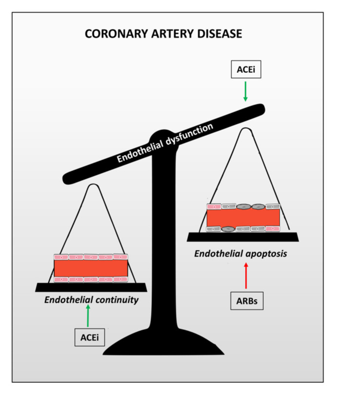
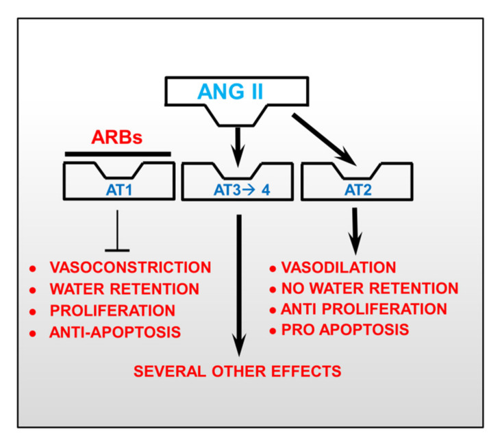
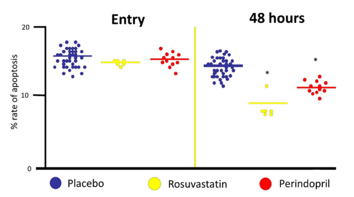
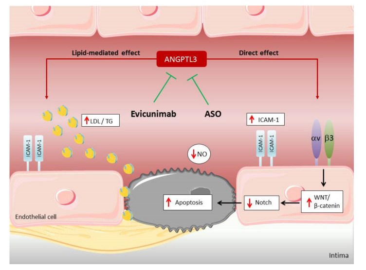
Verification
Claims the paper makes
The claims and statistics of this paper haven’t been checked yet.
Run the check — free, no account needed. It reads the open-access text and checks the sources it cites.
Claims other papers make about this one
Each is checked against this paper’s own text: does it say what the citing paper says it does?
In the citing paper
“CVD, often the top ranked comorbidity after age for severe infection, involves multiple dysfunctions of the vascular endothelium. CVD alters thickness and permeability of the vasculature and those alterations are made worse by endothelium damaging pathogens such as SAR‐CoV‐2, potentially increasing the likelihood of sepsis‐related lung injury (i.e. acute respiratory distress syndrome [ARDS]) (rev”
From On race, human variation, and who gets and dies of sepsis (2022) · cites this paper as Fortini et al., 2021
Self-check
Percentages, sums and reported differences re-computed from the paper’s own numbers.
The self-check needs the paper’s full text.
Citations
Cites
Cell entry mechanisms of SARS-CoV-2
Shang, J., Wan, Y., Luo, C., Ye, G., Geng, Q., Auerbach, A., & Li, F. (2020) · Proceedings of the National Academy of Sciences · doi:10.1073/pnas.2003138117 · Cited by 3,142
ANGPTL3 deficiency and protection against coronary artery disease
Stitziel, N. O., Khera, A. V., Wang, X., Bierhals, A. J., Vourakis, A. C., Sperry, A. E., Natarajan, P., Klarin, D., Emdin, C. A., Zekavat, S. M., et al. (2017) · Journal of the American College of Cardiology · doi:10.1016/j.jacc.2017.02.030 · Cited by 467
KLF2 and KLF4 control endothelial identity and vascular integrity
Sangwung, P., Zhou, G., Nayak, L., Chan, E. R., Kumar, S., Kang, D.-W., Zhang, R., Liao, X., Lu, Y., Sugi, K., et al. (2017) · JCI Insight · doi:10.1172/jci.insight.91700 · Cited by 247
Pouwer, M. G., Pieterman, E. J., Worms, N., Keijzer, N., Jukema, J. W., Gromada, J., Gusarova, V., & Princen, H. M. G. (2020) · Journal of Lipid Research · doi:10.1194/jlr.ra119000419 · Cited by 66
Research works printed but not linked: 89
- Ferrari, 2010, Secondary prevention of CAD with ACE inhibitors: A struggle between life and death of the endothelium, Cardiovasc. Drugs Ther., 24, 331, 10.1007/s10557-010-6244-x · DOI
- Davis, 2004, Shear stress regulates endothelial nitric-oxide synthase promoter activity through nuclear factor kappaB binding, J. Biol. Chem., 279, 163, 10.1074/jbc.M307528200 · DOI
- Walpola, 1995, Expression of ICAM-1 and VCAM-1 and monocyte adherence in arteries exposed to altered shear stress, Arterioscler. Thromb. Vasc. Biol., 15, 2, 10.1161/01.ATV.15.1.2 · DOI
- Sandoo, 2010, The endothelium and its role in regulating vascular tone, Open Cardiovasc. Med. J., 4, 302, 10.2174/1874192401004010302 · DOI
- Cahill, 2016, Vascular endothelium—Gatekeeper of vessel health, Atherosclerosis, 248, 97, 10.1016/j.atherosclerosis.2016.03.007 · DOI · Paper page
- Mollace, 2015, Oxidized LDL attenuates protective autophagy and induces apoptotic cell death of endothelial cells: Role of oxidative stress and LOX-1 receptor expression, Int. J. Cardiol., 184, 152, 10.1016/j.ijcard.2015.02.007 · DOI
- Aquila, 2017, Context-dependent function of ROS in the vascular endothelium: The role of the Notch pathway and shear stress, Biofactors, 43, 475, 10.1002/biof.1359 · DOI
- Aquila, 2018, Heart rate reduction with ivabradine in the early phase of atherosclerosis is protective in the endothelium of ApoE-deficient mice, J. Physiol. Pharmacol., 69, 35
- Fortini, 2019, Notch signaling regulates immune responses in atherosclerosis, Front. Immunol., 10, 1130, 10.3389/fimmu.2019.01130 · DOI
- Souilhol, 2020, Endothelial responses to shear stress in atherosclerosis: A novel role for developmental genes, Nat. Rev. Cardiol., 17, 52, 10.1038/s41569-019-0239-5 · DOI
- Caliceti, 2014, ROS, Notch, and Wnt signaling pathways: Crosstalk between three major regulators of cardiovascular biology, BioMed Res. Int., 2014, 318714, 10.1155/2014/318714 · DOI
- Chapouly, 2020, Desert hedgehog-driven endothelium integrity is enhanced by gas1 (growth arrest-specific 1) but negatively regulated by Cdon (cell adhesion molecule-related/downregulated by oncogenes), Arterioscler. Thromb. Vasc. Biol., 40, e336, 10.1161/ATVBAHA.120.314441 · DOI
- Boopathy, 2019, Role of hippo pathway-YAP/TAZ signaling in angiogenesis, Front. Cell Dev. Biol., 7, 49, 10.3389/fcell.2019.00049 · DOI
- Libby, 2020, COVID-19 is, in the end, an endothelial disease, Eur. Heart J., 41, 3038, 10.1093/eurheartj/ehaa623 · DOI
- Alexander, 2021, Endothelial function in cardiovascular medicine: A consensus paper of the European Society of Cardiology Working Groups on Atherosclerosis and Vascular Biology, Aorta and Peripheral Vascular Diseases, Coronary Pathophysiology and Microcirculation, and Thrombosis, Cardiovasc. Res., 117, 29, 10.1093/cvr/cvaa085 · DOI
- Lee, 2018, Effects of Statins on Coronary Atherosclerotic Plaques: The PARADIGM Study, JACC Cardiovasc. Imaging, 11, 1475, 10.1016/j.jcmg.2018.04.015 · DOI
- Landgraf, 2002, ACE inhibition and atherogenesis, Can. J. Physiol. Pharmacol., 80, 354, 10.1139/y02-038 · DOI
- Rizzo, 2013, The Notch pathway: A crossroad between the life and death of the endothelium, Eur. Heart J., 34, 2504, 10.1093/eurheartj/ehs141 · DOI
- Aquila, 2019, The Notch pathway: A novel therapeutic target for cardiovascular diseases?, Expert Opin. Ther. Targets, 23, 695, 10.1080/14728222.2019.1641198 · DOI
- Mack, 2017, NOTCH1 is a mechanosensor in adult arteries, Nat. Commun., 8, 1620, 10.1038/s41467-017-01741-8 · DOI
- Theodoris, 2015, Human disease modeling reveals integrated transcriptional and epigenetic mechanisms of NOTCH1 haploinsufficiency, Cell, 160, 1072, 10.1016/j.cell.2015.02.035 · DOI · Paper page
- Quillard, 2010, Inflammation dysregulates Notch signaling in endothelial cells: Implication of Notch2 and Notch4 to endothelial dysfunction, Biochem. Pharmacol., 80, 2032, 10.1016/j.bcp.2010.07.010 · DOI
- Fortini, 2017, Estrogen receptor β-dependent Notch1 activation protects vascular endothelium against tumor necrosis factor α (TNFα)-induced apoptosis, J. Biol. Chem., 292, 18178, 10.1074/jbc.M117.790121 · DOI
- Fortini, 2019, Estrogen-mediated protection against coronary heart disease: The role of the Notch pathway, J. Steroid Biochem. Mol. Biol., 189, 87, 10.1016/j.jsbmb.2019.02.008 · DOI
- Quillard, 2008, Impaired Notch4 activity elicits endothelial cell activation and apoptosis: Implication for transplant arteriosclerosis, Arterioscler. Thromb. Vasc. Biol., 28, 2258, 10.1161/ATVBAHA.108.174995 · DOI
- Polacheck, 2017, A non-canonical Notch complex regulates adherens junctions and vascular barrier function, Nature, 552, 258, 10.1038/nature24998 · DOI
- Vieceli Dalla Sega, F., Mastrocola, R., Aquila, G., Fortini, F., Fornelli, C., Zotta, A., Cento, A.S., Perrelli, A., Boda, E., and Pannuti, A. (2019). KRIT1 Deficiency Promotes Aortic Endothelial Dysfunction. Int. J. Mol. Sci., 20. · DOI
- Briot, 2015, Endothelial NOTCH1 is suppressed by circulating lipids and antagonizes inflammation during atherosclerosis, J. Exp. Med., 212, 2147, 10.1084/jem.20150603 · DOI
- Caliceti, C., Aquila, G., Pannella, M., Morelli, M.B., Fortini, C., Pinton, P., Bonora, M., Hrelia, S., Pannuti, A., and Miele, L. (2013). 17β-estradiol enhances signalling mediated by VEGF-A-delta-like ligand 4-notch1 axis in human endothelial cells. PLoS ONE, 8. · DOI
- Norum, 2016, Increased serum levels of the notch ligand DLL1 are associated with diastolic dysfunction, reduced exercise capacity, and adverse outcome in chronic heart failure, J. Card. Fail., 22, 218, 10.1016/j.cardfail.2015.07.012 · DOI
- Rizzo, 2020, COVID-19 in the heart and the lungs: Could we “Notch” the inflammatory storm?, Basic Res. Cardiol., 115, 31, 10.1007/s00395-020-0791-5 · DOI
- Breikaa, 2021, The Notch Pathway: A Link between COVID-19 Pathophysiology and Its Cardiovascular Complications, Front. Cardiovasc. Med., 8, 681948, 10.3389/fcvm.2021.681948 · DOI
- Iaconelli, 2020, First detection of SARS-CoV-2 in untreated wastewaters in Italy, Sci. Total Environ., 736, 139652, 10.1016/j.scitotenv.2020.139652 · DOI
- Vandelli, 2020, Structural analysis of SARS-CoV-2 genome and predictions of the human interactome, Nucleic Acids Res., 48, 11270, 10.1093/nar/gkaa864 · DOI
- Harb, 2021, Notch4 signaling limits regulatory T-cell-mediated tissue repair and promotes severe lung inflammation in viral infections, Immunity, 54, 1186, 10.1016/j.immuni.2021.04.002 · DOI
- Cangiano, 2011, ACE inhibition modulates endothelial apoptosis and renewal via endothelial progenitor cells in patients with acute coronary syndromes, Am. J. Cardiovasc. Drugs, 11, 189, 10.2165/11589400-000000000-00000 · DOI
- Ferrari, 2014, Coronary artery disease: Use of ACE inhibitors in stable CAD—what is the truth?, Nat. Rev. Cardiol., 11, 315, 10.1038/nrcardio.2014.56 · DOI
- Agnoletti, 1999, Serum from patients with severe heart failure downregulates eNOS and is proapoptotic: Role of tumor necrosis factor-alpha, Circulation, 100, 1983, 10.1161/01.CIR.100.19.1983 · DOI
- Valgimigli, 2003, Serum from patients with acute coronary syndromes displays a proapoptotic effect on human endothelial cells: A possible link to pan-coronary syndromes, Circulation, 107, 264, 10.1161/01.CIR.0000045665.57256.86 · DOI
- Bachetti, 1998, Effects of chronic noradrenaline on the nitric oxide pathway in human endothelial cells, Basic Res. Cardiol., 93, 250, 10.1007/s003950050092 · DOI
- Ceconi, 2007, Differences in the effect of angiotensin-converting enzyme inhibitors on the rate of endothelial cell apoptosis: In vitro and in vivo studies, Cardiovasc. Drugs Ther., 21, 423, 10.1007/s10557-007-6068-5 · DOI
- Strauss, 2006, Angiotensin receptor blockers may increase risk of myocardial infarction: Unraveling the ARB-MI paradox, Circulation, 114, 838, 10.1161/CIRCULATIONAHA.105.594986 · DOI
- Ferrari, 2013, The impact of ACE inhibition on all-cause and cardiovascular mortality in contemporary hypertension trials: A review, Expert Rev. Cardiovasc. Ther., 11, 705, 10.1586/erc.13.42 · DOI
- Bangalore, 2016, Angiotensin-converting enzyme inhibitors or angiotensin receptor blockers in patients without heart failure? Insights from 254,301 patients from randomized trials, Mayo Clin. Proc., 91, 51, 10.1016/j.mayocp.2015.10.019 · DOI
- Savarese, 2013, A meta-analysis reporting effects of angiotensin-converting enzyme inhibitors and angiotensin receptor blockers in patients without heart failure, J. Am. Coll. Cardiol., 61, 131, 10.1016/j.jacc.2012.10.011 · DOI
- Cheng, 2014, Effect of angiotensin-converting enzyme inhibitors and angiotensin II receptor blockers on all-cause mortality, cardiovascular deaths, and cardiovascular events in patients with diabetes mellitus: A meta-analysis, JAMA Intern. Med., 174, 773, 10.1001/jamainternmed.2014.348 · DOI
- Thomopoulos, 2015, Effects of blood pressure lowering on outcome incidence in hypertension: 4. Effects of various classes of antihypertensive drugs--overview and meta-analyses, J. Hypertens., 33, 195, 10.1097/HJH.0000000000000447 · DOI
- Messerli, 2017, Angiotensin receptor blockers reduce cardiovascular events, including the risk of myocardial infarction, Circulation, 135, 2085, 10.1161/CIRCULATIONAHA.116.025950 · DOI
- Strauss, 2017, Angiotensin receptor blockers do not reduce risk of myocardial infarction, cardiovascular death, or total mortality: Further evidence for the ARB-MI paradox, Circulation, 135, 2088, 10.1161/CIRCULATIONAHA.117.026112 · DOI
- Ceconi, 2007, ACE inhibition with perindopril and endothelial function. Results of a substudy of the EUROPA study: PERTINENT, Cardiovasc. Res., 73, 237, 10.1016/j.cardiores.2006.10.021 · DOI
- Agnoletti, 2007, Rosuvastatin stimulates eNOS and inhibits apoptosis in HUVECs exposed to sera from cardiovascular diseases patients, J. Mol. Cell. Cardiol., 42, S226, 10.1016/j.yjmcc.2007.03.681 · DOI
- Zacharek, 2009, Simvastatin increases notch signaling activity and promotes arteriogenesis after stroke, Stroke, 40, 254, 10.1161/STROKEAHA.108.524116 · DOI · Paper page
- Musunuru, 2010, Exome sequencing, ANGPTL3 mutations, and familial combined hypolipidemia, N. Engl. J. Med., 363, 2220, 10.1056/NEJMoa1002926 · DOI · Paper page
- Xu, 2018, Role of angiopoietin-like 3 (ANGPTL3) in regulating plasma level of low-density lipoprotein cholesterol, Atherosclerosis, 268, 196, 10.1016/j.atherosclerosis.2017.08.031 · DOI · Paper page
- Camenisch, 2002, ANGPTL3 stimulates endothelial cell adhesion and migration via integrin alpha vbeta 3 and induces blood vessel formation in vivo, J. Biol. Chem., 277, 17281, 10.1074/jbc.M109768200 · DOI
- Yi, 2016, Integrin alphavbeta3 enhances β-catenin signaling in acute myeloid leukemia harboring Fms-like tyrosine kinase-3 internal tandem duplication mutations: Implications for microenvironment influence on sorafenib sensitivity, Oncotarget, 7, 40387, 10.18632/oncotarget.9617 · DOI
- Zheng, 2018, Glycogen synthase kinase 3β inhibition enhances Notch1 recycling, Mol. Biol. Cell, 29, 389, 10.1091/mbc.E17-07-0474 · DOI
- Herrmann, 2006, Angiogenesis in atherogenesis, Arterioscler. Thromb. Vasc. Biol., 26, 1948, 10.1161/01.ATV.0000233387.90257.9b · DOI
- Perico, 2021, Immunity, endothelial injury and complement-induced coagulopathy in COVID-19, Nat. Rev. Nephrol., 17, 46, 10.1038/s41581-020-00357-4 · DOI
- Hamming, 2004, Tissue distribution of ACE2 protein, the functional receptor for SARS coronavirus. A first step in understanding SARS pathogenesis, J. Pathol., 203, 631, 10.1002/path.1570 · DOI · Paper page
- Monteil, 2020, Inhibition of SARS-CoV-2 infections in engineered human tissues using clinical-grade soluble human ACE2, Cell, 181, 905, 10.1016/j.cell.2020.04.004 · DOI · Paper page
- Ackermann, 2020, Pulmonary vascular endothelialitis, thrombosis, and angiogenesis in COVID-19, N. Engl. J. Med., 383, 120, 10.1056/NEJMoa2015432 · DOI · Paper page
- Varga, 2020, Endothelial cell infection and endotheliitis in COVID-19, Lancet, 395, 1417, 10.1016/S0140-6736(20)30937-5 · DOI · Paper page
- Bryce, 2020, Central nervous system involvement by severe acute respiratory syndrome coronavirus-2 (SARS-CoV-2), J. Med. Virol., 92, 699, 10.1002/jmv.25915 · DOI · Paper page
- Colmenero, 2020, SARS-CoV-2 endothelial infection causes COVID-19 chilblains: Histopathological, immunohistochemical and ultrastructural study of seven paediatric cases, Br. J. Dermatol., 183, 729, 10.1111/bjd.19327 · DOI
- Goldsmith, 2020, Electron microscopy of SARS-CoV-2: A challenging task, Lancet, 395, e99, 10.1016/S0140-6736(20)31188-0 · DOI
- Dittmayer, 2020, Why misinterpretation of electron micrographs in SARS-CoV-2-infected tissue goes viral, Lancet, 396, e64, 10.1016/S0140-6736(20)32079-1 · DOI
- Schaefer, 2020, In situ detection of SARS-CoV-2 in lungs and airways of patients with COVID-19, Mod. Pathol., 33, 2104, 10.1038/s41379-020-0595-z · DOI
- Ahmetaj-Shala, B., Peacock, T.P., Baillon, L., Swann, O.C., Gashaw, H., Barclay, W.S., and Mitchell, J.A. (2020). Resistance of endothelial cells to SARS-CoV-2 infection in vitro. bioRxiv. · DOI
- Liu, 2021, SARS-CoV-2 Infects Endothelial Cells, Front. Cell Infect. Microbiol., 11, 701278, 10.3389/fcimb.2021.701278 · DOI
- Teuwen, 2020, COVID-19: The vasculature unleashed, Nat. Rev. Immunol., 20, 389, 10.1038/s41577-020-0343-0 · DOI
- Rauch, 2020, Endotheliopathy is induced by plasma from critically ill patients and associated with organ failure in severe COVID-19, Circulation, 142, 1881, 10.1161/CIRCULATIONAHA.120.050907 · DOI
- Canzano, 2021, Platelet and endothelial activation as potential mechanisms behind the thrombotic complications of COVID-19 patients, JACC Basic Transl. Sci., 6, 202, 10.1016/j.jacbts.2020.12.009 · DOI
- Campo, 2021, Over time relationship between platelet reactivity, myocardial injury and mortality in patients with SARS-CoV-2-associated respiratory failure, Platelets., 32, 560, 10.1080/09537104.2020.1852543 · DOI
- Evans, 2020, Endothelial dysfunction in COVID-19: A position paper of the ESC Working Group for Atherosclerosis and Vascular Biology, and the ESC Council of Basic Cardiovascular Science, Cardiovasc. Res., 116, 2177, 10.1093/cvr/cvaa230 · DOI
- Bonaventura, 2018, The pathophysiological role of neutrophil extracellular traps in inflammatory diseases, Thromb. Haemost., 118, 6, 10.1160/TH17-09-0630 · DOI
- Radermecker, 2020, Neutrophil extracellular traps infiltrate the lung airway, interstitial, and vascular compartments in severe COVID-19, J. Exp. Med., 217, e20201012, 10.1084/jem.20201012 · DOI
- Dupont, 2021, Vascular endothelial damage in the pathogenesis of organ injury in severe COVID-19, Arterioscler. Thromb. Vasc. Biol., 41, 1760, 10.1161/ATVBAHA.120.315595 · DOI
- Fortini, 2021, Time course of endothelial dysfunction markers and mortality in COVID-19 patients: A pilot study, Clin. Transl. Med., 11, e283, 10.1002/ctm2.283 · DOI
- Spadaro, 2021, Markers of endothelial and epithelial pulmonary injury in mechanically ventilated COVID-19 ICU patients, Crit. Care, 25, 74, 10.1186/s13054-021-03499-4 · DOI
- Goshua, 2020, Endotheliopathy in COVID-19-associated coagulopathy: Evidence from a single-centre, cross-sectional study, Lancet Haematol., 7, e575, 10.1016/S2352-3026(20)30216-7 · DOI
- Smadja, 2020, Angiopoietin-2 as a marker of endothelial activation is a good predictor factor for intensive care unit admission of COVID-19 patients, Angiogenesis, 23, 611, 10.1007/s10456-020-09730-0 · DOI
- Guervilly, 2020, Circulating Endothelial Cells as a Marker of Endothelial Injury in Severe COVID-19, J. Infect. Dis., 222, 1789, 10.1093/infdis/jiaa528 · DOI
- Campo, 2017, Biological effects of ticagrelor over clopidogrel in patients with stable coronary artery disease and chronic obstructive pulmonary disease, Thromb. Haemost., 117, 1208, 10.1160/TH16-12-0973 · DOI
- Nagashima, 2020, Endothelial Dysfunction and Thrombosis in Patients With COVID-19-Brief Report, Arterioscler. Thromb. Vasc. Biol., 40, 2404, 10.1161/ATVBAHA.120.314860 · DOI
- Swanson, 2019, The NLRP3 inflammasome: Molecular activation and regulation to therapeutics, Nat. Rev. Immunol., 19, 477, 10.1038/s41577-019-0165-0 · DOI · Paper page
- Rodrigues, 2021, Inflammasomes are activated in response to SARS-CoV-2 infection and are associated with COVID-19 severity in patients, J. Exp. Med., 218, e20201707, 10.1084/jem.20201707 · DOI · Paper page
- Jia, 2019, Endothelial cell pyroptosis plays an important role in Kawasaki disease via HMGB1/RAGE/cathespin B signaling pathway and NLRP3 inflammasome activation, Cell Death. Dis., 10, 778, 10.1038/s41419-019-2021-3 · DOI
- Mitra, 2018, Microparticulate Caspase 1 Regulates Gasdermin D and Pulmonary Vascular Endothelial Cell Injury, Am. J. Respir. Cell Mol. Biol., 59, 56, 10.1165/rcmb.2017-0393OC · DOI
Cited by
Sorted by citations
On race, human variation, and who gets and dies of sepsis
Brinkworth, J. F., & Shaw, J. G. (2022) · American Journal of Biological Anthropology · doi:10.1002/ajpa.24527 · Cited by 9
Metadata
- Authors
- Fortini, F., Vieceli Dalla Sega, F., Marracino, L., Severi, P., Rapezzi, C., Rizzo, P., & Ferrari, R.
- Affiliations
- Maria Cecilia Hospital, GVM Care & Research, 48033 Cotignola, Italy
- Laboratory for Technologies of Advanced Therapies (LTTA), Department of Translational Medicine, University of Ferrara, 44121 Ferrara, Italy
- Venue
- Biomedicines
- Publisher
- MDPI AG
- Published
- 2021-08-11
- Volume
- 9
- Issue
- 8
- Pages
- 997
- ISSN
- 2227-9059
- PMC
- PMC8393382
- Cited by
- 30
- References
- 4
- Language
- en
About these checks
Registry, retraction, Cites and statistics results are computed, not judged, with one exception: a printed reference that no index lists is read once by a model before it counts as a citation error. Claims are extracted by a model and kept only when their quote appears verbatim in the text; each cited source is then checked, with a model’s help, for whether it says what the claim relies on. Those verdicts can be wrong — read the claim’s quote and the cited source before relying on one.
Statistics and arithmetic are recomputed, not judged.
Confidence bands are shown only for fields calibrated on our annotated test set. None is calibrated yet, so no band is shown.
What counts as a claim ›How we measure confidence ›How we check this ›
Are you an author, or do you see a problem with this page? Request a review
