

The DOI matches its Crossref record; 218 of the 218 references it cites were found in a registry or an index.
Oxidative Stress in Human Pathology and Aging: Molecular Mechanisms and Perspectives
Open accessAbstract
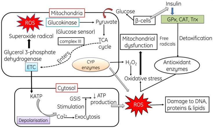
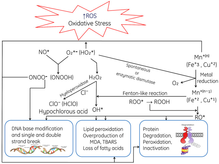
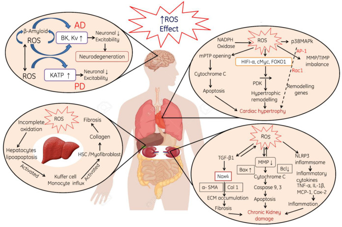
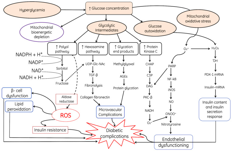
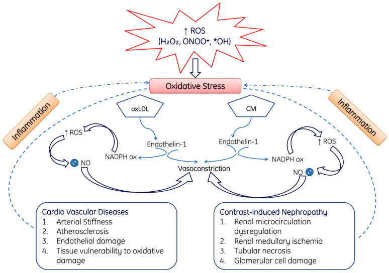
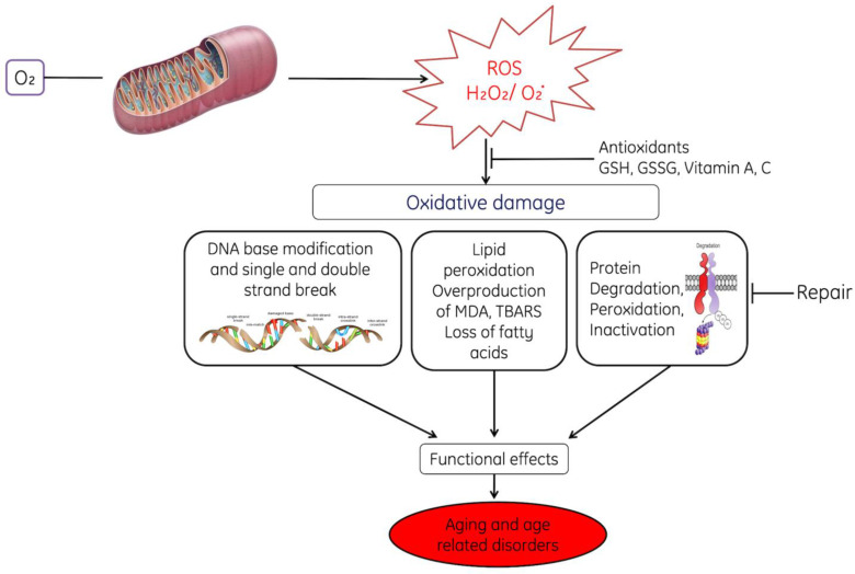
Reactive oxygen and nitrogen species (RONS) are generated through various endogenous and exogenous processes; however, they are neutralized by enzymatic and non-enzymatic antioxidants. An imbalance between the generation and neutralization of oxidants results in the progression to oxidative stress (OS), which in turn gives rise to various diseases, disorders and aging. The characteristics of aging include the progressive loss of function in tissues and organs. The theory of aging explains that age-related functional losses are due to accumulation of reactive oxygen species (ROS), their subsequent damages and tissue deformities. Moreover, the diseases and disorders caused by OS include cardiovascular diseases [CVDs], chronic obstructive pulmonary disease, chronic kidney disease, neurodegenerative diseases and cancer. OS, induced by ROS, is neutralized by different enzymatic and non-enzymatic antioxidants and prevents cells, tissues and organs from damage. However, prolonged OS decreases the content of antioxidant status of cells by reducing the activities of reductants and antioxidative enzymes and gives rise to different pathological conditions. Therefore, the aim of the present review is to discuss the mechanism of ROS-induced OS signaling and their age-associated complications mediated through their toxic manifestations in order to devise effective preventive and curative natural therapeutic remedies.
Overview
The claims and statistics of this paper haven’t been checked yet.
Run the check — free, no account needed. It reads the open-access text and checks the sources it cites.
Integrity signals
Registry
Cites
Statistics
Figures
Verification
Claims the paper makes
The claims and statistics of this paper haven’t been checked yet.
Run the check — free, no account needed. It reads the open-access text and checks the sources it cites.
Claims other papers make about this one
Each is checked against this paper’s own text: does it say what the citing paper says it does?
In the citing paper
“The theory of mitochondrial free radicals in senescence proposes that progressive mitochondrial dysfunction, which occurs with aging, results in an increased production of ROS, which in turn causes further mitochondrial and global cellular damage.”
From The Role of Antioxidants in the Interplay between Oxidative Stress and Senescence (2022) · cites this paper as [46]
Self-check
Percentages, sums and reported differences re-computed from the paper’s own numbers.
The self-check needs the paper’s full text.
Citations
Cites
López-Otín, C., Blasco, M. A., Partridge, L., Serrano, M., & Kroemer, G. (2013) · Cell · doi:10.1016/j.cell.2013.05.039 · Cited by 14,982
Free radicals and antioxidants in normal physiological functions and human disease
Valko, M., Leibfritz, D., Moncol, J., Cronin, M. T. D., Mazur, M., & Telser, J. (2007) · The International Journal of Biochemistry & Cell Biology · doi:10.1016/j.biocel.2006.07.001 · Cited by 14,269
Apoptosis: A Review of Programmed Cell Death
Elmore, S. (2007) · Toxicologic Pathology · doi:10.1080/01926230701320337 · Cited by 13,853
Free Radicals in the Physiological Control of Cell Function
Dröge, W. (2002) · Physiological Reviews · doi:10.1152/physrev.00018.2001 · Cited by 9,855
Oxidants, oxidative stress and the biology of ageing
Finkel, T., & Holbrook, N. J. (2000) · Nature · doi:10.1038/35041687 · Cited by 9,413
The NOX Family of ROS-Generating NADPH Oxidases: Physiology and Pathophysiology
Bedard, K., & Krause, K.-H. (2007) · Physiological Reviews · doi:10.1152/physrev.00044.2005 · Cited by 6,989
ROS function in redox signaling and oxidative stress
Schieber, M., & Chandel, N. S. (2014) · Current Biology · doi:10.1016/j.cub.2014.03.034 · Cited by 6,931
Mitochondrial Dysfunction and Oxidative Stress in Neurodegenerative Diseases
Lin, M. T., & Beal, M. F. (2006) · Nature · doi:10.1038/nature05292 · Cited by 6,455
Nitric Oxide and Peroxynitrite in Health and Disease
Pacher, P., Beckman, J. S., & Liaudet, L. (2007) · Physiological Reviews · doi:10.1152/physrev.00029.2006 · Cited by 6,390
Free radicals, antioxidants and functional foods: impact on human health
Lobo, V, Patil, A., Phatak, A., & Chandra, N. (2010) · Pharmacognosy Reviews · doi:10.4103/0973-7847.70902 · Cited by 5,435
Research works printed but not linked: 1
- Hajam, 2017, Retrieval of reproductive complications by exogenous melatonin treatment in streptozotocin induced diabetic rat model, Res. Rev. J. Zool. Sci., 5, 96
Cited by
Sorted by citations
Items 601–700 of 916
Understanding Cellular Senescence and Senotherapeutics for Nutri-geriatric Solutions
Chawla, S., Bhattacharya, S., & Gupta, A. (2025) · Biomedical & Pharmacology Journal · doi:10.13005/bpj/3214 · Cited by 2
Cell membrane-based nanovesicles as potential drug delivery systems for atherosclerosis therapy
Beltran, O., Costa-Santos, A., Luna, M., Abelenda, Ó., Ogando, A., Topete, A., Juárez, J., Barbosa, S., & Taboada, P. (2025) · Colloids and Surfaces B Biointerfaces · doi:10.1016/j.colsurfb.2025.115190 · Cited by 1
Hossain, M. S., & Alam, A. K. (2025) · Biochemistry · doi:10.5772/intechopen.1012362 · Cited by 1
Suwitono, M. R., Wahyuniari, I. A. I., Luo, H., Suwitono, M. C., & Sulastri, T. (2025) · Ankara Universitesi Eczacilik Fakultesi Dergisi · doi:10.33483/jfpau.1588979 · Cited by 2
Oxidative Stress and Antioxidants in the Aging Brain
Pamu, S., Potdar, M. B., Parameshwar, K., Rajput, D., & Kaushik, M. (2025) · Nutritional neurosciences · doi:10.1007/978-981-95-2790-8_7 · Cited by 1
Loubane, G., Robert, G., Firdaus, S. B., Robidas, R., Comeau, C., Boudreault, P., Komba, J. E., Gagnon, H., Wagner, J. R., Naylor, S., et al. (2025) · Biochimica et Biophysica Acta (BBA) - General Subjects · doi:10.1016/j.bbagen.2025.130882 · Cited by 1
Fan, J., Su, Y., Xing, Y., Hu, K., Ren, J., & Yang, J. (2025) · Current Issues in Molecular Biology · doi:10.3390/cimb47110934 · Cited by 1
“Aging is primarily driven by external stressors, and senescent cells enter a non-dividing state triggered by many factors, including deoxyribonucleic acid (DNA) damage [2], oxidative stress, and chromatin structure changes, leading to accelerated organ and cell degeneration [3].”
Drugs Derived from High Altitude Plants
Swathi, K., Suganya, K., Aswathy, R., Angel, C., Sudha, B., Sumathi, S., & Mandal, S. K. (2025) · Springer eBooks · doi:10.1007/978-3-031-99406-7_11 · Cited by 1
Orchid polysaccharides: Structural characteristics, bioactivities, and applications in biomaterials
Qian, X., Yang, L., Wang, Y., Liu, J., Yang, A., Ding, G., Jacquemyn, H., Chen, W., Ciwangjiumi, & Xing, X. (2025) · Industrial Crops and Products · doi:10.1016/j.indcrop.2025.122237 · Cited by 1
Ma, D., Wu, Y., Yu, J., Xu, C., & Yang, L. (2025) · Nutrition · doi:10.1016/j.nut.2025.113031 · Cited by 1
Singh, V., Anamika, A., Chandra, N., Gupta, V., Gautam, P., Rath, S., Sethi, A. K., Chaurasia, R. N., & Mishra, V. N. (2025) · Elsevier eBooks · doi:10.1016/b978-0-443-36295-8.00001-8 · Cited by 1
A role for long-lived nuclear envelope proteins in cardiac ageing
Shuen, M., Lamberts, R. R., Coffey, S., & Sheard, P. W. (2025) · Mechanisms of Ageing and Development · doi:10.1016/j.mad.2025.112145 · Cited by 1
Sun, Y., Liu, M., Wang, T., Du, M., Cui, X., Wu, T., Yang, K., Liu, X., & Sun, F. (2025) · International Journal of Biological Macromolecules · doi:10.1016/j.ijbiomac.2025.149802 · Cited by 1
Cicio, A., Félix, L. M., Monteiro, S. M., Bruno, M., Zizzo, M. G., & Serio, R. (2025) · Nutraceuticals · doi:10.3390/nutraceuticals6010003 · Cited by 1
“…of reactive oxygen species (ROS) and free radicals can damage cellular components like lipids, proteins, and DNA, often leading to the onset and progression of various diseases, including diabetes, cancer, and neurodegenerative disorders such as Alzheimer’s and Parkinson’s diseases [6].”
Infection at the vascular front: A delicate balance between defense and pathology
Zhou, J., Wang, D., & Yan, B. (2025) · Microbial Pathogenesis · doi:10.1016/j.micpath.2025.108261 · Cited by 1
Xiong, Y., Liu, W., Sun, Q., Wan, S., Song, J., Zhang, Y., Shi, W., Liu, Q., Huo, Z., Hong, L., et al. (2026) · Ecotoxicology and Environmental Safety · doi:10.1016/j.ecoenv.2025.119646 · Cited by 1
de Oliveira, J. M., Carnib, B. L., Destro, A. L. F., Floresta, L. R. S., Lage, T. M., dos Santos, R. M., de Souza, E. V., Muniz, F. C., & Silva, A. C. A. (2026) · Biochemistry · doi:10.5772/intechopen.1013436 · Cited by 1
You, L., Dai, M., Dong, C., Zheng, M., Zhang, K., Ran, H., Liu, J. K., Luo, P., Zhang, Q., Zeng, H., et al. (2026) · Biomaterials Research · doi:10.34133/bmr.0325 · Cited by 1
Luo, S., Zhu, J., Yang, L., Liu, J., & Deng, Y. (2026) · Drug Design Development and Therapy · doi:10.2147/dddt.s563534 · Cited by 1
“109,110 During cerebral ischemia-reperfusion, when blood flow is restored, a large amount of oxygen carried in the blood will quickly enter the tissue.”
Liang, Y., Du, C., Wang, L., Liu, W., Jiang, Y., & Wang, Y. (2026) · Medicine · doi:10.1097/md.0000000000047449 · Cited by 1
“[2] 6] These approaches often produce favorable outcomes in the early stages of treatment.”
Lei, J., Li, Y., Wang, H., Wu, X., Liu, J., Xu, Z., Jiang, Y., Wang, T., Fu, M., Dai, J., et al. (2026) · eFood · doi:10.1002/efd2.70132 · Cited by 1
Exposure to high altitude: a potential driver of accelerated biological aging
Zhang, X., & Shi, Q. (2026) · MedScience · doi:10.1007/s11684-025-1195-6 · Cited by 1
Carvajal, K., Arias‐Chávez, D. J., Mailloux‐Salinas, P., Bravo, G., & Gómez-Viquez, N. L. (2026) · Handbook of experimental pharmacology · doi:10.1007/164_2026_794 · Cited by 1
Preclinical evidence of cannabis-induced oxidative stress: A systematic review and meta-analysis
Sanz-Pérez, A., Pérez, T., & González‐Burgos, E. (2026) · Regulatory Toxicology and Pharmacology · doi:10.1016/j.yrtph.2026.106067 · Cited by 1
dos Santos, H. F., Campos, J. F., Balestieri, J. B. P., Leite, D. F., Oliveira, A. S., Botelho, W. H., da Rocha, P. D. S., Baldivia, D. D. S., Balogun, S. O., Balogun, S. O., et al. (2026) · Pharmaceuticals · doi:10.3390/ph19030433 · Cited by 1
“At the molecular level, oxidative stress causes damage to lipids, proteins, and nucleic acids, events associated with the acceleration of cellular aging and the onset of chronic noncommunicable diseases [5].”
Jiang, K., Huang, S., Li, L., Liu, J., Xi, G., Tang, H., Sun, L., & Xia, E.-Q. (2026) · BMC Chemistry · doi:10.1186/s13065-026-01754-9 · Cited by 1
“Comprehensive oxidative stress biomarkers can be categorized as malondialdehyde (MDA), Protein carbonyls, 8-OHdG, and Lipofuscin [38, 39].”
Fidan, M. (2026) · PLoS ONE · doi:10.1371/journal.pone.0336096 · Cited by 1
“The ability to manage such oxidative damage varies widely among individuals and has been linked to differences in longevity and healthspan [9].”
Hadini, A., Neffa, M., Baddaoui, S., Bendada, M., Moumou, M., Tayebi, A., Aboukhalid, K., Maâtougui, A., Saalaoui, E., D’Elia, M., et al. (2026) · Antioxidants · doi:10.3390/antiox15040416 · Cited by 1
Yu, X., Li, W., Deng, J., He, J., Yang, C., Lin, Q., Bai, J., Cao, Q., Wang, S., & Fei-jun, L. (2026) · Journal of Agricultural and Food Chemistry · doi:10.1021/acs.jafc.6c01353 · Cited by 2
Belenichev, I. F., Popazova, O., Goncharov, O., Bukhtiyarova, N. V., Ryzhenko, V. P., Semenov, D., Oliynyk, S., Petakh, P., & Kamyshnyi, O. (2026) · Biomedicines · doi:10.3390/biomedicines14051018 · Cited by 3
“It is also known that the levels of lipid peroxides and 8-isoprostanes, the main biochemical markers of oxidative stress, are elevated in the plasma and pericardial fluid of CHF patients and positively correlate with disease severity [91].”
Perri, M. R., Cerantonio, A., Cipriani, M. G., Manna, I., Aureli, A., Marziani, B., Andreozzi, F., Mannino, G. C., Mancuso, E., Averta, C., et al. (2026) · Archives of Pharmacal Research · doi:10.1007/s12272-026-01620-1 · Cited by 1
“Several dietary phytochemicals, derived from plant-based foods, could act as powerful scavenging free radicals, modulating oxidative stress and preventing cellular damage linked to chronic inflammatory diseases (Pandey et al. 2009; Pierzchalska et al. 2016; Hajam et al. 2022).”
Valverde-Urrea, M., Otero, C. K., Terradas, M., & Lopez‐Moya, F. (2026) · Plants · doi:10.3390/plants15101485 · Cited by 1
“Antioxidants from marine algae may play an important role in neutralizing reactive oxygen species (ROS), thereby preventing oxidative stress-induced cellular damage—a process implicated in the development of various chronic diseases, including cancer and neurodegenerative disorders [10,11].”
Vitiello, G., Baldassarre, F., Boncristiani, C., Pezzella, A., Luciani, G., Ciccarella, G., & Vergaro, V. (2026) · Materials Advances · doi:10.1039/d5ma01417d · Cited by 1
Contact-electro-catalysis in biological applications
Hua, L., Irwin, M. G., & Wang, S. (2026) · Nano Energy · doi:10.1016/j.nanoen.2026.112052 · Cited by 1
Zhang, T., Zhu, J., Li, G., Jin, Z., Xie, Z., Liu, W., Yang, M., Ding, H., & Chen, Y. (2026) · Discover Oncology · doi:10.1007/s12672-026-05268-7 · Cited by 1
“However, under pathological stress, ROS levels can increase, leading to significant cellular dam-age—a condition known as oxidative stress [14].”
Ben‐Azu, B., Amenotie, A. J., Esuku, D. T., Chijioke, B. S., Adebayo, O. G., Onuelu, J. E., Nekabari, M. K., Lawrence, E. O., & Oyovwi, O. M. (2026) · Brain Disorders · doi:10.1016/j.dscb.2026.100320 · Cited by 1
Metallothionein's role in oxidative stress: A pathway to innovative treatments
Li, Y. C., Shao, Y., & Shi, X. (2026) · Pharmacological Research · doi:10.1016/j.phrs.2026.108282 · Cited by 1
Liu, X., Li, H., Zuo, W., Wang, G., Jiang, J., Li, Z., He, X., Wang, W., Yue, Y., Yang, Z., et al. (2026) · The journal of nutrition health & aging · doi:10.1016/j.jnha.2026.100904 · Cited by 1
“This is largely due to hypobaric hypoxia-induced oxidative stress, which disrupts cellular signaling, and promotes protein damage, thereby contributing to accelerated aging [23].”
da Silva, M. D. F. O., Dias, F. C. R., Magnabosco, A. R. D. S., Farias, C. K. D. S., de Torres, S. M., Alves, A. J., Cadena, P. G., & Silva, V. A. (2026) · Reproductive Toxicology · doi:10.1016/j.reprotox.2026.109297 · Cited by 1
From Defense to Disease: NADPH Oxidase in Cellular Function and Dysregulation
Prasad, A., Kushwaha, R., Alugoju, P., Pospíšil, P., & Rathi, D. (2026) · Journal of Immunology Research · doi:10.1155/jimr/2847417 · Cited by 1
“On the contrary, the intrinsic sources are the membrane-bound organelles, such as mitochondria, peroxisomes, the endoplasmic reticulum, and the Golgi complex [99].”
Ouyang, B., Wu, Q., Zou, Z., Liang, J., Delmas, D., Cao, H., Simal‐Gándara, J., & Xiao, J. (2026) · Phytomedicine · doi:10.1016/j.phymed.2026.158556 · Cited by 1
Redox-responsive LNPs for therapeutics delivery
Zhou, Y., Lu, N., Hasan, M. W., Deng, F., Fan, J., Sang, R., & Chen, W. (2026) · Advanced Drug Delivery Reviews · doi:10.1016/j.addr.2026.115934 · Cited by 1
He, Y., Wu, Y., Wei, Y., Wang, Y., & Feng, C. (2026) · Frontiers in Veterinary Science · doi:10.3389/fvets.2026.1870702 · Cited by 1
“Oxidative stress refers to the physiological response induced in organisms when they are damaged by oxidative stress factors (36).”
Agan, V. (2026) · Black Sea Journal of Engineering and Science · doi:10.34248/bsengineering.1934461 · Cited by 1
Yang, C., Wang, Y.-M., Sun, W., Lv, Y.-Y., Zhai, H.-C., Wei, S., Ma, P.-A., Hu, Y.-S., & Zhang, S.-B. (2026) · Journal of Stored Products Research · doi:10.1016/j.jspr.2026.103186 · Cited by 1
Efficient Production of Antioxidants from Rape Pollen via a Chromatographic Strategy
Jin, L., Liu, H., Liu, X., Gu, K., Wu, M., & Huang, J. (2022) · Separations · doi:10.3390/separations9120445 · Cited by 1
BİRİNCİ, M. C., & Karadeniz, A. (2023) · Kahramanmaraş Sütçü İmam Üniversitesi Tıp Fakültesi Dergisi · doi:10.17517/ksutfd.1080134 · Cited by 1
Epigenetic Signatures of Aging: A Comprehensive Study of Biomarker Discovery
Lee, M. S., Kwon, H., Kim, Y., Min, N. Y., Lee, S. Y., & Lee, I. K. (2023) · Advances in Aging Research · doi:10.4236/aar.2023.122002 · Cited by 2
Токменко, І., Малишевська, Г., & Постернак, Н. (2023) · Art of Medicine · doi:10.21802/artm.2023.2.26.242 · Cited by 1
Birth size and the serum level of biological age markers in men
Żelaźniewicz, A., Nowak, J., & Pawłowski, B. (2023) · Scientific Reports · doi:10.1038/s41598-023-41065-w · Cited by 1
The accelerated organ senescence and proteotoxicity in thyrotoxicosis mice
Qin, F., Xia, W., Feng, Z., Tan, Y., Zhang, Y., Liu, D., & Zhang, G. (2023) · Journal of Cellular Physiology · doi:10.1002/jcp.31108 · Cited by 1
“Increased ROS production is associated with age ‐ related diseases and reduced life expectancy (Hajam et al., 2022; Harman, 2003).”
BackgroundYermukhanova, L. S., Kimatova, K., Nazarbayeva, R. K., Dostanova, Z. A., & Aimbetova, G. Е. (2023) · The Journal of Clinical Pharmacology · doi:10.1002/jcph.2399 · Cited by 1
“12 10 Having studied the work of Sieck, 13 some points regarding changes in the state and functioning of organ systems can be highlighted.”
BackgroundIqbal, R., Asghar, W., Mehmood, Z., & Khalid, N. (2024) · Food Research · doi:10.26656/fr.2017.8(1).073 · Cited by 1
“…for instance, DNA, lipids, and proteins, as well as promoting the deterioration of cellular structures, tissues, and, ultimately, the whole organism, is another avenue for regulation of aging - related phenomena, and consequently, lifespan extension in C. elegans (Hajam et al. , 2022).”
BackgroundThe Role of ADAM17 in PCOS Pathogenesis: A review Paper
Preap, B., Hong, K., Chen, J., & Chen, J. (2024) · Journal of Health Science and Medical Therapy · doi:10.59653/jhsmt.v2i02.632 · Cited by 1
Could cigarette smoke be associated with metaplastic type of meibomian gland dysfunction?
Ali, E., & Sheikh, N. T. (2024) · Journal of the Pakistan Medical Association · doi:10.47391/jpma.10283 · Cited by 1
Elhassaneen, Y. A., & Mahran, M. (2024) · Alexandria Science Exchange Journal · doi:10.21608/asejaiqjsae.2024.347415 · Cited by 1
“Hajam et al .”
BackgroundMolecular Interplay of Oxidative Stress and Gut Microbiome in Aging
Bari, A. B. A., Krishnan, M., & Babu, S. (2024) · Springer eBooks · doi:10.1007/978-981-99-8803-7_7 · Cited by 1
Chen, H., Xu, J., Zhang, Q., Chen, P., Liu, Q., Guo, L., & Xu, B. (2024) · Frontiers in Oncology · doi:10.3389/fonc.2024.1482374 · Cited by 1
Ivanov, E. G., Lebid-Biletska, K., Божков, А. И., & Nikitchenko, Y. V. (2024) · Regulatory Mechanisms in Biosystems · doi:10.15421/022470 · Cited by 1
“…of the concept of oxidative stress (Sies, 2020; Lushchak & Storey, 2021) and the study of its role in the development of pathologies (Alkadi, 2020; Hajam et al., 2022; Jomova et al., 2023) which has greatly expanded our understanding of the regulatory role of free radical reaction products…”
Xia, T., Qian, Y., Zhang, Y., & Shan, G. (2025) · BMC Public Health · doi:10.1186/s12889-024-21249-3 · Cited by 3
Ansarinia, M., Bavili, A. S., Dastmalchi, S., Nayebi, A. M., & H*, R. (2025) · Advances in Clinical Toxicology · doi:10.23880/act-16000323 · Cited by 1
Chaisanam, R., & Wattanathorn, J. (2025) · Foods · doi:10.3390/foods14020277 · Cited by 1
Preventive and therapeutic effects of natural antioxidants against damage caused by X-rays
Jameel, Q. Y., Jameel, T., & Mohammed, N. K. (2025) · Bioactive Compounds in Health and Disease · doi:10.31989/bchd.v8i1.1530 · Cited by 1
“Compared with synthetic compounds, natural antioxidants in plants and organic foods have received significant attention [ 28 ].”
The progression of oxidative stress in early aging and its underlying mechanisms
Hu, H.-L., & Zhang, X. (2025) · New medicine. · doi:10.61958/nmvd5765 · Cited by 1
Baba, B., Yonar, D., Teker, H. T., & Ceylani, T. (2025) · Meandros Medical And Dental Journal · doi:10.69601/meandrosmdj.1583374 · Cited by 1
Gao, Y. T., & Yang, S. (2025) · Drug and Chemical Toxicology · doi:10.1080/01480545.2025.2481873 · Cited by 1
“Oxidative stress plays a significant role in tissue damage (hajam et al., 2022). it results from an imbalance between ROs production and the body’s ability to neutralize or repair the resulting damage (arazi et al., 2021). excessive ROs generation and oxidative stress upregulate proinflammatory…”
Xu, Q., Yao, Z., Zhou, J., & Zou, W. (2025) · Medicine · doi:10.1097/md.0000000000042514 · Cited by 1
“[32] VA and VE, as potent antioxidants, can effectively reduce the accumulation of free radicals and mitigate oxidative damage.”
Wang, Y., Chen, N., Zhang, B., Zhuang, P., Tan, B., Cai, C., He, N., Nie, H., Xiang, S., & Chen, C. (2025) · Scientific Reports · doi:10.1038/s41598-025-05087-w · Cited by 1
Tmprss6 modulates radiation-induced liver injury through the hepcidin axis and PI3K/AKT pathway
Zhou, Y., Wang, Z., Gao, J.-Q., Xu, J., Qin, L. Y., He, Y., Li, M., & He, J. (2025) · International Journal of Radiation Biology · doi:10.1080/09553002.2025.2527894 · Cited by 1
“Their reduction leads to oxidative damage in tissues (Hajam et al. 2022).”
Proteomics Insights Into Gingival Aging: The Role of Oxidative Stress and Key Signaling Pathways
Liu, X., Sun, B., Jiang, Y., Guan, Z., & Li, H. (2025) · International Dental Journal · doi:10.1016/j.identj.2025.100903 · Cited by 1
“4,5 The gingiva, as a critical component of the periodontium, forms a protective barrier against microbial invasion through its biochemical and immunological defenses.”
Singh, N., Khan, S., Khan, M. M., Ahsan, H., & Alam, R. (2022) · Acta Biochimica Indonesiana · doi:10.32889/actabioina.89 · Cited by 1
Bouasla, I., Bouasla, A., Barour, C., ZEKRI, S., HAFIDI, Y., & Hamel, T. (2023) · Applied Ecology and Environmental Research · doi:10.15666/aeer/2102_16971710 · Cited by 1
“…(Print) ● ISSN 1785 0037 (Online) DOI: http://dx.doi.org/10.15666/aeer/2102_16971710 © 2023, ALÖKI Kft., Budapest, Hungary of plants against oxidative stress depend on their contents in bioactive compounds and/or mineral elements (Montagnier et al., 1998; Hajam et al., 2022; Twaij and Hasan, 2022).”
BackgroundInfluentialAntioxidant and antiparasitic activities of Schima wallichii (DC.) Korth. From Mizoram, India
Lalthanpuii, P. B., & Lalchhandama, K. (2023) · Annals of Phytomedicine An International Journal · doi:10.54085/ap.2023.12.2.93 · Cited by 1
Калебина, Т. С., Kulakovskaya, E. V., Rekstina, V. V., Trilisenko, L. V., Ziganshin, R., Marmiy, N. V., Есипов, Д. С., & Kulakovskaya, T. V. (2023) · Biochemistry (Moscow) · doi:10.31857/s0320972523010098 · Cited by 1
Wang, L., Deng, Z., Li, Y., Wu, Y., Yao, R.-Q., Cao, Y., Wang, M., Zhou, F., Zhu, H., & Kang, H. (2023) · Research Square · doi:10.21203/rs.3.rs-3318999/v1 · Cited by 1
Ogunbiyi, O. D., Lemos, L. S., Brinn, R. P., & Quinete, N. (2024) · SSRN Electronic Journal · doi:10.2139/ssrn.4834464 · Cited by 1
Валерьевна, Х. М., Khrapov, S. E., Владимирович, Ч. А., Kozhin, P. M., Romakh, L. P., Евгеньевна, С. А., Викторович, Х. С., Кандалинцева, Н. В., & Брониславовна, М. Е. (2022) · Сибирский научный медицинский журнал · doi:10.18699/ssmj20220502
Cell-free DNA in Embryo Culture Media as Non-invasive Biomarker of the Quality of Embryo Cleavage
Jamil, M., Debbarh, H., Jelloul, H., Kabit, A., Ennaji, M., Zarqaoui, M., Hissane, E. M., Senhaji, W., Louanjli, N., & Cadi, R. (2023) · Annual Research & Review in Biology · doi:10.9734/arrb/2023/v38i230569
Tahseen, Z. S., & Abdoulrahman, K. (2023) · ZANCO Journal of Pure and Applied Sciences · doi:10.21271/zjpas.35.2.14 · Cited by 1
OXIDATIVE STRESS: CLASSIC DOCTRINE AND ITS UPDATE
Vazhnichaya, E. M., Baliuk, O. Y., & Bobrova, N. (2023) · Bulletin of Problems Biology and Medicine · doi:10.29254/2077-4214-2023-2-169-28-33
Ohmori, I., Ouchida, M., Hada, Y., Uchida, H. A., Toyokuni, S., & Mashimo, T. (2023) · bioRxiv (Cold Spring Harbor Laboratory) · doi:10.1101/2023.08.14.553187
Kocabey, H., Bekdaş, M., Çetinkaya, A., Düzcü, S. E., Alışık, M., Dinçel, G. K., & Türel, İ. (2023) · Indian Journal of Experimental Biology · doi:10.56042/ijeb.v61i11.1326 · Cited by 1
Iqbal, M., Vun-Sang, S., & Koh, P. H. (2023) · Journal of Tropical Biology & Conservation (JTBC) · doi:10.51200/jtbc.v20i.4667 · Cited by 1
Hacanlı, Y., ERSÖZ, E. K., Aydın, M. S., Güldür, M. E., Dikme, R., Padak, M., & Büyükfırat, E. (2023) · The Annals of Clinical and Analytical Medicine · doi:10.4328/acam.21840
Antioxidant Activity of Endophytic Fungi Isolated from Cashew (Anacardium occidentale) Leaves
Permatasari, F. P., Elfita, Wıdjajantı, H., Ferlinahayati, Hariani, P. L., & Oktiansyah, R. (2023) · Tropical Journal of Natural Product Research · doi:10.26538/tjnpr/v7i12.18
Lu, C., & Xu, X. (2023) · Stress and Brain · doi:10.26599/sab.2023.9060002
Ramalho, D. L., Silva, J. R., dos Santos, M. E. M. M., Soares, M. E. S., Jesus, A. A. T., Alves, T. H. O., Brugnera, M. F., Moura, S., & Souza, A. O. (2024) · Research Square · doi:10.21203/rs.3.rs-3901012/v1
Fekri, K., Soufilar, M. A., & Alizadeh, Y. (2023) · Biomedical Research Bulletin · doi:10.34172/biomedrb.2023.23
Oxidative Stress and Thrombophilia: Focus on Pregnancy-Related Pathophysiological Mechanisms
Dimitrijević, A., Jakovljevic, V. L., Jovic, N., Djuric, J. M., Andrić, K., & Joksimović, J. (2024) · EABR. Experimental and Applied Biomedical Research · doi:10.2478/eabr-2023-0010
Munćan, J. S., Tsenkova, R. N., Kabayama, S., Watanabe, Y., Iwai, M., & Anantywittayanon, S. (2024) · Preprints.org · doi:10.20944/preprints202402.1692.v1
Historical Aspects and Biology of Aging
Nilsson, P. M. (2024) · Elsevier eBooks · doi:10.1016/b978-0-443-15512-3.00014-3
Tkaczenko, H., Tverdokhlib, O., Honcharenko, V., & Kurhaluk, N. (2024) · Ukrainian Journal of Natural Sciences · doi:10.32782/naturaljournal.7.2024.1 · Cited by 3
Reactivity of TNF-α, NF-κB, and TGF-β in Liver Tissue of Rats Due to the Sand Mussel Consumption
Öztürk, Ş., & İrkin, L. (2023) · Journal of the Hellenic Veterinary Medical Society · doi:10.12681/jhvms.30199
“DNA is constantly exposed to changes throughout life by cellular me - tabolites (ROS; reactive oxygen species) and exoge - nous agents (Hajam et al., 2022).”
BackgroundANAIS DA XXIX SEMANA DE BIOMEDICINA: SAÚDE E DESENVOLVIMENTO
Fontes, A., Mendonça, A. A., de Melo, A. M. M. A., Soares, A. L. B., Bento, B. M., de Oliveira, C. M. F., Santos, D. D. S., Pereira, D. R., Milani, D. R. D. C., de Melo, F. P. T., et al. (2023) · doi:10.47094/978-65-6036-344-1
Investigating the effects of ageing on DNA repair in mesenchymal stromal cells
Frommelt, T. (2024) · Queensland University of Technology · doi:10.5204/thesis.eprints.248588
Zamanian, M. Y., Jahdari, A., Almajidi, Y. Q., Gardanova, Z. R., Hjazi, A. M., Abdulelah, F. M., Noori, S. D., Abosaooda, M., Khalaj, F., Golmohammadi, M., et al. (2024) · doi:10.22541/au.171662307.79731989/v1 · Cited by 1
Recent Advances in Micropropagation of Phoenix dactylifera: A Plant with Antiallergic Properties
El-Dawayati, M. M., & Zayed, E. M. M. (2024) · Springer eBooks · doi:10.1007/978-981-97-1467-4_12
The Role of Oxidative Stress in the Trabecular Meshwork
Beit‐Yannai, E. (2024) · Elsevier eBooks · doi:10.1016/b978-0-443-13820-1.00087-6
Metadata
- Authors
- Hajam, Y. A., Rani, R., Ganie, S. Y., Sheikh, T. A., Javaid, D., Qadri, S. S., Pramodh, S., Alsulimani, A., Alkhanani, M. F., Harakeh, S., Hussain, A., Haque, S., & Reshi, M. S.
- Affiliations
- Career Point University (IN)
- Baba Ghulam Shah Badshah University (IN)
- Zayed University (AE)
- Jazan University (SA)
- Almaarefa University (SA)
- King Abdulaziz University (SA)
- Manipal Academy of Higher Education, Dubai (AE)
- Bursa Uludağ Üni̇versi̇tesi̇ (TR)
- Corresponding author
- Hussain, Arif
- Haque, Shafiul
- Reshi, Mohd Salim
- ORCID
- Venue
- Cells
- Publisher
- MDPI AG
- Published
- 2022-02-05
- Volume
- 11
- Issue
- 3
- Pages
- 552
- ISSN
- 2073-4409 (ISSN-L)
- PMC
- PMC8833991
- OpenAlex
- W4210796776
- Cited by
- 915
- References
- 218
- Citations by year
- 2026: 165 · 2025: 330 · 2024: 226 · 2023: 150 · 2022: 44
- FWCI
- 16.63
- Type
- journal article
- Language
- en
- Open access
- gold
- doi.org
- Licence
- Locations
- Cells (publishedVersion, cc-by)
- PubMed Central (publishedVersion, cc-by)
- PubMed (publishedVersion)
- Bursa Uludag University - AVESIS (submittedVersion, cc-by-nc-nd)
- DOAJ (DOAJ: Directory of Open Access Journals) (submittedVersion, cc-by-sa)
- University of Birmingham Research Portal (University of Birmingham) (submittedVersion, other-oa)
- Funders
- Jazan University
- University Grants Commission (/2018)
- Topics
- Health Sciences › Medicine › Biochemistry (medical) › Antioxidant Activity and Oxidative Stress
- Life Sciences › Biochemistry, Genetics and Molecular Biology › Molecular Biology › Redox biology and oxidative stress
- Health Sciences › Medicine › Physiology (medical) › Exercise and Physiological Responses
- Keywords
- oxidative stress, reactive oxygen species, reactive nitrogen species, antioxidants, aging, age-related diseases, cardiovascular disease, chronic obstructive pulmonary disease, chronic kidney disease, neurodegenerative diseases, cancer, antioxidant enzymes, oxidative stress signaling
- MeSH terms
- Aging, Humans, Reactive Oxygen Species, Oxidative Stress
About these checks
Registry, retraction, Cites and statistics results are computed, not judged, with one exception: a printed reference that no index lists is read once by a model before it counts as a citation error. Claims are extracted by a model and kept only when their quote appears verbatim in the text; each cited source is then checked, with a model’s help, for whether it says what the claim relies on. Those verdicts can be wrong — read the claim’s quote and the cited source before relying on one.
Statistics and arithmetic are recomputed, not judged.
Confidence bands are shown only for fields calibrated on our annotated test set. None is calibrated yet, so no band is shown.
What counts as a claim ›How we measure confidence ›How we check this ›
Are you an author, or do you see a problem with this page? Request a review
