

The DOI matches its Crossref record; 174 of the 176 references it cites were found in a registry or an index.
Primary Cilia Influence Progenitor Function during Cortical Development
Open accessAbstract
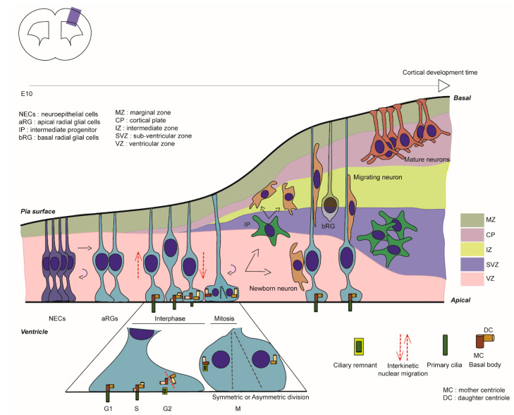
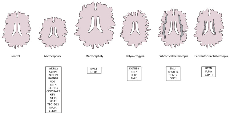
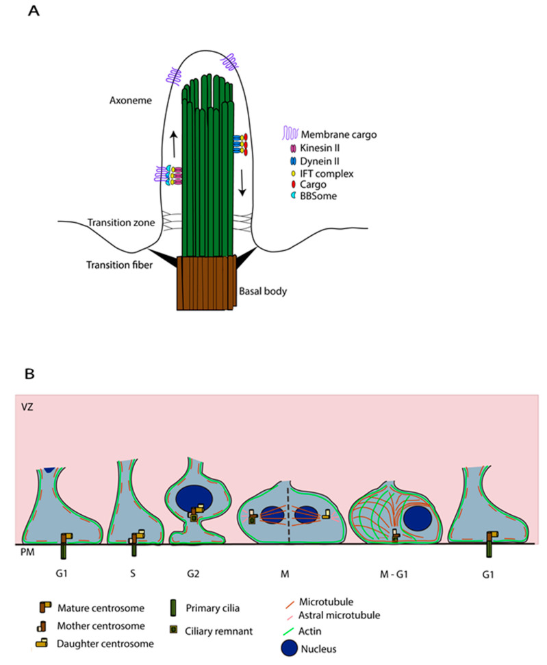
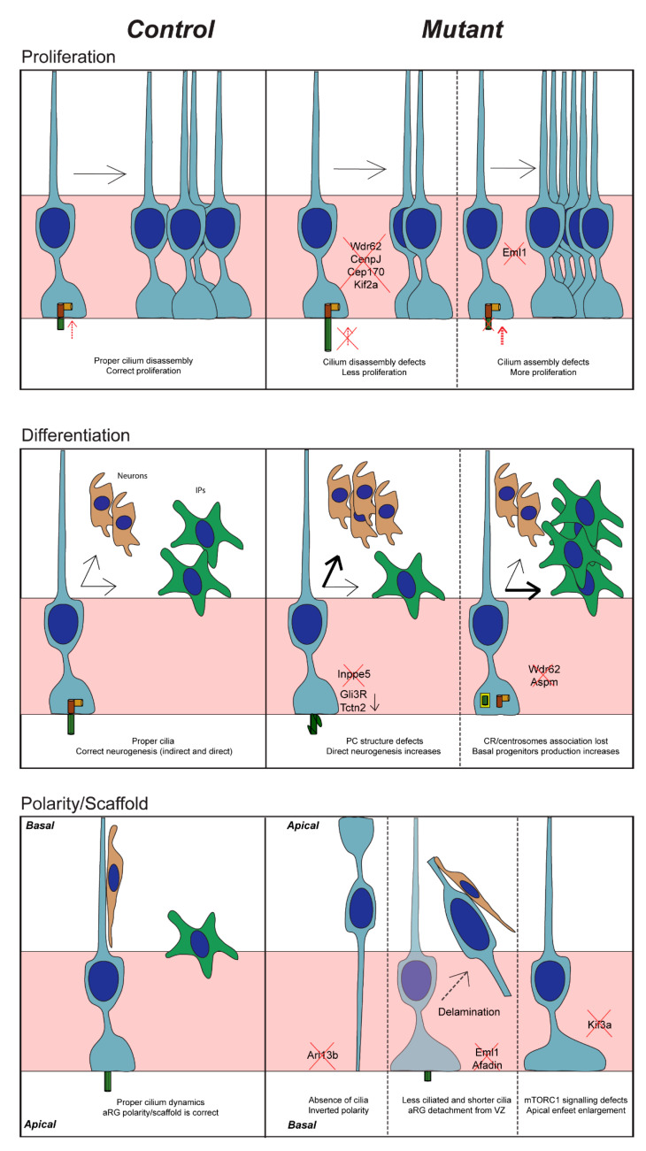
Corticogenesis is an intricate process controlled temporally and spatially by many intrinsic and extrinsic factors. Alterations during this important process can lead to severe cortical malformations. Apical neuronal progenitors are essential cells able to self-amplify and also generate basal progenitors and/or neurons. Apical radial glia (aRG) are neuronal progenitors with a unique morphology. They have a long basal process acting as a support for neuronal migration to the cortical plate and a short apical process directed towards the ventricle from which protrudes a primary cilium. This antenna-like structure allows aRG to sense cues from the embryonic cerebrospinal fluid (eCSF) helping to maintain cell shape and to influence several key functions of aRG such as proliferation and differentiation. Centrosomes, major microtubule organising centres, are crucial for cilia formation. In this review, we focus on how primary cilia influence aRG function during cortical development and pathologies which may arise due to defects in this structure. Reporting and cataloguing a number of ciliary mutant models, we discuss the importance of primary cilia for aRG function and cortical development.
Overview
The claims and statistics of this paper haven’t been checked yet.
Free: it reads the open-access text and checks the first 5 claims that cite a source we can check. Create a free account to check every claim.
Integrity signals
Registry
Cites
Statistics
Figures
Verification
Claims the paper makes
The claims and statistics of this paper haven’t been checked yet.
Free: it reads the open-access text and checks the first 5 claims that cite a source we can check. Create a free account to check every claim.
Claims other papers make about this one
Each is checked against this paper’s own text: does it say what the citing paper says it does?
The source backs part of the claim, not all of it — or backs it for a different population, condition or comparison.
In the citing paper
“Notably, primary cilia function as essential signaling hubs for pathways such as Shh, which is crucial for neural tube patterning and the expansion of neural progenitor populations (Zaidi et al., 2022).”
From Primary cilia in the mature brain: emerging roles in Alzheimer's disease pathogenesis (2025) · cites this paper as Zaidi et al., 2022
Experimental — accuracy not yet measured
Self-check
Percentages, sums and reported differences re-computed from the paper’s own numbers.
The self-check needs the paper’s full text.
Citations
Cites
The Graded Response to Sonic Hedgehog Depends on Cilia Architecture
Caspary, T., Larkins, C. E., & Anderson, K. V. (2007) · Developmental Cell · doi:10.1016/j.devcel.2007.03.004 · Cited by 727
A transition zone complex regulates mammalian ciliogenesis and ciliary membrane composition
Garcia-Gonzalo, F. R., Corbit, K. C., Sirerol-Piquer, M. S., Ramaswami, G., Otto, E. A., Noriega, T. R., Seol, A. D., Robinson, J. F., Bennett, C. L., Josifova, D. J., et al. (2011) · Nature Genetics · doi:10.1038/ng.891 · Cited by 638
Mapping the NPHP-JBTS-MKS Protein Network Reveals Ciliopathy Disease Genes and Pathways
Sang, L., Miller, J. J., Corbit, K. C., Giles, R. H., Brauer, M. J., Otto, E. A., Baye, L. M., Wen, X., Scales, S. J., Kwong, M., et al. (2011) · Cell · doi:10.1016/j.cell.2011.04.019 · Cited by 612
Constitutional aneuploidy and cancer predisposition caused by biallelic mutations in BUB1B
Hanks, S., Coleman, K., Reid, S., Plaja, A., Firth, H., FitzPatrick, D., Kidd, A., Méhes, K., Nash, R., Robin, N., et al. (2004) · Nature Genetics · doi:10.1038/ng1449 · Cited by 605
Jin, H., White, S. R., Shida, T., Schulz, S., Aguiar, M., Gygi, S. P., Bazan, J. F., & Nachury, M. V. (2010) · Cell · doi:10.1016/j.cell.2010.05.015 · Cited by 605
A centrosomal mechanism involving CDK5RAP2 and CENPJ controls brain size
Bond, J., Roberts, E., Springell, K., Lizarraga, S., Scott, S., Higgins, J., Hampshire, D. J., Morrison, E. E., Leal, G. F., Silva, E. O., et al. (2005) · Nature Genetics · doi:10.1038/ng1539 · Cited by 572
Cep164, a novel centriole appendage protein required for primary cilium formation
Graser, S., Stierhof, Y.-D., Lavoie, S. B., Gassner, O. S., Lamla, S., Le Clech, M., & Nigg, E. A. (2007) · The Journal of Cell Biology · doi:10.1083/jcb.200707181 · Cited by 516
Whole-exome sequencing identifies recessive WDR62 mutations in severe brain malformations
Bilgüvar, K., Öztürk, A. K., Louvi, A., Kwan, K. Y., Choi, M., Tatlı, B., Yalnızoğlu, D., Tüysüz, B., Çağlayan, A. O., Gökben, S., et al. (2010) · Nature · doi:10.1038/nature09327 · Cited by 503
Poirier, K., Lebrun, N., Broix, L., Tian, G., Saillour, Y., Boscheron, C., Parrini, E., Valence, S., Pierre, B. S., Oger, M., et al. (2013) · Nature Genetics · doi:10.1038/ng.2613 · Cited by 494
Self-citationSignaling through the Primary Cilium
Wheway, G., Nazlamova, L., & Hancock, J. T. (2018) · Frontiers in Cell and Developmental Biology · doi:10.3389/fcell.2018.00008 · Cited by 483
Cited by
Emerging principles of primary cilia dynamics in controlling tissue organization and function
Gopalakrishnan, J., Feistel, K., Friedrich, B. M., Grapin‐Botton, A., Jurisch‐Yaksi, N., Mass, E., Mick, D. U., Müller, R., May‐Simera, H., Schermer, B., et al. (2023) · The EMBO Journal · doi:10.15252/embj.2023113891 · Cited by 90
“…it is not surprising that primary cilia dysfunction affects NPCs and neurons and, thereby, underlies several neurodevelopmental disorders (Han & Alvarez-Buylla, 2010; Paridaen & Huttner, 2014; Liu et al , 2021; Suciu & Caspary, 2021; Stoufflet & Caill (cid:2) e, 2022; Zaidi et al , 2022).”
Ciliary biology intersects autism and congenital heart disease
Teerikorpi, N., McCluskey, K. E., Bader, E., Lasser, M. C., Wang, S., Nguyen, C. H., Schmidt, J. D., Kostyanovskaya, E., Sun, N., Dea, J., et al. (2025) · Development · doi:10.1242/dev.204295 · Cited by 23
Cilia and Extracellular Vesicles in Brain Development and Disease
Ma, R., Chen, L., Hu, N., Caplan, S., & Hu, G. (2023) · Biological Psychiatry · doi:10.1016/j.biopsych.2023.11.004 · Cited by 18
Kawakami, R., Hiraide, T., Watanabe, K., Miyamoto, S., Hira, K., Komatsu, K., Ishigaki, H., Sakaguchi, K., Maekawa, M., Yamashita, K., et al. (2023) · Journal of Human Genetics · doi:10.1038/s10038-023-01211-8 · Cited by 14
Radial glia progenitor polarity in health and disease
Viola, V., Chinnappa, K., & Francis, F. (2024) · Frontiers in Cell and Developmental Biology · doi:10.3389/fcell.2024.1478283 · Cited by 12
Self-citation
Metadata
- Authors
- Zaidi, D., Chinnappa, K., & Francis, F.
- Affiliations
- Inserm (FR)
- Corresponding author
- Francis, Fiona
- Venue
- Cells
- Publisher
- MDPI AG
- Published
- 2022-09-16
- Volume
- 11
- Issue
- 18
- Pages
- 2895
- ISSN
- 2073-4409 (ISSN-L)
- PMC
- PMC9496791
- OpenAlex
- W4296252021
- Cited by
- 19
- References
- 176
- Citations by year
- 2026: 3 · 2025: 6 · 2024: 4 · 2023: 6
- FWCI
- 0.63
- Type
- journal article
- Language
- en
- Open access
- gold
- doi.org
- Licence
- Locations
- Cells (publishedVersion, cc-by)
- PubMed Central (publishedVersion, cc-by)
- PubMed (publishedVersion)
- HAL (submittedVersion, other-oa)
- HAL (submittedVersion)
- DOAJ (DOAJ: Directory of Open Access Journals) (submittedVersion, cc-by-sa)
- Funders
- French ANR (E-Rare-3, ERARE18-049, EQU202003010323)
- ERA-Net for Research on Rare Diseases (E-Rare-3, ERARE18-049, EQU202003010323)
- French Fondation pour la Recherche Médicale (E-Rare-3, ERARE18-049, EQU202003010323)
- Inserm, Sorbonne University and the Centre national de la recherche scientifique (CNRS) (E-Rare-3, ERARE18-049, EQU202003010323)
- Inserm and Sorbonne University (E-Rare-3, ERARE18-049, EQU202003010323)
- Topics
- Life Sciences › Neuroscience › Developmental Neuroscience › Neurogenesis and neuroplasticity mechanisms
- Life Sciences › Biochemistry, Genetics and Molecular Biology › Genetics › Genetic and Kidney Cyst Diseases
- Health Sciences › Medicine › Pediatrics, Perinatology and Child Health › Fetal and Pediatric Neurological Disorders
- Keywords
- primary cilia, cortical development, neuronal precursors, centrosome, cerebrospinal fluid, malformations of cortical development, neuronal migration, cell proliferation and differentiation
- MeSH terms
- Cell Differentiation, Cerebral Cortex, Cilia, Neurons, Neurogenesis
About these checks
Registry, retraction, Cites and statistics results are computed, not judged, with one exception: a printed reference that no index lists is read once by a model before it counts as a citation error. Claims are extracted by a model and kept only when their quote appears verbatim in the text; each cited source is then checked, with a model’s help, for whether it says what the claim relies on. Those verdicts can be wrong — read the claim’s quote and the cited source before relying on one.
Statistics and arithmetic are recomputed, not judged.
Confidence bands are shown only for fields calibrated on our annotated test set. None is calibrated yet, so no band is shown.
What counts as a claim ›How we measure confidence ›How we check this ›
Are you an author, or do you see a problem with this page? Request a review
How to cite
Cite CoChat for the checks on this page, and the paper for its own findings.
- This page
- CoChat. (2026). Citation check: Primary Cilia Influence Progenitor Function during Cortical Development. https://app.cochat.ai/papers/10.3390/cells11182895
- The paper
- Zaidi, D., Chinnappa, K., & Francis, F. (2022). Primary Cilia Influence Progenitor Function during Cortical Development. Cells. https://doi.org/10.3390/cells11182895
Data sources
Bibliographic data: Semantic Scholar, OpenAlex, Crossref.
Contains information from the Semantic Scholar Academic Graph Datasets, made available under the ODC Attribution License (ODC-BY 1.0).
Semantic Scholar’s data is described in: Kinney, R., et al. (2023). The Semantic Scholar Open Data Platform. arXiv:2301.10140.
