

Primary Cilia Influence Progenitor Function during Cortical Development
Open accessThe DOI matches its Crossref record; 174 of the 176 references it cites were found in a registry or an index.
Abstract
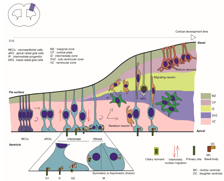
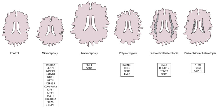
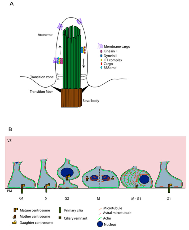
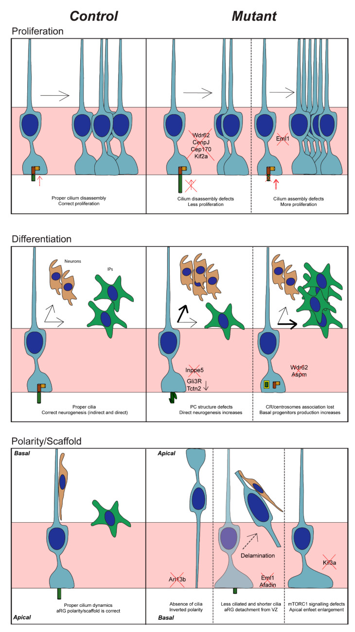
Corticogenesis is an intricate process controlled temporally and spatially by many intrinsic and extrinsic factors. Alterations during this important process can lead to severe cortical malformations. Apical neuronal progenitors are essential cells able to self-amplify and also generate basal progenitors and/or neurons. Apical radial glia (aRG) are neuronal progenitors with a unique morphology. They have a long basal process acting as a support for neuronal migration to the cortical plate and a short apical process directed towards the ventricle from which protrudes a primary cilium. This antenna-like structure allows aRG to sense cues from the embryonic cerebrospinal fluid (eCSF) helping to maintain cell shape and to influence several key functions of aRG such as proliferation and differentiation. Centrosomes, major microtubule organising centres, are crucial for cilia formation. In this review, we focus on how primary cilia influence aRG function during cortical development and pathologies which may arise due to defects in this structure. Reporting and cataloguing a number of ciliary mutant models, we discuss the importance of primary cilia for aRG function and cortical development.
Overview
The claims and statistics of this paper haven’t been checked yet.
Free: it reads the open-access text and checks the first 5 claims that cite a source we can check. Create a free account to check every claim.
Integrity signals
Registry
Cites
Statistics
Figures
Verification
Claims the paper makes
The claims and statistics of this paper haven’t been checked yet.
Free: it reads the open-access text and checks the first 5 claims that cite a source we can check. Create a free account to check every claim.
Claims other papers make about this one
Each is checked against this paper’s own text: does it say what the citing paper says it does?
The source backs part of the claim, not all of it — or backs it for a different population, condition or comparison.
In the citing paper
“Notably, primary cilia function as essential signaling hubs for pathways such as Shh, which is crucial for neural tube patterning and the expansion of neural progenitor populations (Zaidi et al., 2022).”
From Primary cilia in the mature brain: emerging roles in Alzheimer's disease pathogenesis (2025) · cites this paper as Zaidi et al., 2022
Experimental — accuracy not yet measured
Self-check
Percentages, sums and reported differences re-computed from the paper’s own numbers.
The self-check needs the paper’s full text.
Citations
Cites
The Graded Response to Sonic Hedgehog Depends on Cilia Architecture
Caspary, T., Larkins, C. E., & Anderson, K. V. (2007) · Developmental Cell · doi:10.1016/j.devcel.2007.03.004 · Cited by 727
A transition zone complex regulates mammalian ciliogenesis and ciliary membrane composition
Garcia-Gonzalo, F. R., Corbit, K. C., Sirerol-Piquer, M. S., Ramaswami, G., Otto, E. A., Noriega, T. R., Seol, A. D., Robinson, J. F., Bennett, C. L., Josifova, D. J., et al. (2011) · Nature Genetics · doi:10.1038/ng.891 · Cited by 638
Mapping the NPHP-JBTS-MKS Protein Network Reveals Ciliopathy Disease Genes and Pathways
Sang, L., Miller, J. J., Corbit, K. C., Giles, R. H., Brauer, M. J., Otto, E. A., Baye, L. M., Wen, X., Scales, S. J., Kwong, M., et al. (2011) · Cell · doi:10.1016/j.cell.2011.04.019 · Cited by 612
Constitutional aneuploidy and cancer predisposition caused by biallelic mutations in BUB1B
Hanks, S., Coleman, K., Reid, S., Plaja, A., Firth, H., FitzPatrick, D., Kidd, A., Méhes, K., Nash, R., Robin, N., et al. (2004) · Nature Genetics · doi:10.1038/ng1449 · Cited by 605
Jin, H., White, S. R., Shida, T., Schulz, S., Aguiar, M., Gygi, S. P., Bazan, J. F., & Nachury, M. V. (2010) · Cell · doi:10.1016/j.cell.2010.05.015 · Cited by 605
A centrosomal mechanism involving CDK5RAP2 and CENPJ controls brain size
Bond, J., Roberts, E., Springell, K., Lizarraga, S., Scott, S., Higgins, J., Hampshire, D. J., Morrison, E. E., Leal, G. F., Silva, E. O., et al. (2005) · Nature Genetics · doi:10.1038/ng1539 · Cited by 572
Cep164, a novel centriole appendage protein required for primary cilium formation
Graser, S., Stierhof, Y.-D., Lavoie, S. B., Gassner, O. S., Lamla, S., Le Clech, M., & Nigg, E. A. (2007) · The Journal of Cell Biology · doi:10.1083/jcb.200707181 · Cited by 516
Whole-exome sequencing identifies recessive WDR62 mutations in severe brain malformations
Bilgüvar, K., Öztürk, A. K., Louvi, A., Kwan, K. Y., Choi, M., Tatlı, B., Yalnızoğlu, D., Tüysüz, B., Çağlayan, A. O., Gökben, S., et al. (2010) · Nature · doi:10.1038/nature09327 · Cited by 503
Poirier, K., Lebrun, N., Broix, L., Tian, G., Saillour, Y., Boscheron, C., Parrini, E., Valence, S., Pierre, B. S., Oger, M., et al. (2013) · Nature Genetics · doi:10.1038/ng.2613 · Cited by 494
Self-citationSignaling through the Primary Cilium
Wheway, G., Nazlamova, L., & Hancock, J. T. (2018) · Frontiers in Cell and Developmental Biology · doi:10.3389/fcell.2018.00008 · Cited by 483
Asymmetric centrosome inheritance maintains neural progenitors in the neocortex
Wang, X., Tsai, J.-W., Imai, J. H., Lian, W.-N., Vallee, R. B., & Shi, S.-H. (2009) · Nature · doi:10.1038/nature08435 · Cited by 475
Reiter, J. F., Blacque, O. E., & Leroux, M. R. (2012) · EMBO reports · doi:10.1038/embor.2012.73 · Cited by 475
Centriole distal appendages promote membrane docking, leading to cilia initiation
Tanos, B. E., Yang, H.-J., Soni, R., Wang, W.-J., Macaluso, F. P., Asara, J. M., & Tsou, M.-F. B. (2013) · Genes & Development · doi:10.1101/gad.207043.112 · Cited by 417
Autophagy promotes primary ciliogenesis by removing OFD1 from centriolar satellites
Tang, Z., Lin, M. G., Stowe, T. R., Chen, S., Zhu, M., Stearns, T., Franco, B., & Zhong, Q. (2013) · Nature · doi:10.1038/nature12606 · Cited by 414
Bielas, S. L., Silhavy, J. L., Brancati, F., Kisseleva, M. V., Al-Gazali, L., Sztriha, L., Bayoumi, R. A., Zaki, M. S., Abdel-Aleem, A., Rosti, R. O., et al. (2009) · Nature Genetics · doi:10.1038/ng.423 · Cited by 396
Neurogenesis during development of the vertebrate central nervous system
Paridaen, J. T., & Huttner, W. B. (2014) · EMBO reports · doi:10.1002/embr.201438447 · Cited by 388
Centriole ciliation is related to quiescence and DNA synthesis in 3T3 cells
Tucker, R. W., Pardee, A. B., & Fujiwara, K. (1979) · Cell · doi:10.1016/0092-8674(79)90261-7 · Cited by 366
Periventricular heterotopia: phenotypic heterogeneity and correlation with Filamin A mutations
Parrini, E. (2006) · Brain · doi:10.1093/brain/awl125 · Cited by 365
Jacoby, M., Cox, J. J., Gayral, S., Hampshire, D. J., Ayub, M., Blockmans, M., Pernot, E., Kisseleva, M. V., Compère, P., Schiffmann, S. N., et al. (2009) · Nature Genetics · doi:10.1038/ng.427 · Cited by 347
The role of the prefrontal cortex in higher cognitive functions
Frith, C., & Dolan, R. (1996) · Cognitive Brain Research · doi:10.1016/s0926-6410(96)00054-7 · Cited by 343
Filamin A (FLNA) is required for cell–cell contact in vascular development and cardiac morphogenesis
Feng, Y., Chen, M. H., Moskowitz, I. P., Mendonza, A. M., Vidali, L., Nakamura, F., Kwiatkowski, D. J., & Walsh, C. A. (2006) · Proceedings of the National Academy of Sciences · doi:10.1073/pnas.0609628104 · Cited by 323
Paridaen, J. T. M. L., Wilsch-Bräuninger, M., & Huttner, W. B. (2013) · Cell · doi:10.1016/j.cell.2013.08.060 · Cited by 318
The ciliary pocket: an endocytic membrane domain at the base of primary and motile cilia
Molla-Herman, A., Ghossoub, R., Blisnick, T., Meunier, A., Serres, C., Silbermann, F., Emmerson, C., Romeo, K., Bourdoncle, P., Schmitt, A., et al. (2010) · Journal of Cell Science · doi:10.1242/jcs.059519 · Cited by 298
Mutations in the AHI1 Gene, Encoding Jouberin, Cause Joubert Syndrome with Cortical Polymicrogyria
Dixon-Salazar, T., Silhavy, J. L., Marsh, S. E., Louie, C. M., Scott, L. C., Gururaj, A., Al-Gazali, L., Al-Tawari, A. A., Kayserili, H., Sztriha, L., et al. (2004) · The American Journal of Human Genetics · doi:10.1086/425985 · Cited by 292
D'Gama, A. M., Geng, Y., Couto, J. A., Martin, B., Boyle, E. A., LaCoursiere, C. M., Hossain, A., Hatem, N. E., Barry, B. J., Kwiatkowski, D. J., et al. (2015) · Annals of Neurology · doi:10.1002/ana.24357 · Cited by 290
The Hippo Pathway, YAP/TAZ, and the Plasma Membrane
Rausch, V., & Hansen, C. G. (2020) · Trends in Cell Biology · doi:10.1016/j.tcb.2019.10.005 · Cited by 279
Higginbotham, H., Eom, T.-Y., Mariani, L. E., Bachleda, A., Hirt, J., Gukassyan, V., Cusack, C. L., Lai, C., Caspary, T., & Anton, E. S. (2012) · Developmental Cell · doi:10.1016/j.devcel.2012.09.019 · Cited by 253
Nde1-mediated inhibition of ciliogenesis affects cell cycle re-entry
Kim, S., Zaghloul, N. A., Bubenshchikova, E., Oh, E. C., Rankin, S., Katsanis, N., Obara, T., & Tsiokas, L. (2011) · Nature Cell Biology · doi:10.1038/ncb2183 · Cited by 250
Li, A., Saito, M., Chuang, J.-Z., Tseng, Y.-Y., Dedesma, C., Tomizawa, K., Kaitsuka, T., & Sung, C.-H. (2011) · Nature Cell Biology · doi:10.1038/ncb2218 · Cited by 241
CPAP promotes timely cilium disassembly to maintain neural progenitor pool
Gabriel, E., Wason, A., Ramani, A., Gooi, L. M., Keller, P., Pozniakovsky, A., Poser, I., Noack, F., Telugu, N. S., Calegari, F., et al. (2016) · The EMBO Journal · doi:10.15252/embj.201593679 · Cited by 239
Primary Cilia and the Cell Cycle
Plotnikova, O. V., Pugacheva, E. N., & Golemis, E. A. (2009) · Methods in Cell Biology · doi:10.1016/s0091-679x(08)94007-3 · Cited by 236
Stages of ciliogenesis and regulation of ciliary length
Avasthi, P., & Marshall, W. F. (2012) · Differentiation · doi:10.1016/j.diff.2011.11.015 · Cited by 234
Mutations in STIL, Encoding a Pericentriolar and Centrosomal Protein, Cause Primary Microcephaly
Kumar, A., Girimaji, S. C., Duvvari, M. R., & Blanton, S. H. (2009) · The American Journal of Human Genetics · doi:10.1016/j.ajhg.2009.01.017 · Cited by 231
The multipolar stage and disruptions in neuronal migration
LoTurco, J. J., & Bai, J. (2006) · Trends in Neurosciences · doi:10.1016/j.tins.2006.05.006 · Cited by 231
Intraflagellar Transport and Ciliary Dynamics
Ishikawa, H., & Marshall, W. F. (2017) · Cold Spring Harbor Perspectives in Biology · doi:10.1101/cshperspect.a021998 · Cited by 228
The perennial organelle: assembly and disassembly of the primary cilium
Seeley, E. S., & Nachury, M. V. (2010) · Journal of Cell Science · doi:10.1242/jcs.061093 · Cited by 226
Human Mutations in NDE1 Cause Extreme Microcephaly with Lissencephaly
Alkuraya, F. S., Cai, X., Emery, C., Mochida, G. H., Al-Dosari, M. S., Felie, J. M., Hill, R. S., Barry, B. J., Partlow, J. N., Gascon, G. G., et al. (2011) · The American Journal of Human Genetics · doi:10.1016/j.ajhg.2011.04.003 · Cited by 225
Arl13b-regulated cilia activities are essential for polarized radial glial scaffold formation
Higginbotham, H., Guo, J., Yokota, Y., Umberger, N. L., Su, C.-Y., Li, J., Verma, N., Hirt, J., Ghukasyan, V., Caspary, T., et al. (2013) · Nature Neuroscience · doi:10.1038/nn.3451 · Cited by 216
Cell shape and contractility regulate ciliogenesis in cell cycle–arrested cells
Pitaval, A., Tseng, Q., Bornens, M., & Théry, M. (2010) · Journal of Cell Biology · doi:10.1083/jcb.201004003 · Cited by 213
Functional aspects of primary cilia in signaling, cell cycle and tumorigenesis
Basten, S. G., & Giles, R. H. (2013) · Cilia · doi:10.1186/2046-2530-2-6 · Cited by 208
Ostergaard, P., Simpson, M. A., Mendola, A., Vasudevan, P., Connell, F. C., van Impel, A., Moore, A. T., Loeys, B. L., Ghalamkarpour, A., Onoufriadis, A., et al. (2012) · The American Journal of Human Genetics · doi:10.1016/j.ajhg.2011.12.018 · Cited by 203
Kim, J., Jo, H., Hong, H., Kim, M. H., Kim, J. M., Lee, J.-K., Heo, W. D., & Kim, J. (2015) · Nature Communications · doi:10.1038/ncomms7781 · Cited by 202
The regulation of cilium assembly and disassembly in development and disease
Wang, L., & Dynlacht, B. D. (2018) · Development · doi:10.1242/dev.151407 · Cited by 201
Robert, A., Margall-Ducos, G., Guidotti, J.-E., Brégerie, O., Celati, C., Bréchot, C., & Desdouets, C. (2007) · Journal of Cell Science · doi:10.1242/jcs.03366 · Cited by 195
Zhang, W., Yang, S.-L., Yang, M., Herrlinger, S., Shao, Q., Collar, J. L., Fierro, E., Shi, Y., Liu, A., Lu, H., et al. (2019) · Nature Communications · doi:10.1038/s41467-019-10497-2 · Cited by 191
Cilium structure, assembly, and disassembly regulated by the cytoskeleton
Mirvis, M., Stearns, T., & James Nelson, W. (2018) · Biochemical Journal · doi:10.1042/bcj20170453 · Cited by 190
The SIL gene is required for mouse embryonic axial development and left–right specification
Izraeli, S., Lowe, L. A., Bertness, V. L., Good, D. J., Dorward, D. W., Kirsch, I. R., & Kuehn, M. R. (1999) · Nature · doi:10.1038/21429 · Cited by 189
STIL is required for centriole duplication in human cells
Vulprecht, J., David, A., Tibelius, A., Castiel, A., Konotop, G., Liu, F., Bestvater, F., Raab, M. S., Zentgraf, H., Izraeli, S., et al. (2012) · Journal of Cell Science · doi:10.1242/jcs.104109 · Cited by 183
Apical Abscission Alters Cell Polarity and Dismantles the Primary Cilium During Neurogenesis
Das, R. M., & Storey, K. G. (2014) · Science · doi:10.1126/science.1247521 · Cited by 182
A Truncating Mutation of CEP135 Causes Primary Microcephaly and Disturbed Centrosomal Function
Hussain, M. S., Baig, S. M., Neumann, S., Nürnberg, G., Farooq, M., Ahmad, I., Alef, T., Hennies, H. C., Technau, M., Altmüller, J., et al. (2012) · The American Journal of Human Genetics · doi:10.1016/j.ajhg.2012.03.016 · Cited by 181
Ultrastructure of cilia and flagella - back to the future!
Fisch, C., & Dupuis-Williams, P. (2011) · Biology of the Cell · doi:10.1042/bc20100139 · Cited by 179
Mutations in Eml1 lead to ectopic progenitors and neuronal heterotopia in mouse and human
Kielar, M., Tuy, F. P. D., Bizzotto, S., Lebrand, C., de Juan Romero, C., Poirier, K., Oegema, R., Mancini, G. M., Bahi-Buisson, N., Olaso, R., et al. (2014) · Nature Neuroscience · doi:10.1038/nn.3729 · Cited by 174
Self-citationSoluble levels of cytosolic tubulin regulate ciliary length control
Sharma, N., Kosan, Z. A., Stallworth, J. E., Berbari, N. F., & Yoder, B. K. (2011) · Molecular Biology of the Cell · doi:10.1091/mbc.e10-03-0269 · Cited by 173
The Primary Cilium: Keeper of the Key to Cell Division
Pan, J., & Snell, W. (2007) · Cell · doi:10.1016/j.cell.2007.06.018 · Cited by 171
Current topics of functional links between primary cilia and cell cycle
Izawa, I., Goto, H., Kasahara, K., & Inagaki, M. (2015) · Cilia · doi:10.1186/s13630-015-0021-1 · Cited by 168
CDK5RAP2 Regulates Centriole Engagement and Cohesion in Mice
Barrera, J. A., Kao, L.-R., Hammer, R. E., Seemann, J., Fuchs, J. L., & Megraw, T. L. (2010) · Developmental Cell · doi:10.1016/j.devcel.2010.05.017 · Cited by 166
Making the Connection: Ciliary Adhesion Complexes Anchor Basal Bodies to the Actin Cytoskeleton
Antoniades, I., Stylianou, P., & Skourides, P. A. (2014) · Developmental Cell · doi:10.1016/j.devcel.2013.12.003 · Cited by 163
Baudoin, J.-P., Viou, L., Launay, P.-S., Luccardini, C., Espeso Gil, S., Kiyasova, V., Irinopoulou, T., Alvarez, C., Rio, J.-P., Boudier, T., et al. (2012) · Neuron · doi:10.1016/j.neuron.2012.10.027 · Cited by 161
The Essential Role of Centrosomal NDE1 in Human Cerebral Cortex Neurogenesis
Bakircioglu, M., Carvalho, O. P., Khurshid, M., Cox, J. J., Tuysuz, B., Barak, T., Yilmaz, S., Caglayan, O., Dincer, A., Nicholas, A. K., et al. (2011) · The American Journal of Human Genetics · doi:10.1016/j.ajhg.2011.03.019 · Cited by 161
Trichoplein and Aurora A block aberrant primary cilia assembly in proliferating cells
Inoko, A., Matsuyama, M., Goto, H., Ohmuro-Matsuyama, Y., Hayashi, Y., Enomoto, M., Ibi, M., Urano, T., Yonemura, S., Kiyono, T., et al. (2012) · Journal of Cell Biology · doi:10.1083/jcb.201106101 · Cited by 161
Shi, X., Garcia, G., Van De Weghe, J. C., McGorty, R., Pazour, G. J., Doherty, D., Huang, B., & Reiter, J. F. (2017) · Nature Cell Biology · doi:10.1038/ncb3599 · Cited by 159
Jayaraman, D., Kodani, A., Gonzalez, D. M., Mancias, J. D., Mochida, G. H., Vagnoni, C., Johnson, J., Krogan, N., Harper, J. W., Reiter, J. F., et al. (2016) · Neuron · doi:10.1016/j.neuron.2016.09.056 · Cited by 155
Cell cycle progression by the repression of primary cilia formation in proliferating cells
Goto, H., Inoko, A., & Inagaki, M. (2013) · Cellular and Molecular Life Sciences · doi:10.1007/s00018-013-1302-8 · Cited by 154
Ou, Y. Y., Mack, G. J., Zhang, M., & Rattner, J. B. (2002) · Journal of Cell Science · doi:10.1242/jcs.115.9.1825 · Cited by 153
Defective Wnt-dependent cerebellar midline fusion in a mouse model of Joubert syndrome
Lancaster, M. A., Gopal, D. J., Kim, J., Saleem, S. N., Silhavy, J. L., Louie, C. M., Thacker, B. E., Williams, Y., Zaki, M. S., & Gleeson, J. G. (2011) · Nature Medicine · doi:10.1038/nm.2380 · Cited by 153
Miyamoto, T., Hosoba, K., Ochiai, H., Royba, E., Izumi, H., Sakuma, T., Yamamoto, T., Dynlacht, B. D., & Matsuura, S. (2015) · Cell Reports · doi:10.1016/j.celrep.2015.01.003 · Cited by 151
Cerebral cortex development: an outside‐in perspective
Agirman, G., Broix, L., & Nguyen, L. (2017) · FEBS Letters · doi:10.1002/1873-3468.12924 · Cited by 150
Cilia and cell cycle re-entry: More than a coincidence
Kim, S., & Tsiokas, L. (2011) · Cell Cycle · doi:10.4161/cc.10.16.17009 · Cited by 148
The ciliary pocket: a once-forgotten membrane domain at the base of cilia
Ghossoub, R., Molla‐Herman, A., Bastin, P., & Benmerah, A. (2011) · Biology of the Cell · doi:10.1042/bc20100128 · Cited by 147
Developmental disruptions underlying brain abnormalities in ciliopathies
Guo, J., Higginbotham, H., Li, J., Nichols, J., Hirt, J., Ghukasyan, V., & Anton, E. S. (2015) · Nature Communications · doi:10.1038/ncomms8857 · Cited by 146
Genetics and mechanisms leading to human cortical malformations
Romero, D. M., Bahi‐Buisson, N., & Francis, F. (2017) · Seminars in Cell and Developmental Biology · doi:10.1016/j.semcdb.2017.09.031 · Cited by 145
Self-citationBuchsbaum, I. Y., & Cappello, S. (2019) · Development · doi:10.1242/dev.163766 · Cited by 145
Interkinetic nuclear migration: A mysterious process in search of a function
Spear, P. C., & Erickson, C. A. (2012) · Development Growth & Differentiation · doi:10.1111/j.1440-169x.2012.01342.x · Cited by 143
Wu, C.-T., Chen, H.-Y., & Tang, T. K. (2018) · Nature Cell Biology · doi:10.1038/s41556-017-0018-7 · Cited by 142
Carleton, M. O., Mao, M., Biery, M. C., Warrener, P., Kim, S., Buser, C. A., Marshall, C. G., Fernandes, C., Annis, J. E., & Linsley, P. S. (2006) · Molecular and Cellular Biology · doi:10.1128/mcb.26.10.3853-3863.2006 · Cited by 141
Roles of Primary Cilia in the Developing Brain
Park, S. M., Jang, H. J., & Lee, J. H. (2019) · Frontiers in Cellular Neuroscience · doi:10.3389/fncel.2019.00218 · Cited by 128
Cytochalasin D inhibits basal body migration and ciliary elongation in quail oviduct epithelium
Boisvieux‐Ulrich, E., Lainé, M., & Sandoz, D. (1990) · Cell and Tissue Research · doi:10.1007/bf01740770 · Cited by 122
Tuz, K., Bachmann‐Gagescu, R., O’Day, D. R., Hua, K., Isabella, C. R., Phelps, I. G., Stolarski, A. E., O’Roak, B. J., Dempsey, J. C., Lourenço, C. M., et al. (2013) · The American Journal of Human Genetics · doi:10.1016/j.ajhg.2013.11.019 · Cited by 122
Unstable microtubule capture at kinetochores depleted of the centromere‐associated protein CENP‐F
Bomont, P., Maddox, P. S., Shah, J. V., Desai, A. B., & Cleveland, D. W. (2005) · The EMBO Journal · doi:10.1038/sj.emboj.7600848 · Cited by 121
Tucker, R. W., Scher, C. D., & Stiles, C. D. (1979) · Cell · doi:10.1016/0092-8674(79)90219-8 · Cited by 120
Neuronal migration disorders: Focus on the cytoskeleton and epilepsy
Stouffer, M. A., Golden, J. A., & Francis, F. (2016) · Neurobiology of Disease · doi:10.1016/j.nbd.2015.08.003 · Cited by 115
Self-citationMishra-Gorur, K., Çağlayan, A. O., Schaffer, A. E., Chabu, C., Henegariu, O., Vonhoff, F., Akgümüş, G. T., Nishimura, S., Han, W., Tu, S., et al. (2014) · Neuron · doi:10.1016/j.neuron.2014.12.014 · Cited by 114
Park, S. M., Lim, J. S., Ramakrishina, S., Kim, S. H., Kim, W. K., Lee, J., Kang, H.-C., Reiter, J. F., Kim, D. S., Kim, H. (., et al. (2018) · Neuron · doi:10.1016/j.neuron.2018.05.039 · Cited by 114
Cilia, ciliopathies and hedgehog-related forebrain developmental disorders
Andreu-Cervera, A., Catala, M., & Schneider-Maunoury, S. (2021) · Neurobiology of Disease · doi:10.1016/j.nbd.2020.105236 · Cited by 114
Broad-Minded Links Cell Cycle-Related Kinase to Cilia Assembly and Hedgehog Signal Transduction
Ko, H. W., Norman, R. X., Tran, J., Fuller, K. P., Fukuda, M., & Eggenschwiler, J. T. (2010) · Developmental Cell · doi:10.1016/j.devcel.2009.12.014 · Cited by 113
The Centrosome and the Primary Cilium: The Yin and Yang of a Hybrid Organelle
Joukov, V., & Nicolo, A. D. (2019) · Cells · doi:10.3390/cells8070701 · Cited by 112
The centrosome protein AKNA regulates neurogenesis via microtubule organization
Ortega, G. C., Falk, S., Johansson, P. A., Peyre, E., Broix, L., Sahu, S. K., Hirst, W. G., Schlichthaerle, T., Romero, C. D. J., Draganova, K., et al. (2019) · Nature · doi:10.1038/s41586-019-0962-4 · Cited by 111
Self-citationBasolateral rather than apical primary cilia on neuroepithelial cells committed to delamination
Wilsch-Bräuninger, M., Peters, J., Paridaen, J. T. M. L., & Huttner, W. B. (2012) · Development · doi:10.1242/dev.069294 · Cited by 109
Mechanism of ciliary disassembly
Liang, Y., Meng, D., Zhu, B., & Pan, J. (2016) · Cellular and Molecular Life Sciences · doi:10.1007/s00018-016-2148-7 · Cited by 107
A meckelin–filamin A interaction mediates ciliogenesis
Adams, M., Simms, R. J., Abdelhamed, Z., Dawe, H. R., Szymanska, K., Logan, C. V., Wheway, G., Pitt, E., Gull, K., Knowles, M. A., et al. (2012) · Human Molecular Genetics · doi:10.1093/hmg/ddr557 · Cited by 106
Cortical progenitor biology: key features mediating proliferation versus differentiation
Uzquiano, A., Gladwyn-Ng, I. E., Nguyen, L., Reiner, O., Götz, M., Matsuzaki, F., & Francis, F. (2018) · Journal of Neurochemistry · doi:10.1111/jnc.14338 · Cited by 102
Self-citationJuric‐Sekhar, G., Adkins, J. R., Doherty, D., & Hevner, R. F. (2012) · Acta Neuropathologica · doi:10.1007/s00401-012-0951-2 · Cited by 100
Genes and Mechanisms Involved in the Generation and Amplification of Basal Radial Glial Cells
Penisson, M., Ladewig, J., Belvindrah, R., & Francis, F. (2019) · Frontiers in Cellular Neuroscience · doi:10.3389/fncel.2019.00381 · Cited by 100
Self-citationCiliary Genes TBC1D32/C6orf170 and SCLT1 are Mutated in Patients with OFD Type IX
Adly, N., Alhashem, A., Ammari, A., & Alkuraya, F. S. (2014) · Human Mutation · doi:10.1002/humu.22477 · Cited by 98
Katanin p80 Regulates Human Cortical Development by Limiting Centriole and Cilia Number
Hu, W. F., Pomp, O., Ben-Omran, T., Kodani, A., Henke, K., Mochida, G. H., Yu, T. W., Woodworth, M. B., Bonnard, C., Raj, G. S., et al. (2014) · Neuron · doi:10.1016/j.neuron.2014.12.017 · Cited by 98
The kinetochore protein, CENPF , is mutated in human ciliopathy and microcephaly phenotypes
Waters, A. M., Asfahani, R., Carroll, P., Bicknell, L. S., Lescai, F., Bright, A., Chanudet, E., Brooks, A., Christou-Savina, S., Osman, G. A., et al. (2015) · Journal of Medical Genetics · doi:10.1136/jmedgenet-2014-102691 · Cited by 98
Cellular Mechanisms of Ciliary Length Control
Keeling, J., Tsiokas, L., & Maskey, D. (2016) · Cells · doi:10.3390/cells5010006 · Cited by 98
Centrosome anchoring regulates progenitor properties and cortical formation
Shao, W., Yang, J., He, M., Yu, X.-Y., Lee, C. H., Yang, Z., Joyner, A. L., Anderson, K. V., Zhang, J., Tsou, M.-F. B., et al. (2020) · Nature · doi:10.1038/s41586-020-2139-6 · Cited by 98
Hsiao, Y.-C., Tong, Z. J., Westfall, J. E., Ault, J. G., Page-McCaw, P. S., & Ferland, R. J. (2009) · Human Molecular Genetics · doi:10.1093/hmg/ddp335 · Cited by 97
Mechanisms of ciliogenesis suppression in dividing cells
Goto, H., Inaba, H., & Inagaki, M. (2016) · Cellular and Molecular Life Sciences · doi:10.1007/s00018-016-2369-9 · Cited by 97
Microtubule stabilization drives 3D centrosome migration to initiate primary ciliogenesis
Pitaval, A., Senger, F., Letort, G., Gidrol, X., Guyon, L., Sillibourne, J., & Théry, M. (2017) · The Journal of Cell Biology · doi:10.1083/jcb.201610039 · Cited by 91
Primary cilium loss in mammalian cells occurs predominantly by whole-cilium shedding
Mirvis, M., Siemers, K. A., Nelson, W. J., & Stearns, T. P. (2019) · PLOS Biology · doi:10.1371/journal.pbio.3000381 · Cited by 91
RTTN Mutations Link Primary Cilia Function to Organization of the Human Cerebral Cortex
Kia, S. K., Verbeek, E., Engelen, E., Schot, R., Poot, R. A., de Coo, I. F. M., Lequin, M. H., Poulton, C. J., Pourfarzad, F., Grosveld, F. G., et al. (2012) · The American Journal of Human Genetics · doi:10.1016/j.ajhg.2012.07.008 · Cited by 90
Neuronal Migration Dynamics in the Developing Ferret Cortex
Gertz, C. C., & Kriegstein, A. R. (2015) · The Journal of Neuroscience · doi:10.1523/jneurosci.2198-15.2015 · Cited by 90
The transition zone protein Rpgrip1l regulates proteasomal activity at the primary cilium
Gerhardt, C., Lier, J. M., Burmühl, S., Struchtrup, A., Deutschmann, K., Vetter, M., Leu, T., Reeg, S., Grune, T., & Rüther, U. (2015) · Journal of Cell Biology · doi:10.1083/jcb.201408060 · Cited by 88
The Zebra fish cassiopeia Mutant Reveals that SIL Is Required for Mitotic Spindle Organization
Pfaff, K. L., Straub, C. T., Chiang, K., Bear, D. M., Zhou, Y., & Zon, L. I. (2007) · Molecular and Cellular Biology · doi:10.1128/mcb.00175-07 · Cited by 87
Lian, G., Lu, J., Hu, J., Zhang, J., Cross, S. H., Ferland, R. J., & Sheen, V. L. (2012) · Journal of Neuroscience · doi:10.1523/jneurosci.0894-12.2012 · Cited by 86
Role for the IFT-A Complex in Selective Transport to the Primary Cilium
Fu, W., Wang, L., Kim, S., Li, J., & Dynlacht, B. D. (2016) · Cell Reports · doi:10.1016/j.celrep.2016.10.018 · Cited by 86
Mutations of KIF14 cause primary microcephaly by impairing cytokinesis
Moawia, A., Shaheen, R., Rasool, S., Waseem, S. S., Ewida, N., Budde, B. S., Kawalia, A., Motameny, S., Khan, K., Fatima, A., et al. (2017) · Annals of Neurology · doi:10.1002/ana.25044 · Cited by 85
Kajii, T., Ikeuchi, T., Yang, Z.-Q., Nakamura, Y., Tsuji, Y., Yokomori, K., Kawamura, M., Fukuda, S., Horita, S., & Asamoto, A. (2001) · American journal of medical genetics · doi:10.1002/ajmg.1580 · Cited by 84
Habbig, S., Bartram, M. P., Sägmüller, J. G., Griessmann, A., Franke, M., Müller, R.-U., Schwarz, R., Hoehne, M., Bergmann, C., Tessmer, C., et al. (2012) · Human Molecular Genetics · doi:10.1093/hmg/dds408 · Cited by 80
Identification of drugs that restore primary cilium expression in cancer cells
Khan, N. A., Willemarck, N., Talebi, A., Marchand, A., Binda, M. M., Dehairs, J., Rueda-Rincon, N., Daniels, V. W., Bagadi, M., Raj, D. B. T. G., et al. (2016) · Oncotarget · doi:10.18632/oncotarget.7198 · Cited by 80
Liu, W. A., Chen, S., Li, Z., Lee, C. H., Mirzaa, G., Dobyns, W. B., Ross, M. E., Zhang, J., & Shi, S. (2018) · Genes & Development · doi:10.1101/gad.313171.118 · Cited by 78
Dauber, A. N., LAFRANCHI, S. H., Maliga, Z., Lui, J. C. K., Moon, J. E., McDeed, C., Henke, K., Zonana, J., Kingman, G. A. R., Pers, T. H., et al. (2012) · The Journal of Clinical Endocrinology & Metabolism · doi:10.1210/jc.2012-2150 · Cited by 77
Spatial centrosome proteome of human neural cells uncovers disease-relevant heterogeneity
O’Neill, A. C., Uzbaş, F., Antognolli, G., Merino, F., Draganova, K., Jäck, A., Zhang, S., Pedini, G., Schessner, J. P., Cramer, K., et al. (2022) · Science · doi:10.1126/science.abf9088 · Cited by 75
mTORC1 signaling and primary cilia are required for brain ventricle morphogenesis
Foerster, P., Daclin, M., Asm, S., Faucourt, M., Boletta, A., Genovesio, A., & Spassky, N. (2016) · Development · doi:10.1242/dev.138271 · Cited by 74
Morris-Rosendahl, D. J., & Kaindl, A. M. (2015) · Molecular and Cellular Probes · doi:10.1016/j.mcp.2015.05.015 · Cited by 73
Siller, S. S., Sharma, H., Li, S., Yang, J. F., Zhang, Y., Holtzman, M. J., Winuthayanon, W., Colognato, H., Holdener, B. C., Li, F., et al. (2017) · PLoS Genetics · doi:10.1371/journal.pgen.1007128 · Cited by 71
Ferent, J., Zaidi, D., & Francis, F. (2020) · Frontiers in Cell and Developmental Biology · doi:10.3389/fcell.2020.578341 · Cited by 67
Self-citationThe multifaceted roles of primary cilia in the development of the cerebral cortex
Hasenpusch-Theil, K., & Theil, T. (2021) · Frontiers in Cell and Developmental Biology · doi:10.3389/fcell.2021.630161 · Cited by 66
Birtel, J., Gliem, M., Mangold, E., Tebbe, L., Spier, I., Müller, P. L., Holz, F. G., Neuhaus, C., Wolfrum, U., Bolz, H. J., et al. (2017) · Investigative Ophthalmology & Visual Science · doi:10.1167/iovs.17-21679 · Cited by 64
Rotatin is a novel gene required for axial rotation and left–right specification in mouse embryos
Faisst, A. M., Álvarez-Bolado, G., Treichel, D., & Gruß, P. (2002) · Mechanisms of Development · doi:10.1016/s0925-4773(02)00003-5 · Cited by 63
The Ciliary Protein Arl13b Functions Outside of the Primary Cilium in Shh-Mediated Axon Guidance
Ferent, J., Constable, S., Gigante, E. D., Yam, P. T., Mariani, L. E., Legué, E., Liem, K. F., Caspary, T., & Charron, F. (2019) · Cell Reports · doi:10.1016/j.celrep.2019.11.015 · Cited by 63
Patzke, S., Hauge, H., Sioud, M., Finne, E. F., Sivertsen, E. A., Delabie, J., Stokke, T., & Aasheim, H. (2004) · Oncogene · doi:10.1038/sj.onc.1208267 · Cited by 62
Field, M. J., Scheffer, I. E., Gill, D. S., Wilson, M., Christie, L., Shaw, M. A., Gardner, A. E., Glubb, G. C., Hobson, L., Corbett, M. A., et al. (2012) · European Journal of Human Genetics · doi:10.1038/ejhg.2012.9 · Cited by 60
Miyamoto, T., Porazinski, S., Wang, H., Borovina, A., Ciruna, B., Shimizu, A., Kajii, T., Kikuchi, A., Furutani-Seiki, M., & Matsuura, S. (2011) · Human Molecular Genetics · doi:10.1093/hmg/ddr090 · Cited by 60
Yanagida, M., Miyoshi, R., Toyokuni, R., Zhu, Y., & Murakami, F. (2012) · Proceedings of the National Academy of Sciences · doi:10.1073/pnas.1209166109 · Cited by 58
Andreu‐Cervera, A., Anselme, I., Karam, A., Laclef, C., Catala, M., & Schneider‐Maunoury, S. (2019) · Journal of Neuroscience · doi:10.1523/jneurosci.2199-18.2019 · Cited by 57
Mirzaa, G., Enyedi, L. B., Parsons, G., Collins, S., Medne, L., Adams, C., Ward, T., Davitt, B. V., Bicknese, A. R., Zackai, E. H., et al. (2014) · American Journal of Medical Genetics Part A · doi:10.1002/ajmg.a.36707 · Cited by 56
Kif14 Mutation Causes Severe Brain Malformation and Hypomyelination
Fujikura, K., Setsu, T., Tanigaki, K., Abe, T., Kiyonari, H., Terashima, T., & Sakisaka, T. (2013) · PLoS ONE · doi:10.1371/journal.pone.0053490 · Cited by 55
A Cilia Independent Role of Ift88/Polaris during Cell Migration
Boehlke, C., Janusch, H., Hamann, C., Powelske, C., Mergen, M., Herbst, H., Kotsis, F., Nitschke, R., & Kuehn, E. W. (2015) · PLOS ONE · doi:10.1371/journal.pone.0140378 · Cited by 55
Cenpj Regulates Cilia Disassembly and Neurogenesis in the Developing Mouse Cortex
Ding, W., Wu, Q., Sun, L., Pan, N., & Wang, X. (2019) · Journal of Neuroscience · doi:10.1523/jneurosci.1849-18.2018 · Cited by 55
Mutations in the Heterotopia Gene Eml1/EML1 Severely Disrupt the Formation of Primary Cilia
Uzquiano, A., Cifuentes-Díaz, C., Jabali, A., Romero, D. M., Houllier, A., Dingli, F., Maillard, C., Boland, A., Deleuze, J., Loew, D., et al. (2019) · Cell Reports · doi:10.1016/j.celrep.2019.06.096 · Cited by 53
Self-citationMorphological and functional aspects of progenitors perturbed in cortical malformations
Bizzotto, S., & Francis, F. (2015) · Frontiers in Cellular Neuroscience · doi:10.3389/fncel.2015.00030 · Cited by 51
Self-citationHu, H.-B., Song, Z.-Q., Song, G.-P., Li, S., Tu, H.-Q., Wu, M., Zhang, Y.-C., Yuan, J.-F., Li, T.-T., Li, P.-Y., et al. (2021) · Nature Communications · doi:10.1038/s41467-021-20986-y · Cited by 50
A novel mechanism of microtubule length-dependent force to pull centrosomes toward the cell center
Kimura, K., & Kimura, A. (2011) · BioArchitecture · doi:10.4161/bioa.1.2.15549 · Cited by 49
Hong, H., Kim, J., & Kim, J. (2015) · Biochemical and Biophysical Research Communications · doi:10.1016/j.bbrc.2015.04.028 · Cited by 49
Broix, L., Asselin, L., Silva, C. G., Ivanova, E. L., Tilly, P., Gilet, J., Lebrun, N., Jagline, H., Muraca, G., Saillour, Y., et al. (2017) · Human Molecular Genetics · doi:10.1093/hmg/ddx384 · Cited by 49
Self-citationEml1 loss impairs apical progenitor spindle length and soma shape in the developing cerebral cortex
Bizzotto, S., Uzquiano, A., Dingli, F., Ershov, D., Houllier, A., Arras, G., Richards, M. W., Loew, D., Minc, N., Croquelois, A., et al. (2017) · Scientific Reports · doi:10.1038/s41598-017-15253-4 · Cited by 48
Self-citationCilia in hereditary cerebral anomalies
Thomas, S., Boutaud, L., Reilly, M. L., & Benmerah, A. (2019) · Biology of the Cell · doi:10.1111/boc.201900012 · Cited by 47
Neuronal Delamination and Outer Radial Glia Generation in Neocortical Development
Kawaguchi, A. (2021) · Frontiers in Cell and Developmental Biology · doi:10.3389/fcell.2020.623573 · Cited by 46
TSC1 and TSC2 regulate cilia length and canonical Hedgehog signaling via different mechanisms
Rosengren, T., Larsen, L. J., Pedersen, L. B., Christensen, S. T., & Møller, L. B. (2018) · Cellular and Molecular Life Sciences · doi:10.1007/s00018-018-2761-8 · Cited by 45
Shohayeb, B., Ho, U. Y., Yeap, Y. Y. C., Parton, R. G., Millard, S. S., Xu, Z., Piper, M., & Ng, D. C. H. (2019) · Human Molecular Genetics · doi:10.1093/hmg/ddz281 · Cited by 45
Gli2 Rescues Delays in Brain Development Induced by Kif3a Dysfunction
Chen, J.-L., Chang, C.-H., & Tsai, J. (2017) · Cerebral Cortex · doi:10.1093/cercor/bhx356 · Cited by 44
Primary Cilia and Centrosomes in Neocortex Development
Wilsch‐Bräuninger, M., & Huttner, W. Β. (2021) · Frontiers in Neuroscience · doi:10.3389/fnins.2021.755867 · Cited by 43
Ofd1 Controls Dorso-Ventral Patterning and Axoneme Elongation during Embryonic Brain Development
D'Angelo, A., Angelis, A. D., Avallone, B., Piscopo, I., Tammaro, R., Studer, M., & Franco, B. (2012) · PLoS ONE · doi:10.1371/journal.pone.0052937 · Cited by 41
Heterogeneous clinical phenotypes and cerebral malformations reflected by rotatin cellular dynamics
Vandervore, L. V., Schot, R., Kasteleijn, E., Oegema, R., Stouffs, K., Gheldof, A., Grochowska, M. M., van der Sterre, M. L. T., van Unen, L., Wilke, M., et al. (2019) · Brain · doi:10.1093/brain/awz045 · Cited by 40
Tuz, K., Hsiao, Y.-C., Juárez, O. X., Shi, B., Harmon, E. Y., Phelps, I. G., Lennartz, M. R., Glass, I. A., Doherty, D., & Ferland, R. J. (2013) · Journal of Biological Chemistry · doi:10.1074/jbc.m112.420786 · Cited by 38
Sclt1 deficiency causes cystic kidney by activating ERK and STAT3 signaling
Li, J., Lü, D., Liu, H., Williams, B. O., Overbeek, P. A., Lee, B., Zheng, L., & Yang, T. (2017) · Human Molecular Genetics · doi:10.1093/hmg/ddx183 · Cited by 36
Human cerebral organoids reveal progenitor pathology in EML1‐linked cortical malformation
Jabali, A., Hoffrichter, A., Uzquiano, A., Marsoner, F., Wilkens, R., Siekmann, M. T., Bohl, B., Rossetti, A. C., Horschitz, S., Koch, P., et al. (2022) · EMBO Reports · doi:10.15252/embr.202154027 · Cited by 36
Self-citationmTORC1 signaling and primary cilia are required for brain ventricle morphogenesis
Foerster, P., Daclin, M., Shihavuddin, A., Faucourt, M., Boletta, A., Genovesio, A., & Spassky, N. (2017) · Journal of Cell Science · doi:10.1242/jcs.201640 · Cited by 35
Extensive branching of radially‐migrating neurons in the mammalian cerebral cortex
Martínez‐Martínez, M. Á., Ciceri, G., Espinós, A., Fernández, V., Marín, Ó., & Borrell, V. (2018) · The Journal of Comparative Neurology · doi:10.1002/cne.24597 · Cited by 35
Loss of CPAP in developing mouse brain and its functional implication for human primary microcephaly
Lin, Y., Lee, Y.-S., Li, S.-K., & Tang, T. K. (2020) · Journal of Cell Science · doi:10.1242/jcs.243592 · Cited by 35
Nagasaka, A., Shinoda, T., Kawaue, T., Suzuki, M., NAGAYAMA, K., MATSUMOTΟ, T., Ueno, N., Kawaguchi, A., & Miyata, T. (2016) · Frontiers in Cell and Developmental Biology · doi:10.3389/fcell.2016.00139 · Cited by 32
Unique spatiotemporal requirements for intraflagellar transport genes during forebrain development
Snedeker, J., Schock, E. N., Struve, J. N., Chang, C., Cionni, M., Tran, P. V., Brugmann, S. A., & Stottmann, R. W. (2017) · PLoS ONE · doi:10.1371/journal.pone.0173258 · Cited by 32
EML1‐associated brain overgrowth syndrome with ribbon‐like heterotopia
Oegema, R., McGillivray, G., Leventer, R. J., Moing, A. G. L., Bahi‐Buisson, N., Barnicoat, A. J., Mandelstam, S. A., Francis, D. I., Francis, F., Mancini, G. M. S., et al. (2019) · American Journal of Medical Genetics Part C Seminars in Medical Genetics · doi:10.1002/ajmg.c.31751 · Cited by 31
Self-citationKIF14 controls ciliogenesis via regulation of Aurora A and is important for Hedgehog signaling
Pejšková, P., Reilly, M. L., Binó, L., Bernatík, O., Dolanská, L., Ganji, R. S., Zdráhal, Z., Benmerah, A., & Čajánek, L. (2020) · The Journal of Cell Biology · doi:10.1083/jcb.201904107 · Cited by 31
Wang, C., Li, J., Takemaru, K., Jiang, X., Xu, G., & Wang, B. (2018) · Development · doi:10.1242/dev.164236 · Cited by 30
Hasenpusch-Theil, K., Laclef, C., Colligan, M., Fitzgerald, E., Howe, K., Carroll, E., Abrams, S. R., Reiter, J. F., Schneider-Maunoury, S., & Theil, T. (2020) · eLife · doi:10.7554/elife.58162 · Cited by 30
Simmons, A., Park, R., Sterling, N. A., Jang, M., van Deursen, J. M. A., Yen, T. J., Cho, S., & Kim, S. (2019) · Human Molecular Genetics · doi:10.1093/hmg/ddz022 · Cited by 28
Afadin controls cell polarization and mitotic spindle orientation in developing cortical radial glia
Rakotomamonjy, J., Brunner, M., Jüschke, C., Zang, K., Huang, E. J., Reichardt, L. F., & Chenn, A. (2017) · Neural Development · doi:10.1186/s13064-017-0085-2 · Cited by 27
Hsiao, C.-J., Chang, C.-H., Ibrahim, R. B., Lin, I.-H., Wang, C., Wang, W., & Tsai, J. (2018) · Journal of Cell Science · doi:10.1242/jcs.221218 · Cited by 27
Conditional switching of KIF2A mutation provides new insights into cortical malformation pathogeny
Gilet, J., Ivanova, E. L., Trofimova, D., Rudolf, G., Méziane, H., Broix, L., Drouot, N., Courraud, J., Skory, V., Voulleminot, P., et al. (2019) · Human Molecular Genetics · doi:10.1093/hmg/ddz316 · Cited by 27
Zalenski, A. A., Majumder, S., De, K., & Venere, M. (2020) · Scientific Reports · doi:10.1038/s41598-020-70787-4 · Cited by 23
The neuroanatomy of Eml1 knockout mice, a model of subcortical heterotopia
Collins, S. C., Uzquiano, A., Selloum, M., Wendling, O., Gaborit, M., Osipenko, M., Birling, M., Yalcin, B., & Francis, F. (2019) · Journal of Anatomy · doi:10.1111/joa.13013 · Cited by 22
Self-citationZoubovsky, S. P., Oh, E. C. T., Cash-Padgett, T. V., Johnson, A. W., Hou, Z., Mori, S., Gallagher, M., Katsanis, N., Sawa, A., & Jaaro-Peled, H. (2015) · Neuroscience Research · doi:10.1016/j.neures.2015.02.002 · Cited by 21
Alfieri, M., Iaconis, D., Tammaro, R., Perone, L., Calì, G., Nitsch, L., Dougherty, G. W., Ragnini‐Wilson, A., & Franco, B. (2020) · Tissue and Cell · doi:10.1016/j.tice.2020.101369 · Cited by 21
Tectonic Proteins Are Important Players in Non-Motile Ciliopathies
Gong, S., Ji, F., Wang, B., Zhang, Y., Xu, X., & Sun, M. (2018) · Cellular Physiology and Biochemistry · doi:10.1159/000494017 · Cited by 19
Morleo, M., & Franco, B. (2021) · Cell Stress · doi:10.15698/cst2021.03.244 · Cited by 18
González‐Martínez, J., Cwetsch, A. W., Martínez‐Alonso, D., López-Sainz, L. R., Almagro, J., Melati, A., Gómez, J., Pérez‐Martínez, M., Megías, D., Boskovic, J., et al. (2021) · JCI Insight · doi:10.1172/jci.insight.146364 · Cited by 16
Cil primaire, cycle cellulaire et prolifération
Delgehyr, N., & Spassky, N. (2014) · médecine/sciences · doi:10.1051/medsci/20143011011 · Cited by 15
Malformations in the Murine Kidney Caused by Loss of CENP‐F Function
Haley, C. O., Waters, A. M., & Bader, D. M. (2018) · The Anatomical Record · doi:10.1002/ar.24018 · Cited by 14
Mishra-Gorur, K., Çağlayan, A. O., Schaffer, A. E., Chabu, C. Y., Henegariu, O., Vonhoff, F., Akgumus, G. T., Nishimura, S., Han, W., Tu, S., et al. (2015) · Neuron · doi:10.1016/j.neuron.2014.12.046 · Cited by 10
Biallelic variants in CENPF causing a phenotype distinct from Strømme syndrome
Cappuccio, G., Brillante, S., Tammaro, R., Pinelli, M., Bernardi, M. L. D., Gensini, M. G., Bijlsma, E. K., Koopmann, T. T., Hoffer, M. J. V., McDonald, K., et al. (2022) · American Journal of Medical Genetics Part C Seminars in Medical Genetics · doi:10.1002/ajmg.c.31973 · Cited by 8
Cited by
Emerging principles of primary cilia dynamics in controlling tissue organization and function
Gopalakrishnan, J., Feistel, K., Friedrich, B. M., Grapin‐Botton, A., Jurisch‐Yaksi, N., Mass, E., Mick, D. U., Müller, R., May‐Simera, H., Schermer, B., et al. (2023) · The EMBO Journal · doi:10.15252/embj.2023113891 · Cited by 90
“…it is not surprising that primary cilia dysfunction affects NPCs and neurons and, thereby, underlies several neurodevelopmental disorders (Han & Alvarez-Buylla, 2010; Paridaen & Huttner, 2014; Liu et al , 2021; Suciu & Caspary, 2021; Stoufflet & Caill (cid:2) e, 2022; Zaidi et al , 2022).”
Ciliary biology intersects autism and congenital heart disease
Teerikorpi, N., McCluskey, K. E., Bader, E., Lasser, M. C., Wang, S., Nguyen, C. H., Schmidt, J. D., Kostyanovskaya, E., Sun, N., Dea, J., et al. (2025) · Development · doi:10.1242/dev.204295 · Cited by 23
Cilia and Extracellular Vesicles in Brain Development and Disease
Ma, R., Chen, L., Hu, N., Caplan, S., & Hu, G. (2023) · Biological Psychiatry · doi:10.1016/j.biopsych.2023.11.004 · Cited by 18
Kawakami, R., Hiraide, T., Watanabe, K., Miyamoto, S., Hira, K., Komatsu, K., Ishigaki, H., Sakaguchi, K., Maekawa, M., Yamashita, K., et al. (2023) · Journal of Human Genetics · doi:10.1038/s10038-023-01211-8 · Cited by 14
Radial glia progenitor polarity in health and disease
Viola, V., Chinnappa, K., & Francis, F. (2024) · Frontiers in Cell and Developmental Biology · doi:10.3389/fcell.2024.1478283 · Cited by 13
Self-citation
Metadata
- Authors
- Zaidi, D., Chinnappa, K., & Francis, F.
- Affiliations
- Inserm (FR)
- Corresponding author
- Francis, Fiona
- Venue
- Cells
- Publisher
- MDPI AG
- Published
- 2022-09-16
- Volume
- 11
- Issue
- 18
- Pages
- 2895
- ISSN
- 2073-4409 (ISSN-L)
- PMC
- PMC9496791
- OpenAlex
- W4296252021
- Cited by
- 19
- References
- 176
- Citations by year
- 2026: 3 · 2025: 6 · 2024: 4 · 2023: 6
- FWCI
- 0.63
- Type
- journal article
- Language
- en
- Open access
- gold
- doi.org
- Licence
- Locations
- Cells (publishedVersion, cc-by)
- PubMed Central (publishedVersion, cc-by)
- PubMed (publishedVersion)
- HAL (submittedVersion, other-oa)
- HAL (submittedVersion)
- DOAJ (DOAJ: Directory of Open Access Journals) (submittedVersion, cc-by-sa)
- Funders
- French ANR (E-Rare-3, ERARE18-049, EQU202003010323)
- ERA-Net for Research on Rare Diseases (E-Rare-3, ERARE18-049, EQU202003010323)
- French Fondation pour la Recherche Médicale (E-Rare-3, ERARE18-049, EQU202003010323)
- Inserm, Sorbonne University and the Centre national de la recherche scientifique (CNRS) (E-Rare-3, ERARE18-049, EQU202003010323)
- Inserm and Sorbonne University (E-Rare-3, ERARE18-049, EQU202003010323)
- Topics
- Life Sciences › Neuroscience › Developmental Neuroscience › Neurogenesis and neuroplasticity mechanisms
- Life Sciences › Biochemistry, Genetics and Molecular Biology › Genetics › Genetic and Kidney Cyst Diseases
- Health Sciences › Medicine › Pediatrics, Perinatology and Child Health › Fetal and Pediatric Neurological Disorders
- Keywords
- primary cilia, cortical development, neuronal precursors, centrosome, cerebrospinal fluid, malformations of cortical development, neuronal migration, cell proliferation and differentiation
- MeSH terms
- Cell Differentiation, Cerebral Cortex, Cilia, Neurons, Neurogenesis
About these checks
Registry, retraction, Cites and statistics results are computed, not judged, with one exception: a printed reference that no index lists is read once by a model before it counts as a citation error. Claims are extracted by a model and kept only when their quote appears verbatim in the text; each cited source is then checked, with a model’s help, for whether it says what the claim relies on. Those verdicts can be wrong — read the claim’s quote and the cited source before relying on one.
Statistics and arithmetic are recomputed, not judged.
Confidence bands are shown only for fields calibrated on our annotated test set. None is calibrated yet, so no band is shown.
What counts as a claim ›How we measure confidence ›How we check this ›
Are you an author, or do you see a problem with this page? Request a review
Data sources
Bibliographic data: Semantic Scholar, OpenAlex, Crossref.
Contains information from the Semantic Scholar Academic Graph Datasets, made available under the ODC Attribution License (ODC-BY 1.0).
Semantic Scholar’s data is described in: Kinney, R., et al. (2023). The Semantic Scholar Open Data Platform. arXiv:2301.10140.
