

The DOI matches its Crossref record; 3 of the 3 references it cites were found in a registry or an index.
miR-154-5p Is a Novel Endogenous Ligand for TLR7 Inducing Microglial Activation and Neuronal Injury
Abstract
Toll-like receptors (TLRs) are a collection of pattern recognition sensors that form a first line of defence by detecting pathogen- or damage-associated molecular patterns and initiating an inflammatory response. TLR activation in microglia, the major immune cells in the brain, can trigger the release of inflammatory molecules, which may contribute to various CNS diseases including Alzheimer’s disease. Recently, some microRNAs were shown to serve as signalling molecules for TLRs. Here, we present miR-154-5p as a novel TLR7 ligand. Exposing microglia to miR-154-5p results in cytokine release and alters expression of the TLR signalling pathway dependent on TLR7. Additionally, miR-154-5p causes neuronal injury in enriched cortical neuron cultures and additive toxicity in the presence of microglia. Finally, intrathecal injection of miR-154-5p into mice leads to neuronal injury and accumulation of microglia in the cerebral cortex dependent on TLR7 expression. In conclusion, this study establishes miR-154-5p as a direct activator of TLR7 that can cause neuroinflammation and neuronal injury, which may contribute to CNS disease.
Overview
The claims and statistics of this paper haven’t been checked yet.
Run the check — free, no account needed. It reads the open-access text and checks the sources it cites.
Integrity signals
Registry
Cites
Statistics
Figures
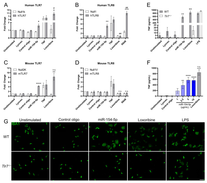
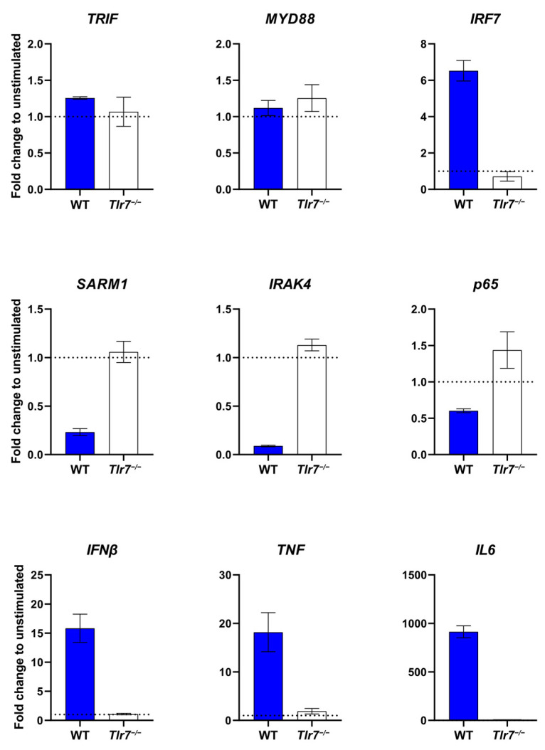
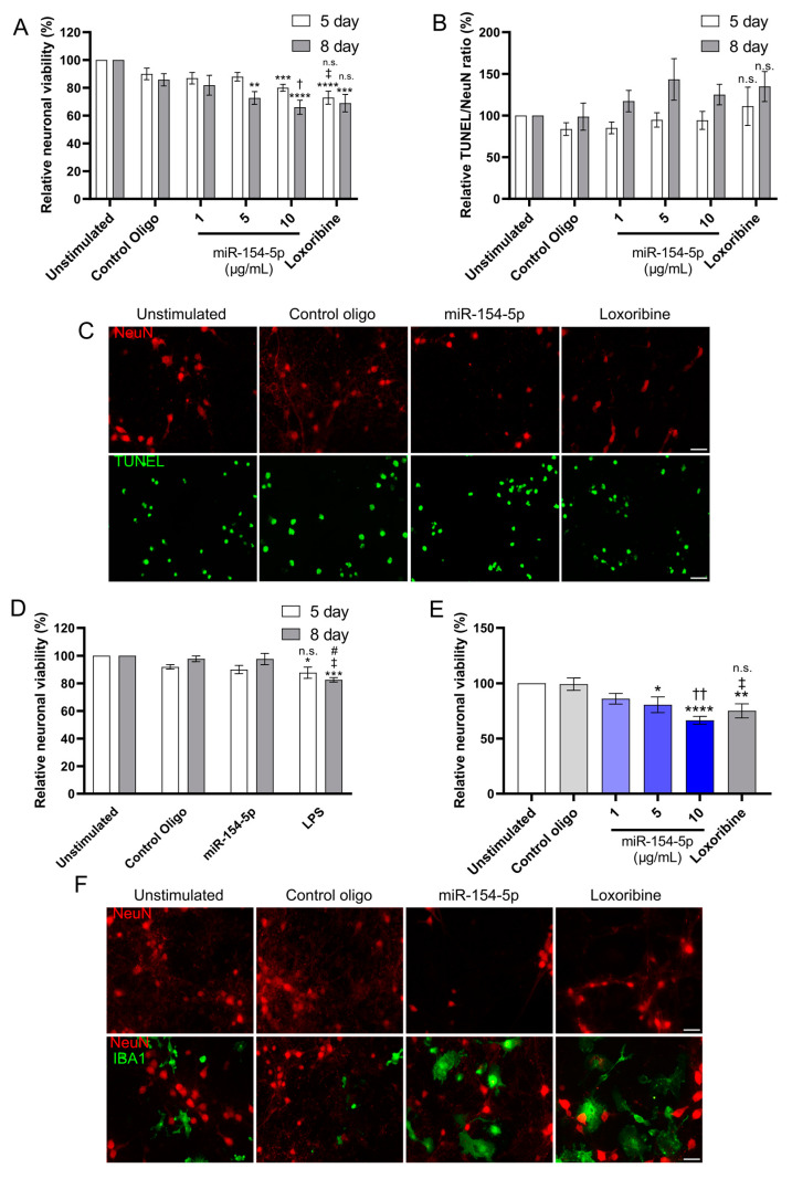
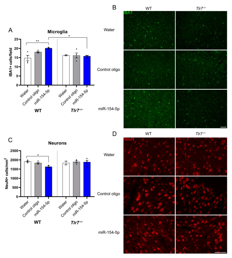
Verification
Claims the paper makes
The claims and statistics of this paper haven’t been checked yet.
Run the check — free, no account needed. It reads the open-access text and checks the sources it cites.
Claims other papers make about this one
Each is checked against this paper’s own text: does it say what the citing paper says it does?
The cited source says what the claim attributes to it.
In the citing paper
“the model of intrathecal injection has been established over the past years in our laboratory to evaluate the effects of extracellular single-stranded RNA in the CNS in vivo [4, 7, 8, 76, 77]”
From Neurodegenerative disease-associated microRNAs acting as signaling molecules modulate CNS neuron structure and viability (2025) · cites this paper as [76]
Self-check
Percentages, sums and reported differences re-computed from the paper’s own numbers.
The self-check needs the paper’s full text.
Citations
Cites
Zhang, Z., Ohto, U., Shibata, T., Krayukhina, E., Taoka, M., Yamauchi, Y., Tanji, H., Isobe, T., Uchiyama, S., Miyake, K., et al. (2016) · Immunity · doi:10.1016/j.immuni.2016.09.011 · Cited by 461
Arcuri, C., Mecca, C., Bianchi, R., Giambanco, I., & Donato, R. (2017) · Frontiers in Molecular Neuroscience · doi:10.3389/fnmol.2017.00191 · Cited by 279
Validation of differentially expressed brain‐enriched microRNAs in the plasma of PD patients
Ravanidis, S., Bougea, A., Papagiannakis, N., Koros, C., Simitsi, A. M., Pachi, I., Breza, M., Stefanis, L., & Doxakis, E. (2020) · Annals of Clinical and Translational Neurology · doi:10.1002/acn3.51146 · Cited by 63
Research works printed but not linked: 53
- Khakpour, 2015, Vascular endothelial cell Toll-like receptor pathways in sepsis, Innate Immun., 21, 827, 10.1177/1753425915606525 · DOI
- Hanke, 2011, Toll-like receptors in health and disease in the brain: Mechanisms and therapeutic potential, Clin. Sci. Lond. Engl. 1979, 121, 367
- Gao, 2017, Inhibition of toll-like receptor signaling as a promising therapy for inflammatory diseases: A journey from molecular to nano therapeutics, Front. Physiol., 8, 508, 10.3389/fphys.2017.00508 · DOI
- Li, 2021, Role of Toll-Like Receptors in Neuroimmune Diseases: Therapeutic Targets and Problems, Front. Immunol., 12, 777606, 10.3389/fimmu.2021.777606 · DOI
- Majidi, 2015, Toll-Like Receptors in the Pathogenesis of Autoimmune Diseases, Adv. Pharm. Bull., 5, 605, 10.15171/apb.2015.082 · DOI
- Lehmann, 2012, An unconventional role for miRNA: Let-7 activates Toll-like receptor 7 and causes neurodegeneration, Nat. Neurosci., 15, 827, 10.1038/nn.3113 · DOI · Paper page
- Lehmann, 2012, Extracellularly Delivered Single-Stranded Viral RNA Causes Neurodegeneration Dependent on TLR7, J. Immunol., 189, 1448, 10.4049/jimmunol.1201078 · DOI · Paper page
- Luo, Z., Su, R., Wang, W., Liang, Y., Zeng, X., Shereen, M.A., Bashir, N., Zhang, Q., Zhao, L., and Wu, K. (2019). EV71 infection induces neurodegeneration via activating TLR7 signaling and IL-6 production. PLoS Pathog., 15. · DOI
- Deng, L., Gao, R., Chen, H., Jiao, B., Zhang, C., Wei, L., Yan, C., Ye-Lehmann, S., Zhu, T., and Chen, C. (2023). Let-7b-TLR7 Signaling Axis Contributes to the Anesthesia/Surgery-Induced Cognitive Impairment. Mol. Neurobiol. · DOI
- Liu, 2017, Tlr7 Deletion Selectively Ameliorates Spatial Learning but does not Influence beta Deposition and Inflammatory Response in an Alzheimers Disease Mouse Model, Neuropsychiatry, 7, 509
- Ha, 2014, Regulation of microRNA biogenesis, Nat. Rev. Mol. Cell Biol., 15, 509, 10.1038/nrm3838 · DOI · Paper page
- Peng, 2016, The role of microRNAs in human cancer, Signal Transduct. Target. Ther., 1, 15004, 10.1038/sigtrans.2015.4 · DOI
- Peters, 2020, Small Things Matter: Relevance of MicroRNAs in Cardiovascular Disease, Front. Physiol., 11, 793, 10.3389/fphys.2020.00793 · DOI
- Pauley, 2009, MicroRNA in autoimmunity and autoimmune diseases, J. Autoimmun., 32, 189, 10.1016/j.jaut.2009.02.012 · DOI
- Kamal, 2015, Current Update on Synopsis of miRNA Dysregulation in Neurological Disorders, CNS Neurol. Disord. Drug Targets, 14, 492, 10.2174/1871527314666150225143637 · DOI
- Liang, 2019, Extracellular microRNAs initiate immunostimulation via activating toll-like receptor signaling pathways, ExRNA, 1, 9, 10.1186/s41544-019-0009-x · DOI
- Wu, 2021, Sequence-specific extracellular microRNAs activate TLR7 and induce cytokine secretion and leukocyte migration, Mol. Cell Biochem., 476, 4139, 10.1007/s11010-021-04220-3 · DOI
- Wallach, 2021, MicroRNA-100-5p and microRNA-298-5p released from apoptotic cortical neurons are endogenous Toll-like receptor 7/8 ligands that contribute to neurodegeneration, Mol. Neurodegener., 16, 80, 10.1186/s13024-021-00498-5 · DOI · Paper page
- Wallach, T., Wetzel, M., Dembny, P., Staszewski, O., Krüger, C., Buonfiglioli, A., Prinz, M., and Lehnardt, S. (2020). Identification of CNS Injury-Related microRNAs as Novel Toll-Like Receptor 7/8 Signaling Activators by Small RNA Sequencing. Cells, 9. · DOI · Paper page
- Woodbury, 2015, miR-155 Is Essential for Inflammation-Induced Hippocampal Neurogenic Dysfunction, J. Neurosci., 35, 9764, 10.1523/JNEUROSCI.4790-14.2015 · DOI
- Kho, 2016, Dynamic Modulation of Microglia/Macrophage Polarization by miR-124 after Focal Cerebral Ischemia, J. Neuroimmune Pharmacol., 11, 733, 10.1007/s11481-016-9700-y · DOI
- Chen, 2023, Exosomes from ADSCs ameliorate nerve damage in the hippocampus caused by post traumatic brain injury via the delivery of circ-Scmh1 promoting microglial M2 polarization, Injury, 54, 110927, 10.1016/j.injury.2023.110927 · DOI
- Chi, 2019, SNHG5 Promotes Breast Cancer Proliferation by Sponging the miR-154-5p/PCNA Axis, Mol. Ther.-Nucleic Acids, 17, 138, 10.1016/j.omtn.2019.05.013 · DOI
- Xin, 2014, miR-154 suppresses colorectal cancer cell growth and motility by targeting TLR2, Mol. Cell Biochem., 387, 271, 10.1007/s11010-013-1892-3 · DOI
- Wei, 2019, LncRNA X inactive specific transcript contributes to neuropathic pain development by sponging miR-154-5p via inducing toll-like receptor 5 in CCI rat models, J. Cell Biochem., 120, 1271, 10.1002/jcb.27088 · DOI
- Wang, 2017, MiRNA-154-5p inhibits cell proliferation and metastasis by targeting PIWIL1 in glioblastoma, Brain Res., 1676, 69, 10.1016/j.brainres.2017.08.014 · DOI
- Guedes, 2016, MicroRNA deregulation and chemotaxis and phagocytosis impairment in Alzheimer’s disease, Alzheimers Dement. Diagn. Assess. Dis. Monit., 3, 7
- Han, 2022, Circulating exo-miR-154-5p regulates vascular dementia through endothelial progenitor cell-mediated angiogenesis, Front. Cell Neurosci., 16, 881175, 10.3389/fncel.2022.881175 · DOI
- Zhao, 2020, SP1 activated-lncRNA SNHG1 mediates the development of epilepsy via miR-154-5p/TLR5 axis, Epilepsy Res., 168, 106476, 10.1016/j.eplepsyres.2020.106476 · DOI
- Chen, 2020, Long Noncoding RNA SNHG5 Knockdown Alleviates Neuropathic Pain by Targeting the miR-154-5p/CXCL13 Axis, Neurochem. Res., 45, 1566, 10.1007/s11064-020-03021-2 · DOI
- Hoffmann, 2007, TLR2 Mediates Neuroinflammation and Neuronal Damage, J. Immunol., 178, 6476, 10.4049/jimmunol.178.10.6476 · DOI
- 2008, TLR7 and TLR8 as targets in cancer therapy, Oncogene, 27, 190, 10.1038/sj.onc.1210913 · DOI
- Raden, 2021, Structure-aware machine learning identifies microRNAs operating as Toll-like receptor 7/8 ligands, RNA Biol., 18, 268, 10.1080/15476286.2021.1940697 · DOI · Paper page
- Lax, 2020, Systemic microbial TLR2 agonists induce neurodegeneration in Alzheimer’s disease mice, J. Neuroinflammation, 17, 55, 10.1186/s12974-020-01738-z · DOI
- Buchanan, 2010, Toll-like receptor 4 in CNS pathologies, J. Neurochem., 114, 13, 10.1111/j.1471-4159.2010.06736.x · DOI
- Ifuku, 2020, Activation of Toll-like receptor 5 in microglia modulates their function and triggers neuronal injury, Acta Neuropathol. Commun., 8, 1, 10.1186/s40478-020-01031-3 · DOI
- Derkow, K., Krüger, C., Dembny, P., and Lehnardt, S. (2015). Microglia Induce Neurotoxic IL-17+ γδ T Cells Dependent on TLR2, TLR4, and TLR9 Activation. PLoS ONE, 10. · DOI
- Frederiksen, H.R., Haukedal, H., and Freude, K. (2019). Cell Type Specific Expression of Toll-Like Receptors in Human Brains and Implications in Alzheimer’s Disease. BioMed Res. Int., 2019. · DOI · Paper page
- Santovito, 2020, Noncanonical inhibition of caspase-3 by a nuclear microRNA confers endothelial protection by autophagy in atherosclerosis, Sci. Transl. Med., 12, eaaz2294, 10.1126/scitranslmed.aaz2294 · DOI
- Yang, 2021, MicroRNA Biophysically Modulates Cardiac Action Potential by Direct Binding to Ion Channel, Circulation, 143, 1597, 10.1161/CIRCULATIONAHA.120.050098 · DOI
- Fabbri, 2012, MicroRNAs bind to Toll-like receptors to induce prometastatic inflammatory response, Proc. Natl. Acad. Sci. USA, 109, E2110, 10.1073/pnas.1209414109 · DOI · Paper page
- Heppner, 2015, Immune attack: The role of inflammation in Alzheimer disease, Nat. Rev. Neurosci., 16, 358, 10.1038/nrn3880 · DOI · Paper page
- Tansey, 2022, Inflammation and immune dysfunction in Parkinson disease, Nat. Rev. Immunol., 22, 657, 10.1038/s41577-022-00684-6 · DOI · Paper page
- Sriram, U., Xu, J., Chain, R.W., Varghese, L., Chakhtoura, M., Bennett, H.L., Zoltick, P.W., and Gallucci, S. (2014). IL-4 Suppresses the Responses to TLR7 and TLR9 Stimulation and Increases the Permissiveness to Retroviral Infection of Murine Conventional Dendritic Cells. PLoS ONE, 9. · DOI
- Kim, 2021, MiR-154-5p-MCP1 Axis Regulates Allergic Inflammation by Mediating Cellular Interactions, Front. Immunol., 12, 663726, 10.3389/fimmu.2021.663726 · DOI
- Jurk, 2002, Human TLR7 or TLR8 independently confer responsiveness to the antiviral compound R-848, Nat. Immunol., 3, 499, 10.1038/ni0602-499 · DOI · Paper page
- Hemmi, 2002, Small anti-viral compounds activate immune cells via the TLR7 MyD88–dependent signaling pathway, Nat. Immunol., 3, 196, 10.1038/ni758 · DOI
- Liu, 2013, TLR7 Negatively Regulates Dendrite Outgrowth through the Myd88–c-Fos–IL-6 Pathway, J. Neurosci., 33, 11479, 10.1523/JNEUROSCI.5566-12.2013 · DOI · Paper page
- Liu, 2023, Activation of TLR7-mediated autophagy increases epileptic susceptibility via reduced KIF5A-dependent GABAA receptor transport in a murine model, Exp. Mol. Med., 55, 1159, 10.1038/s12276-023-01000-5 · DOI
- Menassa, 2018, Microglial dynamics during human brain development, Front. Immunol., 9, 1014, 10.3389/fimmu.2018.01014 · DOI
- Paolicelli, 2011, Synaptic pruning by microglia is necessary for normal brain development, Science, 333, 1456, 10.1126/science.1202529 · DOI · Paper page
- Cornell, 2022, Microglia regulation of synaptic plasticity and learning and memory, Neural Regen. Res., 17, 705, 10.4103/1673-5374.322423 · DOI · Paper page
- Custodero, 2022, Role of inflammatory markers in the diagnosis of vascular contributions to cognitive impairment and dementia: A systematic review and meta-analysis, GeroScience, 44, 1373, 10.1007/s11357-022-00556-w · DOI
Cited by
Sorted by citations
Kumbol, V., Ivanov, A., McGurran, H., Schüler, J., Zhai, Y., Ludwik, K., Hinkelmann, L., Brehm, M., Krüger, C., Küchler, J., et al. (2025) · Cell Communication and Signaling · doi:10.1186/s12964-025-02199-8 · Cited by 8
Self-citation
Metadata
- Authors
- McGurran, H., Kumbol, V., Krüger, C., Wallach, T., & Lehnardt, S.
- Affiliations
- Charité—Universitätsmedizin Berlin, Einstein Center for Neurosciences Berlin, 10117 Berlin, Germany
- Institute of Cell Biology and Neurobiology, Charité—Universitätsmedizin Berlin, Corporate Member of Freie Universität Berlin, Humboldt-Universität zu Berlin and Berlin Institute of Health, 10117 Berlin, Germany
- Venue
- Cells
- Publisher
- MDPI AG
- Published
- 2024-02-26
- Volume
- 13
- Issue
- 5
- Pages
- 407
- ISSN
- 2073-4409
- PMC
- PMC10930870
- Cited by
- 12
- References
- 3
- Language
- en
- Funders
- Deutsche Forschungsgemeinschaft (SFB-TRR 167/B03, LE 2420/7-1)
About these checks
Registry, retraction, Cites and statistics results are computed, not judged, with one exception: a printed reference that no index lists is read once by a model before it counts as a citation error. Claims are extracted by a model and kept only when their quote appears verbatim in the text; each cited source is then checked, with a model’s help, for whether it says what the claim relies on. Those verdicts can be wrong — read the claim’s quote and the cited source before relying on one.
Statistics and arithmetic are recomputed, not judged.
Confidence bands are shown only for fields calibrated on our annotated test set. None is calibrated yet, so no band is shown.
What counts as a claim ›How we measure confidence ›How we check this ›
Are you an author, or do you see a problem with this page? Request a review
