

The DOI matches its Crossref record; 127 of the 128 references it cites were found in a registry or an index.
Cell Death in Chondrocytes, Osteoblasts, and Osteocytes
Open accessAbstract
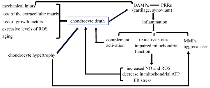
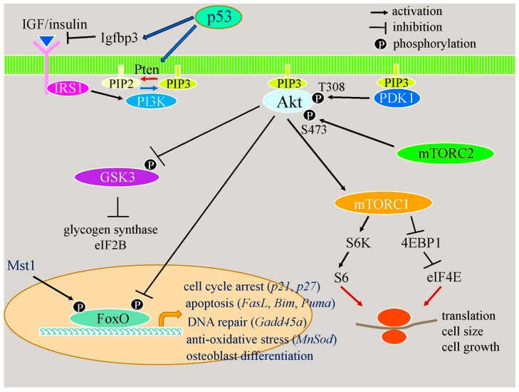
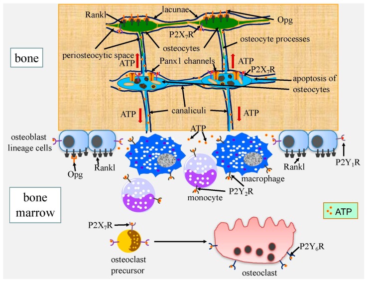
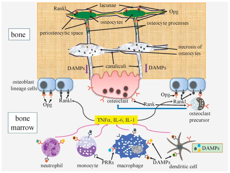
Cell death in skeletal component cells, including chondrocytes, osteoblasts, and osteocytes, plays roles in skeletal development, maintenance, and repair as well as in the pathogenesis of osteoarthritis and osteoporosis. Chondrocyte proliferation, differentiation, and apoptosis are important steps for endochondral ossification. Although the inactivation of P53 and RB is involved in the pathogenesis of osteosarcomas, the deletion of p53 and inactivation of Rb are insufficient to enhance chondrocyte proliferation, indicating the presence of multiple inhibitory mechanisms against sarcomagenesis in chondrocytes. The inflammatory processes induced by mechanical injury and chondrocyte death through the release of danger-associated molecular patterns (DAMPs) are involved in the pathogenesis of posttraumatic osteoarthritis. The overexpression of BCLXL increases bone volume with a normal structure and maintains bone during aging by inhibiting osteoblast apoptosis. p53 inhibits osteoblast proliferation and enhances osteoblast apoptosis, thereby reducing bone formation, but also exerts positive effects on osteoblast differentiation through the Akt–FoxOs pathway. Apoptotic osteocytes release ATP, which induces the receptor activator of nuclear factor κ-B ligand (Rankl) expression and osteoclastogenesis, from pannexin 1 channels. Osteocyte death ultimately results in necrosis; DAMPs are released to the bone surface and promote the production of proinflammatory cytokines, which induce Rankl expression, and osteoclastogenesis is further enhanced.
Overview
The claims and statistics of this paper haven’t been checked yet.
Run the check — free, no account needed. It reads the open-access text and checks the sources it cites.
Integrity signals
Registry
Cites
Statistics
Figures
Verification
Claims the paper makes
The claims and statistics of this paper haven’t been checked yet.
Run the check — free, no account needed. It reads the open-access text and checks the sources it cites.
Claims other papers make about this one
Each is checked against this paper’s own text: does it say what the citing paper says it does?
The cited source says what the claim attributes to it.
In the citing paper
“Necrotic osteocytes release damage-associated molecular pattern (DAMP) proteins after the cell membrane is disrupted.61”
From Bone remodeling: an operational process ensuring survival and bone mechanical competence (2022) · cites this paper as [61]
In the citing paper
“In vivo, osteocyte apoptosis is fundamental for initiating bone remodeling,61 independent of the stimulus causing osteocyte death such as estrogen loss,62 fatigue,63 or unloading.64”
From Bone remodeling: an operational process ensuring survival and bone mechanical competence (2022) · cites this paper as [61]
Self-check
Percentages, sums and reported differences re-computed from the paper’s own numbers.
The self-check needs the paper’s full text.
Citations
Cites
Stöckl, S., Göttl, C., Grifka, J. K., & Grässel, S. (2013) · Cellular Physiology and Biochemistry · doi:10.1159/000350089 · Cited by 4,670
Osteoarthritis: A disease of the joint as an organ
Loeser, R. F., Goldring, S. R., Scanzello, C. R., & Goldring, M. B. (2012) · Arthritis & Rheumatism · doi:10.1002/art.34453 · Cited by 3,234 · cited 1×
“The signaling pathway through PRRs induces synovitis, transduces inflammation, increases the expression of matrix metalloproteinases (MMPs) and aggrecanases, and induces oxidative stress and cell death [55,56].”
BackgroundHigh-mobility group box 1 protein (HMGB1): nuclear weapon in the immune arsenal
Lotze, M. T., & Tracey, K. J. (2005) · Nature reviews. Immunology · doi:10.1038/nri1594 · Cited by 2,490
Regulation of Rate of Cartilage Differentiation by Indian Hedgehog and PTH-Related Protein
Vortkamp, A., Lee, K., Lanske, B., Segre, G. V., Kronenberg, H. M., & Tabin, C. J. (1996) · Science · doi:10.1126/science.273.5275.613 · Cited by 1,999
Evidence for osteocyte regulation of bone homeostasis through RANKL expression
Nakashima, T., Hayashi, M., Fukunaga, T., Kurata, K., Oh-hora, M., Feng, J. Q., Bonewald, L. F., Kodama, T., Wutz, A., Wagner, E. F., et al. (2011) · Nature Medicine · doi:10.1038/nm.2452 · Cited by 1,778 · cited 1×
“The osteocyte-specific deletion of Rankl results in the inhibition of bone resorption, indicating that osteocytes are a major source of Rankl [121,122].”
BackgroundWeinstein, R. S., Jilka, R. L., Parfitt, A. M., & Manolagas, S. C. (1998) · Journal of Clinical Investigation · doi:10.1172/jci2799 · Cited by 1,753
St‐Jacques, B., Hammerschmidt, M., & McMahon, A. P. (1999) · Genes & Development · doi:10.1101/gad.13.16.2072 · Cited by 1,742 · cited 1×
“Ihh enhances chondrocyte proliferation [3].”
Park, J. S., Svetkauskaite, D., He, Q., Kim, J., Strassheim, D., Ishizaka, A., & Abraham, E. H. (2004) · Journal of Biological Chemistry · doi:10.1074/jbc.m306793200 · Cited by 1,582
p53 mutant mice that display early ageing-associated phenotypes
Tyner, S. D., Venkatachalam, S., Choi, J., Jones, S., Ghebranious, N., Igelmann, H., Lu, X., Soron, G., Cooper, B., Brayton, C., et al. (2002) · Nature · doi:10.1038/415045a · Cited by 1,459 · cited 1×
“Mice with activated mutations in p53 exhibit osteopenia [86].”
BackgroundCyclin D as a therapeutic target in cancer
Musgrove, E. A., Caldon, C. E., Barraclough, J., Stone, A., & Sutherland, R. L. (2011) · Nature Reviews Cancer · doi:10.1038/nrc3090 · Cited by 1,370 · cited 2×
“This leads to the transcriptional activation of E2f-responsive genes that are essential for DNA synthesis, including cyclins E and A [16].”
Background
Cited by
Sorted by citations
Items 101–176 of 176
Kawata-Matsuura, V. K. S., Yoshida, C. A., Komori, H., Sakane, C., Yamana, K., Jiang, Q., & Komori, T. (2020) · International Journal of Molecular Sciences · doi:10.3390/ijms21082682 · Cited by 10
“…pathogenesis of osteoarthritis and the inflammatory processes induced by mechanical injury lead to the development of post-traumatic osteoarthritis [30–33], Hck may also be involved in the development of osteoarthritis, especially in osteophyte formation because of the capacity to promote…”
Self-citationModic changes: From potential molecular mechanisms to future research directions (Review)
Zhu, W., Yang, Z., Zhou, S., Zhang, J., Xu, Z., Xiong, W. H., & Liu, P. (2025) · Molecular Medicine Reports · doi:10.3892/mmr.2025.13455 · Cited by 10
“Similar to osteoarthritis, necrotic cells in MC may release DAMPs, which may acti‑ vate the classical complement pathway, based on reasonable inference of similar conditions (34,39,108).”
Wu, W., Cui, Y., Wu, Y., Ni, Y., Zhao, C., Sun, W., & Yi, Q. (2025) · International Journal of Molecular Medicine · doi:10.3892/ijmm.2025.5556 · Cited by 9
“6), as they play crucial roles in regulating bone cell growth, influ‑ encing bone density and quality, affecting bone formation and repair processes as well as maintaining the balance of bone tissue (147).”
Wang, L., & Pan, S. (2023) · Frontiers in Genetics · doi:10.3389/fgene.2023.1154299 · Cited by 9
“…et al., 2022) have indicated that upregulation of p53 enhances atypical ferroptosis in the course of osteosarcoma, whereas other studies (Komori, 2016; Van Maerken et al., 2014; Pang et al., 2020; Saraf et al., 2018; Yang and Zhang, 2013; Leroy et al., 2017) have reported that…”
BackgroundResultInfluentialBone Cells Metabolic Changes Induced by Ageing
Cardoneanu, A., Rezuș, C., Tamba, B.-I., & Rezuş, E. (2023) · Sub-cellular biochemistry/Subcellular biochemistry · doi:10.1007/978-3-031-26576-1_2 · Cited by 9
Moriishi, T., Kawai, Y., Fukuyama, R., Matsuo, Y., He, Y.-W., Akiyama, H., Asahina, I., & Komori, T. (2023) · International Journal of Molecular Sciences · doi:10.3390/ijms242417319 · Cited by 9
“Osteoblast apoptosis was increased in Bcl2l1 fl/flCre mice, and the deletion of Bcl2l1 in vitro increased osteoblast apoptosis and ATP, which is released from apoptotic osteoblasts and induces Tnfsf11 expression and osteoclastogenesis [28], in the medium.”
BackgroundSelf-citationImpact of FasL Stimulation on Sclerostin Expression and Osteogenic Profile in IDG-SW3 Osteocytes
Kratochvílová, A., Ramešová, A., Veselá, B., Švandová, E., Lesot, H., Gruber, R., & Matalová, E. (2021) · Biology · doi:10.3390/biology10080757 · Cited by 9
“Ihh is important in bone development, particularly in the endochondral type, together with PTHrP and Runx2 [32].”
BackgroundCai, T., Chen, S., Wu, C., Lou, C., Wang, W., Lin, C., Jiang, H., & Xu, X. (2023) · BMC Musculoskeletal Disorders · doi:10.1186/s12891-023-07028-y · Cited by 8
Li, W., Liu, Y., Wei, M., Yang, Z., Tang, H., & Huang, W. (2025) · International Immunopharmacology · doi:10.1016/j.intimp.2025.114841 · Cited by 8
Functional micro-RNA drugs acting as a fate manipulator in the regulation of osteoblastic death
Cai, Z., Liu, F., Li, Y., Bai, L., Feng, M., Li, S., Ma, W., & Shi, S. (2023) · Nanoscale · doi:10.1039/d3nr02318d · Cited by 8
Wang, Q., Yang, J., Pan, R., & Zha, Z. (2024) · Immunobiology · doi:10.1016/j.imbio.2024.152799 · Cited by 11
Li, L., Wei, X., Geng, X., Duan, Z., Wang, X., Li, P., Wang, C., & Lei, W. (2018) · Swiss Medical Weekly · doi:10.4414/smw.2018.14607 · Cited by 8
Osteocyte dysregulation in periodontitis: Pathological mechanisms and therapeutic potential
Si, L., Tian, X., Tian, L., & Yang, K. (2025) · Cellular Signalling · doi:10.1016/j.cellsig.2025.112062 · Cited by 8
Li, M., Wang, X., Guo, M., Zhang, W., Li, T., & Zheng, J. (2024) · BMC Musculoskeletal Disorders · doi:10.1186/s12891-024-07349-6 · Cited by 7
“Programmed cell death is widely involved in skeletal repair, maintenance, and development, and is closely related to the pathogenesis of osteoarthritis and OP [10].”
BackgroundKaplan, M., Kalajzić, Ž., Choi, T., Maleeh, I., Ricupero, C. L., Skelton, M. N., Daily, M. L., Chen, J., & Wadhwa, S. (2021) · Progress in Orthodontics · doi:10.1186/s40510-021-00366-4 · Cited by 7
Евгеньевич, К. Р., Matveeva, N. Y., & Калиниченко, С. Г. (2022) · Bulletin of Experimental Biology and Medicine · doi:10.1007/s10517-022-05513-3 · Cited by 7
“On the other hand, high levels of TNFα at a late stage contribute to accelerated death of chondrocytes, the formation of fibrous tissue between the bone fragments, and a decrease in biomechanical stability [6,19].”
BackgroundHMGB1 as an emerging key modulator of bone remodeling: a narrative review
Tang, J., Li, M., & Huang, L. C. (2025) · Stem Cell Research & Therapy · doi:10.1186/s13287-025-04543-8 · Cited by 7
“Osteocyte apoptosis is the initiation of bone remodeling in vivo [112].”
He, M., Tian, J.-Q., He, X., Yang, P. H., Lin, T., Zhang, Q., He, W., & Wei, Q. (2023) · Frontiers in Cell and Developmental Biology · doi:10.3389/fcell.2023.1250070 · Cited by 6
“Furthermore, proinflammatory cytokines, including tumor necrosis factor-α (TNF-α) and interleukin (IL)-6, which induce Rankl expression in osteoblasts and lead to osteoclastogenesis, are released from lacunae to the bone surface through canaliculi (Komori, 2016).”
BackgroundLiang, W., Li, X., Hu, L., Ding, S., Kang, J., Shen, J., Zheng, C., Li, C., Ye, H., & Asakawa, T. (2018) · Journal of Ethnopharmacology · doi:10.1016/j.jep.2018.10.022 · Cited by 6
Wang, C., Xie, Q., Sun, W., Zhou, Y. C., & Liu, Y. (2021) · Experimental and Therapeutic Medicine · doi:10.3892/etm.2021.10166 · Cited by 6
Shalmiev, A., Nadeau, G., Aaron, M., Ouimet-Grennan, E., Drouin, S., Bertout, L., Beaulieu, P., St-Onge, P., Veilleux, L., Rauch, F., et al. (2021) · The Pharmacogenomics Journal · doi:10.1038/s41397-021-00252-6 · Cited by 6
Icariin promotes osteogenic differentiation by upregulating alpha-enolase expression
Xie, D., Xu, Y., Cai, W., Zhuo, J., Zhu, Z., Zhang, H., Zhang, Y., Lan, X., & Yan, H. (2023) · Biochemistry and Biophysics Reports · doi:10.1016/j.bbrep.2023.101471 · Cited by 6
Wang, Y., Liu, J., Huang, B., Long, X., Su, X., & Sun, D. (2023) · Frontiers in Cell and Developmental Biology · doi:10.3389/fcell.2023.1288431 · Cited by 5
“Studies have shown that chondrocyte apoptosis plays a key role in the occurrence and development of osteoarthritis, and its number is positively correlated with the severity of osteoarthritis (Gu et al., 2016; Toshihisa, 2016).”
Cabozantinib prevents AGEs-induced degradation of type 2 collagen and aggrecan in human chondrocytes
Dong, Y., Lin, L., Ji, Y., Xu, C., & Zhang, Z. (2023) · Aging · doi:10.18632/aging.205186 · Cited by 5
“The ECM of cartilage is complex, and mainly composed of collagen fibers, water, electrolytes, and proteoglycans [25].”
BackgroundWei, J., Chen, S., Huang, C., Guo, W., Yang, S., Feng, B., & Chu, J. (2017) · Biochemical and Biophysical Research Communications · doi:10.1016/j.bbrc.2017.10.051 · Cited by 5
Sághy, T., Gupta, P., Horkeby, K., Henning, P., Ohlsson, C., Corciulo, C., Lagerquist, M. K., Chagin, A. S., & Engdahl, C. (2025) · Cell Death and Disease · doi:10.1038/s41419-025-08303-7 · Cited by 4
“Apoptotic bone lining cells and osteoblasts from microfractures are known to stimulate osteoclasts by releasing cytokines and TGF-β 1 [53].”
The growth plate: Zonal architecture, plasticity, and endocrine control of linear growth
Barba‐Ostria, C., & Yakar, S. (2026) · Growth Hormone & IGF Research · doi:10.1016/j.ghir.2026.101679 · Cited by 7
The ghrelin paradox in the control of equine chondrocyte function: The good and the bad
Ceriotti, S., Lange-Consiglio, A., Casati, L., Cremonesi, F., Sibilia, V., & Ferrucci, F. (2018) · Peptides · doi:10.1016/j.peptides.2018.03.003 · Cited by 4
Cell Transdifferentiation: A Challenging Strategy with Great Potential
Wang, F., Li, R., Zhang, L., Nie, X., Wang, L., & Chen, L. (2023) · Cellular Reprogramming · doi:10.1089/cell.2023.0015 · Cited by 4
Gutgesell, R. M., Jamshed, L., Frank, R., Hewitt, L. M., Thomas, P. J., & Holloway, A. C. (2022) · Journal of Applied Toxicology · doi:10.1002/jat.4370 · Cited by 3
“Osteoblasts synthesize new bone matrix on bone-forming surfaces as they differentiate from osteoblast progenitor cells during the formation phase of the remodeling cycle (Komori, 2016).”
BackgroundJahn, J., Ehlen, Q. T., Kaplan, L. D., Best, T. M., Meng, Z., & Huang, C.-Y. C. (2024) · Bioengineering · doi:10.3390/bioengineering11100972 · Cited by 3
“This process is typically achieved by triggering apoptosis using intracellular pathways incorporating p53 and caspases, to maintain tissue homeostasis through the removal of damaged-beyond-repair cells [35,36].”
BackgroundCell Apoptosis and Glucocorticoid‐Induced Osteonecrosis
Tang, X., Pang, Y., Zhu, P., Zhang, Z., Guo, S., Wei, Z., Wang, W., & Ning, B. (2025) · Med Research · doi:10.1002/mdr2.70016 · Cited by 3
Cui, Y., Yu, G., Li, D., Fu, X., Yang, Z., Yang, W., & Yang, F. (2026) · Bone · doi:10.1016/j.bone.2026.117958 · Cited by 3
Wang, D., Cao, L., Zhang, H., Wang, X., & You, W. (2024) · International Immunopharmacology · doi:10.1016/j.intimp.2024.113417 · Cited by 3
The Mdm2-p53 axis links cementocyte survival to cellular cementum volume
Tian, X., Yang, G., Zheng, H., Pi, Y., Cao, Z., Duan, P., Chen, Z., & Yuan, G. (2025) · Journal of Bone and Mineral Research · doi:10.1093/jbmr/zjaf025 · Cited by 3
Калиниченко, С. Г., Matveeva, N. Y., Евгеньевич, К. Р., & Edranov, S. S. (2020) · Bio-Medical Materials and Engineering · doi:10.3233/bme-201119 · Cited by 3
“These factors together with TGF-𝛽 inhibit the activation of osteoclasts and initiate their apoptotic death [13,14].”
Matveeva, N. Y., Калиниченко, С. Г., & Евгеньевич, К. Р. (2021) · Bulletin of Experimental Biology and Medicine · doi:10.1007/s10517-021-05269-2 · Cited by 3
FOXP1 promotes osteoblast differentiation via regulation of TGF-β/ALK-5 pathway
Zhang, M., Ma, T., Hu, B., & Xiang, W. (2022) · ScienceAsia · doi:10.2306/scienceasia1513-1874.2022.071 · Cited by 3
Rapid and inexpensive method of studying dedifferentiation in rabbit articular chondrocytesPreprint
Shah, F. H., Eom, Y. S., Ko, B. S., Han, Y., & Kim, S. J. (2021) · Research Square · doi:10.21203/rs.3.pex-1618/v1 · Cited by 3
Uehara, K., Katō, K., Tsuji, S., Yukata, K., Takemoto, K., Sakamoto, A., Seto, T., Yamada, Y., Yoshimura, M., Togawa, G., et al. (2026) · Biochemical and Biophysical Research Communications · doi:10.1016/j.bbrc.2026.153591 · Cited by 2
Wang, Y.-X., Xia, L., Xiao, Y., Wu, M., Zhang, R., Rexiti, P., & Zhang, H. (2026) · Journal of Bone and Mineral Metabolism · doi:10.1007/s00774-026-01710-6 · Cited by 2
“…of Nuclear Factor κB Ligand) and VEGF (Vascular Endothelial Growth Factor) in older mice hinder osteoblast maturation and induce apoptosis [6], while Moriishi et al. show that the anti-apoptotic protein Bcl-xL (B-cell lymphoma-extra-large) reduces osteo-blast apoptosis and supports bone…”
Roux, C. H. (2020) · EMC - Podología · doi:10.1016/s1762-827x(20)44250-6 · Cited by 2
Hong, S., Li, Y., Su, D.-C., Mo, L., Han, B., Fang, Q., Hu, Z., Li, K., Chen, X., & Jia, J. (2019) · PubMed Central · Cited by 2
Bone Cells: Osteoblast/Osteoclast/Osteocyte
Rucci, N., & Teti, A. M. (2017) · Elsevier eBooks · doi:10.1016/b978-0-12-801238-3.64347-7 · Cited by 3
Ferrari, B., Ferreira, W. C., de Morais, S. R. L., & Branco, B. H. M. (2023) · Retos · doi:10.47197/retos.v52.101991 · Cited by 1
Yan, Y., Zhang, J., Li, N., Li, J., Wen, Y., Wang, Y., & Huang, Y. (2026) · Frontiers in Immunology · doi:10.3389/fimmu.2026.1782149 · Cited by 2
“These cytokines further induce RANKL expression in osteoblast lineage cells or directly promote osteoclastogenesis (99).”
Aydın, N. E., & Çeti̇ner, S. (2026) · BMC Oral Health · doi:10.1186/s12903-026-08956-6 · Cited by 1
“These structural changes negatively affect the regenerative capacity of bone tissue and impair the healing of bone defects [5].”
Land, W. G. (2018) · Springer eBooks · doi:10.1007/978-3-319-78655-1_9 · Cited by 1
“So it is not surprising that these cells are also equipped with various PRMs including TLRs that can sense DAMPs to initiate inflammatory responses [136, 137] .”
BackgroundThamm, J. R., Jounaïdi, Y., Kolk, A., Rosen, V., Troulis, M. J., & Guastaldi, F. P. S. (2022) · Oral Surgery Oral Medicine Oral Pathology and Oral Radiology · doi:10.1016/j.oooo.2022.08.005 · Cited by 1
Bone Complications in Allogenic Hematopoietic Stem Cell Transplantation
Saowapa, S., Js, I., & Salehian, B. (2019) · Journal of Cancer Biology and Therapeutics · doi:10.18314/gjct.v5i1.1926 · Cited by 1
“GCs negatively impact bone remodeling by increasing osteoclast activity, inhibiting proliferation of osteoblasts, and increasing apoptosis of osteoblasts and mature osteocytes [37].”
BackgroundThe role of forkhead box class O1 during implant osseointegration
Zhou, F., Yi, Z., Wu, Y., & Xiong, Y. (2021) · European Journal Of Oral Sciences · doi:10.1111/eos.12822 · Cited by 1
Iron accumulation in ferroptosis and the role of lipid peroxidation in osteoarthritis
Shang, T. (2023) · International Journal of Frontiers in Medicine · doi:10.25236/ijfm.2023.050314
“ROS generation and subsequent lipid peroxidation are related to the antioxidant capacity of chondrocytes and play a key role in cartilage degradation and chondrocyte death [5-7].”
BackgroundSilva, B. F., Ferreira, W. C., & Branco, B. H. M. (2022) · SSRN Electronic Journal · doi:10.2139/ssrn.4180814 · Cited by 1
“To analyze the cellular aging process, the literature presents that local stress events can promote the early aging of joint structures as chondrocytes (Komori, 2016; Leong & Sun, 2011; Rahmati et al., 2017).”
BackgroundRath, P. C. (2017) · Healthy ageing and longevity · doi:10.1007/978-3-319-63001-4_13 · Cited by 1
Modulating Aged Macrophages and Osteoprogenitors with a Calcium Phosphate Drug Delivery System
Alhamdi, J. R. (2018) · OpenCommons at University of Connecticut (University of Connecticut)
Roux, C. H. (2019) · EMC - Aparato Locomotor · doi:10.1016/s1286-935x(19)42131-x
Roux, C. H. (2021) · EMC - Kinesiterapia - Medicina Física · doi:10.1016/s1293-2965(21)45448-4
Roux, C. H. (2021) · EMC - Medicina Riabilitativa · doi:10.1016/s1283-078x(21)45493-3
How does the viability of bone-restricted osteocytes regulate bone atrophy?
Bözec, A., Andreev, D., & Alexander, F. (2021) · The Project Repository Journal · doi:10.54050/prj10064065
DAMPs in Systemic Autoimmune Diseases
Land, W. G. (2023) · Springer eBooks · doi:10.1007/978-3-031-21776-0_7
BIOCHEMICAL MEDIATORS IN OSTEOPOROSIS
Neves, P. K. F., da Silva, G. B. R., Barbosa, J. A. P., Lima, M. C. V., & Santana, M. A. N. (2023) · International Journal of Health Science · doi:10.22533/at.ed.1593322328048
Xiao, Y., Wang, Y., Xu, X., Jiao, X., & Huo, Y. (2023) · Electronic Journal of Biotechnology · doi:10.1016/j.ejbt.2023.05.002
Phase diagrams of bone remodeling using a 3D stochastic cellular automaton
Heller, A.-D., Valleriani, A., & Cipitria, A. (2024) · PLoS ONE · doi:10.1371/journal.pone.0304694
“Possible spikes of stimulation by osteo-cyte death to initiate Resorption (recruit osteoclasts [39, 40]) are not yet captured by the current model.”
BackgroundA Mathematical Model for Fibrous Dysplasia: The Role of the Flow of Mutant Cells
Soloviova, M., Beltran-Vargas, J. C., de Castro, L. F., Belmonte-Beitia, J., Pérez‐García, V. M., & Caballero, M. (2024) · Bulletin of Mathematical Biology · doi:10.1007/s11538-024-01336-7
“It is known that at all differentiation stages osteoprogenitors undergo apoptosis, Komori (2016), and this phenomenon is driven by the competition for space in the trabecula, Bolamperti et al. (2022); Khosla (2013).”
InfluentialBone cells: Osteoblast/osteoclast/osteocyte
Ucci, A., & Rucci, N. (2024) · Elsevier eBooks · doi:10.1016/b978-0-443-13825-6.00158-8
Liu, W., Jiang, T., Zheng, W., Zhang, J., Li, A., Lu, C., & Lin, Z. (2023) · Figshare · doi:10.6084/m9.figshare.22619786
Cai, T., Chen, S., Wu, C., Lou, C., Wang, W., Lin, C., Jiang, H., & Xu, X. (2024) · Figshare · doi:10.6084/m9.figshare.26641804
The Multifaceted Role of p53 in Musculoskeletal Diseases: A Comprehensive Review
Hossain, M., Hao, S., Yeasin, A., & Wang, Y. (2025) · International Journal of Rheumatic Diseases · doi:10.1111/1756-185x.70523 · Cited by 1
“Osteoblast differentiation p53/miR-145a/Cbfb axis [18] Osteoclast differentiation PI3K-AKT pathway [21] Bone resorption RANK/RANKL pathway [23] Bone formation p53- Nedd4-Runx2 axis [21] Role of p53 in bone remodeling Disruption of bone homeostasis Activates osteoclasts and inhibits osteoblast…”
Models of myeloma bone disease: In vivo and in vitro approaches.
Wang, J., Zweegman, S., & Groen, R. (2026) · Bone · doi:10.1016/j.bone.2026.117789 · Cited by 1
Autophagy: A Novel Therapeutic Target for Osteoporosis
Meng, L., Zhang, Z., Huang, Y., Zheng, C., Wang, L., Li, Z., Wang, Y., Li, Y., Liu, M., Zhao, B., et al. (2026) · Phenomics · doi:10.1007/s43657-025-00244-7
Guo, A., Tillotson, C. V., Sena, C., Muthukumar, A., Pantrangi, M., Ganapathi, M., Gewartowska, O., Krawczyk, P. S., Thaker, V., & Semenova, E. (2026) · JBMR Plus · doi:10.1093/jbmrpl/ziag029
“25,26 While bisphosphonates reduce fracture risk and increase bone density, their inhibition of osteoclast activity may suppress remodeling and pr omote microdamage accumulation in low-turnover states.”
Liu, C., Nie, X., Xing, F., Wang, D., Liu, C., Feng, L., Zhang, J., Zhao, Y., Cao, R., Luo, S., et al. (2026) · International Immunopharmacology · doi:10.1016/j.intimp.2026.117174
Murray, J. S., Kim, P. S., Lehmann, P., Mitov, M. I., & Deevska, G. M. (2026) · Lipidology · doi:10.3390/lipidology3040026
Aydın, N. E., Özden, K., Kırkpunar, A., & Çeti̇ner, S. (2026) · Biomedicines · doi:10.3390/biomedicines14102204
Metadata
- Authors
- Komori, T.
- Affiliations
- Nagasaki University (JP)
- Corresponding author
- Komori, Toshihisa
- ORCID
- Komori, Toshihisa
- Venue
- International Journal of Molecular Sciences
- Publisher
- MDPI AG
- Published
- 2016-12-06
- Volume
- 17
- Issue
- 12
- Pages
- 2045
- ISSN
- 1422-0067 (ISSN-L)
- PMC
- PMC5187845
- OpenAlex
- W2560057190
- Cited by
- 176
- References
- 128
- Citations by year
- 2026: 12 · 2025: 16 · 2024: 17 · 2023: 36 · 2022: 24 · 2021: 19 · 2020: 21 · 2019: 9 · 2018: 14 · 2017: 8
- FWCI
- 0.93
- Type
- journal article
- Language
- en
- Open access
- gold
- doi.org
- Licence
- Locations
- International Journal of Molecular Sciences (publishedVersion, cc-by)
- PubMed Central (publishedVersion, cc-by)
- PubMed (publishedVersion)
- Funders
- Japanese Ministry of Education, Culture, Sports, Science and Technology, Japan (26221310)
- Topics
- Life Sciences › Biochemistry, Genetics and Molecular Biology › Molecular Biology › Bone Metabolism and Diseases
- Life Sciences › Biochemistry, Genetics and Molecular Biology › Molecular Biology › Cell death mechanisms and regulation
- Health Sciences › Medicine › Rheumatology › Osteoarthritis Treatment and Mechanisms
- Keywords
- cell death, chondrocytes, osteoblasts, osteocytes, apoptosis, osteoarthritis, osteoporosis, endochondral ossification, p53, retinoblastoma protein, osteosarcoma, post-traumatic osteoarthritis, damage-associated molecular patterns, Bcl-xL, RANKL, osteoclastogenesis, pannexin-1 channels, ATP release, skeletal development, bone preservation
- MeSH terms
- Alarmins, Animals, Humans, Osteoblasts, Osteocytes, Apoptosis, Chondrocytes
About these checks
Registry, retraction, Cites and statistics results are computed, not judged, with one exception: a printed reference that no index lists is read once by a model before it counts as a citation error. Claims are extracted by a model and kept only when their quote appears verbatim in the text; each cited source is then checked, with a model’s help, for whether it says what the claim relies on. Those verdicts can be wrong — read the claim’s quote and the cited source before relying on one.
Statistics and arithmetic are recomputed, not judged.
Confidence bands are shown only for fields calibrated on our annotated test set. None is calibrated yet, so no band is shown.
What counts as a claim ›How we measure confidence ›How we check this ›
Are you an author, or do you see a problem with this page? Request a review
