

The DOI matches its Crossref record; 127 of the 128 references it cites were found in a registry or an index.
Cell Death in Chondrocytes, Osteoblasts, and Osteocytes
Open accessAbstract
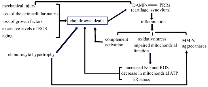
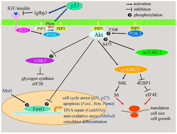
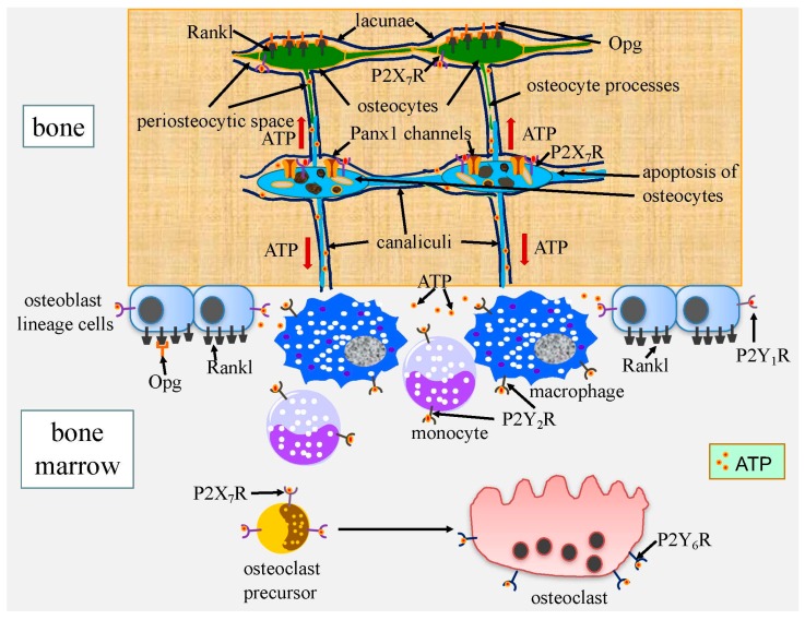
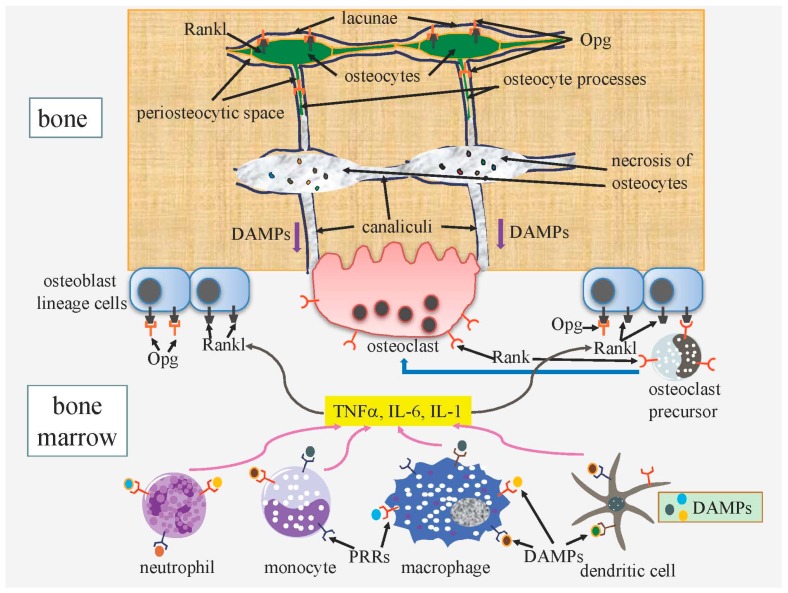
Cell death in skeletal component cells, including chondrocytes, osteoblasts, and osteocytes, plays roles in skeletal development, maintenance, and repair as well as in the pathogenesis of osteoarthritis and osteoporosis. Chondrocyte proliferation, differentiation, and apoptosis are important steps for endochondral ossification. Although the inactivation of P53 and RB is involved in the pathogenesis of osteosarcomas, the deletion of p53 and inactivation of Rb are insufficient to enhance chondrocyte proliferation, indicating the presence of multiple inhibitory mechanisms against sarcomagenesis in chondrocytes. The inflammatory processes induced by mechanical injury and chondrocyte death through the release of danger-associated molecular patterns (DAMPs) are involved in the pathogenesis of posttraumatic osteoarthritis. The overexpression of BCLXL increases bone volume with a normal structure and maintains bone during aging by inhibiting osteoblast apoptosis. p53 inhibits osteoblast proliferation and enhances osteoblast apoptosis, thereby reducing bone formation, but also exerts positive effects on osteoblast differentiation through the Akt–FoxOs pathway. Apoptotic osteocytes release ATP, which induces the receptor activator of nuclear factor κ-B ligand (Rankl) expression and osteoclastogenesis, from pannexin 1 channels. Osteocyte death ultimately results in necrosis; DAMPs are released to the bone surface and promote the production of proinflammatory cytokines, which induce Rankl expression, and osteoclastogenesis is further enhanced.
Overview
The claims and statistics of this paper haven’t been checked yet.
Run the check — free, no account needed. It reads the open-access text and checks the sources it cites.
Integrity signals
Registry
Cites
Statistics
Figures
Verification
Claims the paper makes
The claims and statistics of this paper haven’t been checked yet.
Run the check — free, no account needed. It reads the open-access text and checks the sources it cites.
Claims other papers make about this one
Each is checked against this paper’s own text: does it say what the citing paper says it does?
The cited source says what the claim attributes to it.
In the citing paper
“Necrotic osteocytes release damage-associated molecular pattern (DAMP) proteins after the cell membrane is disrupted.61”
From Bone remodeling: an operational process ensuring survival and bone mechanical competence (2022) · cites this paper as [61]
In the citing paper
“In vivo, osteocyte apoptosis is fundamental for initiating bone remodeling,61 independent of the stimulus causing osteocyte death such as estrogen loss,62 fatigue,63 or unloading.64”
From Bone remodeling: an operational process ensuring survival and bone mechanical competence (2022) · cites this paper as [61]
Self-check
Percentages, sums and reported differences re-computed from the paper’s own numbers.
The self-check needs the paper’s full text.
Citations
Cites
Stöckl, S., Göttl, C., Grifka, J. K., & Grässel, S. (2013) · Cellular Physiology and Biochemistry · doi:10.1159/000350089 · Cited by 4,670
Osteoarthritis: A disease of the joint as an organ
Loeser, R. F., Goldring, S. R., Scanzello, C. R., & Goldring, M. B. (2012) · Arthritis & Rheumatism · doi:10.1002/art.34453 · Cited by 3,234 · cited 1×
“The signaling pathway through PRRs induces synovitis, transduces inflammation, increases the expression of matrix metalloproteinases (MMPs) and aggrecanases, and induces oxidative stress and cell death [55,56].”
BackgroundHigh-mobility group box 1 protein (HMGB1): nuclear weapon in the immune arsenal
Lotze, M. T., & Tracey, K. J. (2005) · Nature reviews. Immunology · doi:10.1038/nri1594 · Cited by 2,490
Regulation of Rate of Cartilage Differentiation by Indian Hedgehog and PTH-Related Protein
Vortkamp, A., Lee, K., Lanske, B., Segre, G. V., Kronenberg, H. M., & Tabin, C. J. (1996) · Science · doi:10.1126/science.273.5275.613 · Cited by 1,999
Evidence for osteocyte regulation of bone homeostasis through RANKL expression
Nakashima, T., Hayashi, M., Fukunaga, T., Kurata, K., Oh-hora, M., Feng, J. Q., Bonewald, L. F., Kodama, T., Wutz, A., Wagner, E. F., et al. (2011) · Nature Medicine · doi:10.1038/nm.2452 · Cited by 1,778 · cited 1×
“The osteocyte-specific deletion of Rankl results in the inhibition of bone resorption, indicating that osteocytes are a major source of Rankl [121,122].”
BackgroundWeinstein, R. S., Jilka, R. L., Parfitt, A. M., & Manolagas, S. C. (1998) · Journal of Clinical Investigation · doi:10.1172/jci2799 · Cited by 1,753
St‐Jacques, B., Hammerschmidt, M., & McMahon, A. P. (1999) · Genes & Development · doi:10.1101/gad.13.16.2072 · Cited by 1,742 · cited 1×
“Ihh enhances chondrocyte proliferation [3].”
Park, J. S., Svetkauskaite, D., He, Q., Kim, J., Strassheim, D., Ishizaka, A., & Abraham, E. H. (2004) · Journal of Biological Chemistry · doi:10.1074/jbc.m306793200 · Cited by 1,582
p53 mutant mice that display early ageing-associated phenotypes
Tyner, S. D., Venkatachalam, S., Choi, J., Jones, S., Ghebranious, N., Igelmann, H., Lu, X., Soron, G., Cooper, B., Brayton, C., et al. (2002) · Nature · doi:10.1038/415045a · Cited by 1,459 · cited 1×
“Mice with activated mutations in p53 exhibit osteopenia [86].”
BackgroundCyclin D as a therapeutic target in cancer
Musgrove, E. A., Caldon, C. E., Barraclough, J., Stone, A., & Sutherland, R. L. (2011) · Nature Reviews Cancer · doi:10.1038/nrc3090 · Cited by 1,370 · cited 2×
“This leads to the transcriptional activation of E2f-responsive genes that are essential for DNA synthesis, including cyclins E and A [16].”
Background
Cited by
Sorted by citations
Molecular mechanosensors in osteocytes
Qin, L., Liu, W., Cao, H., & Xiao, G. (2020) · Bone Research · doi:10.1038/s41413-020-0099-y · Cited by 484
“These apoptotic osteocytes further induced Rankl expression and osteoclastogenesis, which resulted in increased bone resorption and bone mass loss.(176) Interestingly, in experimental animals, treatment with an apoptosis inhibitor (the pancaspase inhibitor QVD-OPh) rescued osteocyte cell loss and bone mass reduction d…”
BackgroundBone remodeling: an operational process ensuring survival and bone mechanical competence
Bolamperti, S., Villa, I., & Rubinacci, A. (2022) · Bone Research · doi:10.1038/s41413-022-00219-8 · Cited by 482
“Necrotic osteocytes release damage-associated molecular pattern (DAMP) proteins after the cell membrane is disrupted.(61) DAMP proteins flow through the canalicular network and reach the bone/marrow interface, where they bind to pattern recognition receptors (PRRs) on bone marrow cells.”
BackgroundThe Regulation of Bone Metabolism and Disorders by Wnt Signaling
Maeda, K., Kobayashi, Y., Koide, M., Uehara, S., Okamoto, M., Ishihara, A., Kayama, T., Saito, M., & Marumo, K. (2019) · International Journal of Molecular Sciences · doi:10.3390/ijms20225525 · Cited by 360
“OA is a degenerative disease associated with articular cartilage damage, osteophyte formation, and hardening of the subchondral bone [152,153].”
BackgroundRegulation and Role of Transcription Factors in Osteogenesis
Chan, W. C. W., Tan, Z., To, M. K.-T., & Chan, D. T.-M. (2021) · International Journal of Molecular Sciences · doi:10.3390/ijms22115445 · Cited by 276
“p53 and FoxOs have similar functions in inhibiting cell cycles and inducing cell death in osteoblasts [141].”
BackgroundOsteocyte necrosis triggers osteoclast-mediated bone loss through macrophage-inducible C-type lectin
Andreev, D., Liu, M., Weidner, D., Kachler, K., Faas, M., Grüneboom, A., Schlötzer-Schrehardt, U., Muñoz, L. E., Steffen, U., Grötsch, B., et al. (2020) · Journal of Clinical Investigation · doi:10.1172/jci134214 · Cited by 188
“However, because phagocytes cannot reach and engulf apoptotic osteocytes in their isolated lacunae, these cells automatically undergo secondary necrosis, losing their membrane integrity and releasing damage-associated molecular patterns (DAMPs) into their environment (10).”
Background
Metadata
- Authors
- Komori, T.
- Affiliations
- Nagasaki University (JP)
- Corresponding author
- Komori, Toshihisa
- ORCID
- Komori, Toshihisa
- Venue
- International Journal of Molecular Sciences
- Publisher
- MDPI AG
- Published
- 2016-12-06
- Volume
- 17
- Issue
- 12
- Pages
- 2045
- ISSN
- 1422-0067 (ISSN-L)
- PMC
- PMC5187845
- OpenAlex
- W2560057190
- Cited by
- 176
- References
- 128
- Citations by year
- 2026: 12 · 2025: 16 · 2024: 17 · 2023: 36 · 2022: 24 · 2021: 19 · 2020: 21 · 2019: 9 · 2018: 14 · 2017: 8
- FWCI
- 0.93
- Type
- journal article
- Language
- en
- Open access
- gold
- doi.org
- Licence
- Locations
- International Journal of Molecular Sciences (publishedVersion, cc-by)
- PubMed Central (publishedVersion, cc-by)
- PubMed (publishedVersion)
- Funders
- Japanese Ministry of Education, Culture, Sports, Science and Technology, Japan (26221310)
- Topics
- Life Sciences › Biochemistry, Genetics and Molecular Biology › Molecular Biology › Bone Metabolism and Diseases
- Life Sciences › Biochemistry, Genetics and Molecular Biology › Molecular Biology › Cell death mechanisms and regulation
- Health Sciences › Medicine › Rheumatology › Osteoarthritis Treatment and Mechanisms
- Keywords
- cell death, chondrocytes, osteoblasts, osteocytes, apoptosis, osteoarthritis, osteoporosis, endochondral ossification, p53, retinoblastoma protein, osteosarcoma, post-traumatic osteoarthritis, damage-associated molecular patterns, Bcl-xL, RANKL, osteoclastogenesis, pannexin-1 channels, ATP release, skeletal development, bone preservation
- MeSH terms
- Alarmins, Animals, Humans, Osteoblasts, Osteocytes, Apoptosis, Chondrocytes
About these checks
Registry, retraction, Cites and statistics results are computed, not judged, with one exception: a printed reference that no index lists is read once by a model before it counts as a citation error. Claims are extracted by a model and kept only when their quote appears verbatim in the text; each cited source is then checked, with a model’s help, for whether it says what the claim relies on. Those verdicts can be wrong — read the claim’s quote and the cited source before relying on one.
Statistics and arithmetic are recomputed, not judged.
Confidence bands are shown only for fields calibrated on our annotated test set. None is calibrated yet, so no band is shown.
What counts as a claim ›How we measure confidence ›How we check this ›
Are you an author, or do you see a problem with this page? Request a review
