

The DOI matches its Crossref record; 127 of the 128 references it cites were found in a registry or an index.
Cell Death in Chondrocytes, Osteoblasts, and Osteocytes
Open accessAbstract
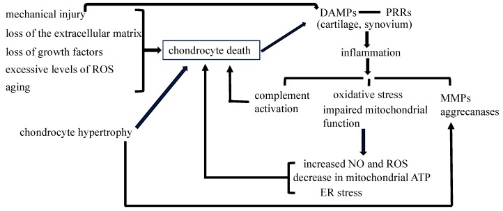
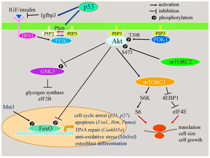
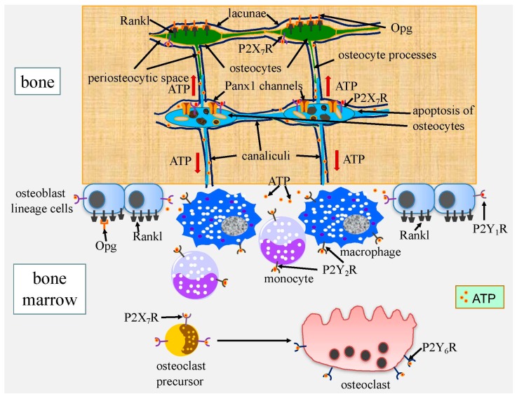
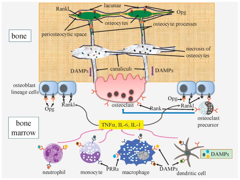
Cell death in skeletal component cells, including chondrocytes, osteoblasts, and osteocytes, plays roles in skeletal development, maintenance, and repair as well as in the pathogenesis of osteoarthritis and osteoporosis. Chondrocyte proliferation, differentiation, and apoptosis are important steps for endochondral ossification. Although the inactivation of P53 and RB is involved in the pathogenesis of osteosarcomas, the deletion of p53 and inactivation of Rb are insufficient to enhance chondrocyte proliferation, indicating the presence of multiple inhibitory mechanisms against sarcomagenesis in chondrocytes. The inflammatory processes induced by mechanical injury and chondrocyte death through the release of danger-associated molecular patterns (DAMPs) are involved in the pathogenesis of posttraumatic osteoarthritis. The overexpression of BCLXL increases bone volume with a normal structure and maintains bone during aging by inhibiting osteoblast apoptosis. p53 inhibits osteoblast proliferation and enhances osteoblast apoptosis, thereby reducing bone formation, but also exerts positive effects on osteoblast differentiation through the Akt–FoxOs pathway. Apoptotic osteocytes release ATP, which induces the receptor activator of nuclear factor κ-B ligand (Rankl) expression and osteoclastogenesis, from pannexin 1 channels. Osteocyte death ultimately results in necrosis; DAMPs are released to the bone surface and promote the production of proinflammatory cytokines, which induce Rankl expression, and osteoclastogenesis is further enhanced.
Overview
The claims and statistics of this paper haven’t been checked yet.
Run the check — free, no account needed. It reads the open-access text and checks the sources it cites.
Integrity signals
Registry
Cites
Statistics
Figures
Verification
Claims the paper makes
The claims and statistics of this paper haven’t been checked yet.
Run the check — free, no account needed. It reads the open-access text and checks the sources it cites.
Claims other papers make about this one
Each is checked against this paper’s own text: does it say what the citing paper says it does?
The cited source says what the claim attributes to it.
In the citing paper
“Necrotic osteocytes release damage-associated molecular pattern (DAMP) proteins after the cell membrane is disrupted.61”
From Bone remodeling: an operational process ensuring survival and bone mechanical competence (2022) · cites this paper as [61]
In the citing paper
“In vivo, osteocyte apoptosis is fundamental for initiating bone remodeling,61 independent of the stimulus causing osteocyte death such as estrogen loss,62 fatigue,63 or unloading.64”
From Bone remodeling: an operational process ensuring survival and bone mechanical competence (2022) · cites this paper as [61]
Self-check
Percentages, sums and reported differences re-computed from the paper’s own numbers.
The self-check needs the paper’s full text.
Citations
Cites
Stöckl, S., Göttl, C., Grifka, J. K., & Grässel, S. (2013) · Cellular Physiology and Biochemistry · doi:10.1159/000350089 · Cited by 4,670
Osteoarthritis: A disease of the joint as an organ
Loeser, R. F., Goldring, S. R., Scanzello, C. R., & Goldring, M. B. (2012) · Arthritis & Rheumatism · doi:10.1002/art.34453 · Cited by 3,234 · cited 1×
“The signaling pathway through PRRs induces synovitis, transduces inflammation, increases the expression of matrix metalloproteinases (MMPs) and aggrecanases, and induces oxidative stress and cell death [55,56].”
BackgroundHigh-mobility group box 1 protein (HMGB1): nuclear weapon in the immune arsenal
Lotze, M. T., & Tracey, K. J. (2005) · Nature reviews. Immunology · doi:10.1038/nri1594 · Cited by 2,490
Regulation of Rate of Cartilage Differentiation by Indian Hedgehog and PTH-Related Protein
Vortkamp, A., Lee, K., Lanske, B., Segre, G. V., Kronenberg, H. M., & Tabin, C. J. (1996) · Science · doi:10.1126/science.273.5275.613 · Cited by 1,999
Evidence for osteocyte regulation of bone homeostasis through RANKL expression
Nakashima, T., Hayashi, M., Fukunaga, T., Kurata, K., Oh-hora, M., Feng, J. Q., Bonewald, L. F., Kodama, T., Wutz, A., Wagner, E. F., et al. (2011) · Nature Medicine · doi:10.1038/nm.2452 · Cited by 1,778 · cited 1×
“The osteocyte-specific deletion of Rankl results in the inhibition of bone resorption, indicating that osteocytes are a major source of Rankl [121,122].”
BackgroundWeinstein, R. S., Jilka, R. L., Parfitt, A. M., & Manolagas, S. C. (1998) · Journal of Clinical Investigation · doi:10.1172/jci2799 · Cited by 1,753
St‐Jacques, B., Hammerschmidt, M., & McMahon, A. P. (1999) · Genes & Development · doi:10.1101/gad.13.16.2072 · Cited by 1,742 · cited 1×
“Ihh enhances chondrocyte proliferation [3].”
Park, J. S., Svetkauskaite, D., He, Q., Kim, J., Strassheim, D., Ishizaka, A., & Abraham, E. H. (2004) · Journal of Biological Chemistry · doi:10.1074/jbc.m306793200 · Cited by 1,582
p53 mutant mice that display early ageing-associated phenotypes
Tyner, S. D., Venkatachalam, S., Choi, J., Jones, S., Ghebranious, N., Igelmann, H., Lu, X., Soron, G., Cooper, B., Brayton, C., et al. (2002) · Nature · doi:10.1038/415045a · Cited by 1,459 · cited 1×
“Mice with activated mutations in p53 exhibit osteopenia [86].”
BackgroundCyclin D as a therapeutic target in cancer
Musgrove, E. A., Caldon, C. E., Barraclough, J., Stone, A., & Sutherland, R. L. (2011) · Nature Reviews Cancer · doi:10.1038/nrc3090 · Cited by 1,370 · cited 2×
“This leads to the transcriptional activation of E2f-responsive genes that are essential for DNA synthesis, including cyclins E and A [16].”
Background
Cited by
Sorted by citations
Molecular mechanosensors in osteocytes
Qin, L., Liu, W., Cao, H., & Xiao, G. (2020) · Bone Research · doi:10.1038/s41413-020-0099-y · Cited by 484
“These apoptotic osteocytes further induced Rankl expression and osteoclastogenesis, which resulted in increased bone resorption and bone mass loss.(176) Interestingly, in experimental animals, treatment with an apoptosis inhibitor (the pancaspase inhibitor QVD-OPh) rescued osteocyte cell loss and bone mass reduction d…”
BackgroundBone remodeling: an operational process ensuring survival and bone mechanical competence
Bolamperti, S., Villa, I., & Rubinacci, A. (2022) · Bone Research · doi:10.1038/s41413-022-00219-8 · Cited by 482
“Necrotic osteocytes release damage-associated molecular pattern (DAMP) proteins after the cell membrane is disrupted.(61) DAMP proteins flow through the canalicular network and reach the bone/marrow interface, where they bind to pattern recognition receptors (PRRs) on bone marrow cells.”
BackgroundThe Regulation of Bone Metabolism and Disorders by Wnt Signaling
Maeda, K., Kobayashi, Y., Koide, M., Uehara, S., Okamoto, M., Ishihara, A., Kayama, T., Saito, M., & Marumo, K. (2019) · International Journal of Molecular Sciences · doi:10.3390/ijms20225525 · Cited by 360
“OA is a degenerative disease associated with articular cartilage damage, osteophyte formation, and hardening of the subchondral bone [152,153].”
BackgroundRegulation and Role of Transcription Factors in Osteogenesis
Chan, W. C. W., Tan, Z., To, M. K.-T., & Chan, D. T.-M. (2021) · International Journal of Molecular Sciences · doi:10.3390/ijms22115445 · Cited by 276
“p53 and FoxOs have similar functions in inhibiting cell cycles and inducing cell death in osteoblasts [141].”
BackgroundOsteocyte necrosis triggers osteoclast-mediated bone loss through macrophage-inducible C-type lectin
Andreev, D., Liu, M., Weidner, D., Kachler, K., Faas, M., Grüneboom, A., Schlötzer-Schrehardt, U., Muñoz, L. E., Steffen, U., Grötsch, B., et al. (2020) · Journal of Clinical Investigation · doi:10.1172/jci134214 · Cited by 188
“However, because phagocytes cannot reach and engulf apoptotic osteocytes in their isolated lacunae, these cells automatically undergo secondary necrosis, losing their membrane integrity and releasing damage-associated molecular patterns (DAMPs) into their environment (10).”
BackgroundCell death regulation: A new way for natural products to treat osteoporosis
Li, Z., Li, D., Chen, R., Gao, S., Xu, Z., & Li, N. (2022) · Pharmacological Research · doi:10.1016/j.phrs.2022.106635 · Cited by 183
The role of TGF-beta3 in cartilage development and osteoarthritis
Du, X., Cai, L., Xie, J., & Zhou, X. (2023) · Bone Research · doi:10.1038/s41413-022-00239-4 · Cited by 176
“Chondrocyte death Dynamic and accurate regulation of cell death in chondrocytes is pivotal in skeletal development, maintenance, and repair as well as in the pathological course of OA and osteoporosis (OP).(132,133) Different types of cell death in cartilage have been reported, including three types of physiological/p…”
BackgroundExercise for osteoporosis: A literature review of pathology and mechanism
Zhang, L., Zheng, Y.-L., Wang, R., Wang, X., & Zhang, H. (2022) · Frontiers in Immunology · doi:10.3389/fimmu.2022.1005665 · Cited by 161
“Hughes et al. (13) reported that 17b-estradiol increased the apoptosis rate of rat OCs from 0.5% to 2.7%, and estrogen deficiency prolonged the survival period of OCs, increasing the number of cells by 2–3 times, and leading to enlargement of the bone absorption cavity.”
BackgroundInfluentialRunx2 is essential for the transdifferentiation of chondrocytes into osteoblasts
Qin, X., Jiang, Q., Nagano, K., Moriishi, T., Miyazaki, T., Komori, H., Ito, K., von der Mark, K., Sakane, C., Kaneko, H., et al. (2020) · PLoS Genetics · doi:10.1371/journal.pgen.1009169 · Cited by 126
“…apoptosis of terminal hypertrophic chondrocytes in Runx2 fl/flCre mice may have promoted the vascular invasion into the cartilage because apoptotic terminal hypertrophic chondrocytes may finally die by necrosis without phagocytosis and release DAMPs, which induce osteoclastogenesis [23, 31].”
Self-citationThe roles of osteocytes in alveolar bone destruction in periodontitis
Huang, X., Xie, M., Xie, Y., Feng, M., Lu, X., Li, X., & Chen, L. (2020) · Journal of Translational Medicine · doi:10.1186/s12967-020-02664-7 · Cited by 122
“Apoptotic osteocytes can also release adenosine triphosphate (ATP) through activated pannexin 1 channels, and ATP acts on osteocytes, osteoblasts, osteoclasts and macrophages via ATP receptor-gated (P2) channels and consequently induces enhanced production of RANKL in bystander osteocytes and osteoblasts, aggregation…”
BackgroundCorrado, A., Cici, D., Rotondo, C., Maruotti, N., & Cantatore, F. P. (2020) · International Journal of Molecular Sciences · doi:10.3390/ijms21103679 · Cited by 101
“The increase in osteoblast apoptosis has been proposed among the mechanisms of the reduced bone-forming capacity of senescent bone [75].”
BackgroundScaffold-Based Tissue Engineering Strategies for Osteochondral Repair
Fu, J.-N., Wang, X., Yang, M., Chen, Y., Zhang, J., Deng, R., Zhang, Z., Yu, J., & Yuan, F. (2022) · Frontiers in Bioengineering and Biotechnology · doi:10.3389/fbioe.2021.812383 · Cited by 96
Zhang, S., Xu, J., Si, H., Wu, Y., Zhou, S., & Shen, B. (2022) · Antioxidants · doi:10.3390/antiox11091668 · Cited by 85
“Reactive oxygen species (ROS) production and subsequent lipid peroxidation are related to the antioxidant capacity of chondrocytes, playing key roles in cartilage degradation and chondrocyte death [5,6].”
BackgroundRiegger, J., & Brenner, R. E. (2020) · International Journal of Molecular Sciences · doi:10.3390/ijms21051560 · Cited by 75
Osteoprotective Roles of Green Tea Catechins
Huang, H., Cheng, T., Lin, S., Ho, C.-J., Chyu, J., Yang, R., Chen, C., & Shen, C. (2020) · Antioxidants · doi:10.3390/antiox9111136 · Cited by 70
“In addition, the increase in osteoblast apoptosis is a possible mechanism for the reduced bone-forming ability of senescent bone [18].”
BackgroundZhang, H., Wang, A., Li, G., Zhai, Q., Huang, Z., Wang, X., Cao, Z., Liu, L., Liu, G., Chen, B., et al. (2023) · Free Radical Biology and Medicine · doi:10.1016/j.freeradbiomed.2023.01.026 · Cited by 70
ECM signaling in cartilage development and endochondral ossification
Prein, C., & Beier, F. (2018) · Current topics in developmental biology/Current Topics in Developmental Biology · doi:10.1016/bs.ctdb.2018.11.003 · Cited by 68
IL-33/IL-31 Axis in Osteoporosis
Martinis, M. D., Sirufo, M. M., Suppa, M., & Ginaldi, L. (2020) · International Journal of Molecular Sciences · doi:10.3390/ijms21041239 · Cited by 68
“Release of alarmins or DAMPs (danger-associated molecular patterns), including IL-33, from damaged cells is a relevant mechanism by which immune cells can be alerted of tissue damage, and alarmins play a key role in the development of acute or chronic inflammatory diseases, cancer and osteoporosis [89,90].”
BackgroundDNMT aberration-incurred GPX4 suppression prompts osteoblast ferroptosis and osteoporosis
Ruan, B., Dong, J. P., Wei, F., Huang, Z., Yang, B., Zhang, L., Li, C., Dong, H., Cao, W., Wang, H., et al. (2024) · Bone Research · doi:10.1038/s41413-024-00365-1 · Cited by 71
“8,9 Although past researches have established that various forms of regulated cell death, such as apoptosis, 10 autophagic cell death, 11 pyroptosis, 12 necroptosis.”
Xu, T., Chen, P., Zhang, C., Shaukat, A., Lin, L., Yue, K., Ding, W., Tong, X., Liu, K., He, Y., et al. (2023) · npj Biofilms and Microbiomes · doi:10.1038/s41522-022-00360-6 · Cited by 73
Emerging roles of long noncoding RNA in chondrogenesis, osteogenesis, and osteoarthritis.
Sun, H., Peng, G., Ning, X., Wang, J., Yang, H., & Deng, J. (2019) · Europe PMC (PubMed Central) · Cited by 63
Chondrocyte death involvement in osteoarthritis
Salucci, S., Falcieri, E., & Battistelli, M. (2022) · Cell and Tissue Research · doi:10.1007/s00441-022-03639-4 · Cited by 63
“Necrosis is a pathological and non-programmed form of cell death, caspase- and energy-independent (Zhang et al. 2018; Komori 2016; Martin and Henry 2013).”
BackgroundAguirre, J. I., Castillo, E. J., & Kimmel, D. B. (2021) · Bone · doi:10.1016/j.bone.2021.116168 · Cited by 62
“The accumulated necrotic osteocytes generate an accrued amount of DAMPs that further stimulate inflammation.”
BackgroundInfluentialGuided Bone Regeneration Using Barrier Membrane in Dental Applications
Kim, K., Su, Y., Kucine, A. J., Cheng, K., & Zhu, D. (2023) · ACS Biomaterials Science & Engineering · doi:10.1021/acsbiomaterials.3c00690 · Cited by 61
Zhu, S., Zhuang, J., Wu, Q., Liu, Z., Liao, C., Luo, S., Chen, J., & Zhong, Z. (2018) · Aging Cell · doi:10.1111/acel.12764 · Cited by 55
“Osteoblast apoptosis increases with age (Komori, 2016; Tower, 2015) and is responsible for reduction in osteoblast number and activity, which contribute to decreased bone formation (Jilka & O’Brien, 2016; Jilka, Weinstein, Parfitt & Manolagas, 2007; Weinstein & Manolagas, 2000).”
BackgroundIcariin: A Potential Molecule for Treatment of Knee Osteoarthritis
Zhang, J., Fan, F., Liu, A., Zhang, C., Li, Q., Zhang, C., He, F., & Shang, M. (2022) · Frontiers in Pharmacology · doi:10.3389/fphar.2022.811808 · Cited by 58
“Degradation of the extracellular matrix promotes chondrocyte apoptosis at the same time (Komori 2016).”
BackgroundOsteoimmunology in Periodontitis: Local Proteins and Compounds to Alleviate Periodontitis
Sirisereephap, K., Maekawa, T., Tamura, H., Hiyoshi, T., Domon, H., Isono, T., Terao, Y., Maeda, T., & Tabeta, K. (2022) · International Journal of Molecular Sciences · doi:10.3390/ijms23105540 · Cited by 57
Ferroptosis Plays a Role in Human Chondrocyte of Osteoarthritis Induced by IL-1β In Vitro
Xu, W., Zhang, B., Xi, C., Qin, Y., Lin, X., Wang, B., Kong, P., & Yan, J. (2023) · Cartilage · doi:10.1177/19476035221142011 · Cited by 56
Yang, S., Sun, Y., Капилевич, Л. В., Zhang, X., & Huang, Y. (2023) · Frontiers in Pharmacology · doi:10.3389/fphar.2023.1249418 · Cited by 52
“A large body of evidence suggests that the apoptosis of osteoblasts under pathological conditions leads to an imbalance in bone formation–resorption, which is closely associated with the development of OP (Komori, 2016; Chen et al., 2018).”
BackgroundTargeting regulated chondrocyte death in osteoarthritis therapy
Zhu, R., Wang, Y., Ouyang, Z., Hao, W., Zhou, F., Lin, Y., Cheng, Y., Zhou, R., & Hu, W. M. (2023) · Biochemical Pharmacology · doi:10.1016/j.bcp.2023.115707 · Cited by 52
Geniposide ameliorates glucocorticoid-induced osteoblast apoptosis by activating autophagy
Huang, J., Ye, Y., Xiao, Y., Ren, Q., Zhou, Q., Zhong, M., Jiao, L., & Wu, L. (2022) · Biomedicine & Pharmacotherapy · doi:10.1016/j.biopha.2022.113829 · Cited by 47
McCutcheon, S., Majeska, R. J., Spray, D. C., Schaffler, M. B., & Vázquez, M. (2020) · Journal of Bone and Mineral Research · doi:10.1002/jbmr.3954 · Cited by 46
“In the current studies we found that acute ATP exposure turned on osteocyte RANKL expression, consistent with previous studies showing that ATP can stimulate RANKL expression.((40,41)) We further discovered that with osteocyte apoptosis, ATP was the essential signal driving the increase in bystander osteocyte RANKL ex…”
ResultZheng, L., Pi, C., Zhang, J., Fan, Y., Cui, C., Zhou, Y., Sun, J., Yuan, Q., Xu, X., Ye, L., et al. (2018) · Bone Research · doi:10.1038/s41413-018-0027-6 · Cited by 46
“Articular chondrocytes exhibited an agerelated decline in proliferation and synthetic capacity.(46) As age increased, the calcified cartilage layer became thicker.”
BackgroundFabrication of chondroitin sulfate calcium complex and its chondrocyte proliferation in vitro
Shen, Q., Zhang, C., Mo, H., Zhang, H., Qin, X., Li, J., Zhang, Z., & Richel, A. (2020) · Carbohydrate Polymers · doi:10.1016/j.carbpol.2020.117282 · Cited by 45
LIM domain proteins Pinch1/2 regulate chondrogenesis and bone mass in mice
Lei, Y., Fu, X., Li, P., Lin, S., Yan, Q., Lai, Y., Liu, X., Wang, Y., Bai, X., Liu, C.-J., et al. (2020) · Bone Research · doi:10.1038/s41413-020-00108-y · Cited by 45
“In the absence of Pinch, TGF-β/Smad2/3 signaling is impaired due to reduced expression and activation and accelerated degradation of Smad2/3, which reduces chondrocyte proliferation and survival and upregulates Runx2.”
BackgroundInfluentialCao, S., Wei, Y., Xu, H., Weng, J., Qi, T., Yu, F., Liu, S., Xiong, A., Liu, P., & Zeng, H. (2023) · Frontiers in Immunology · doi:10.3389/fimmu.2023.1202436 · Cited by 42
Non-bone metastatic cancers promote osteocyte-induced bone destruction
Pin, F., Prideaux, M., Huot, J. R., Essex, A. L., Plotkin, L. I., Bonetto, A., & Bonewald, L. F. (2021) · Cancer Letters · doi:10.1016/j.canlet.2021.06.030 · Cited by 36
Zhang, W., Zhong, B., Zhang, C., Luo, C., & Zhan, Y. (2017) · FEBS Open Bio · doi:10.1002/2211-5463.12345 · Cited by 35
Li, L., Wei, X., Wang, D., Lv, Z., Geng, X., Li, P., Lu, J., Wang, K., Wang, X., Sun, J. M., et al. (2020) · Arthritis & Rheumatology · doi:10.1002/art.41230 · Cited by 36
“Decreases in chondrocytes and in the cartilage proliferative capacity contribute to the degradation of the age- related OA cartilage matrix (2,3).”
BackgroundLymphatic platelet thrombosis limits bone repair by precluding lymphatic transporting DAMPs
Zheng, Y., Cong, L., Zhao, L., Wang, P., Xing, L., Liu, J., Xu, H., Li, N., Zhao, Y., Yuan, L., et al. (2025) · Nature Communications · doi:10.1038/s41467-025-56147-8 · Cited by 36
“Pro-in fl ammatory factors (TNF-α , IL-1, and IL-6) and growth factors (TGF β 1 and PDGF) are vitalligands of OB apoptosis and BMSC proliferation 20,33 .”
Roper, P. M., Shao, C. G., & Novack, D. V. (2020) · Cells · doi:10.3390/cells9102157 · Cited by 32
“Other DAMPs, such as S100 proteins and high-mobility group box 1 (HMGB1), likely activate other PRRs and contribute additional osteolytic stimuli [67].”
BackgroundFTO-mediated m6A demethylation of pri-miR-3591 alleviates osteoarthritis progression
Liu, W., Jiang, T., Zheng, W., Zhang, J., Li, A., Lu, C., & Lin, Z. (2023) · Arthritis Research & Therapy · doi:10.1186/s13075-023-03035-5 · Cited by 31
Osteocytes: Their Lacunocanalicular Structure and Mechanoresponses
Moriishi, T., & Komori, T. (2022) · International Journal of Molecular Sciences · doi:10.3390/ijms23084373 · Cited by 36
“Thus, osteocyte ablation by diphtheria toxin enhanced bone resorption and inhibited bone formation [29,35] (Figures 2 and 4).”
BackgroundSelf-citationHu, H., Zhang, Z.-Y., Luo, Z. X., Ti, H., Wu, J., Nie, H., Yuan, Z., Wu, X. L., Zhang, K.-Y., Shi, S., et al. (2025) · Cell Death Discovery · doi:10.1038/s41420-025-02633-3 · Cited by 40
“The process of cell death within skeletal component cells is crucial for bone development, maintenance, repair, and overall functional integrity [12].”
Comparison of Necroptosis With Apoptosis for OVX-Induced Osteoporosis
He, B., Zhu, Y., Cui, H., Sun, B., Su, T., & Wen, P. (2021) · Frontiers in Molecular Biosciences · doi:10.3389/fmolb.2021.790613 · Cited by 31
“These stimulate the generation of multiple immunostimulatory cytokines and proinflammatory molecules, the aggregation and energization of immune cells (e.g., macrophages, monocytes, and neutrophils), which also induce the secretion of RANKL and bone resorption (Komori, 2013; Komori, 2016).”
BackgroundJia, L., Ma, T., Lv, L., Yu, Y., Zhao, M., Chen, H., & Gao, L. (2023) · Ecotoxicology and Environmental Safety · doi:10.1016/j.ecoenv.2023.114517 · Cited by 29
Zhao, X., Wang, T., Cai, B., Wang, X., Feng, W. S., Han, Y., Li, D., Li, S., & Liu, J. (2019) · PubMed Central · Cited by 29
The role of the complement system in disc degeneration and Modic changes
Heggli, I., Teixeira, G. Q., Iatridis, J. C., Neidlinger‐Wilke, C., & Dudli, S. (2024) · JOR Spine · doi:10.1002/jsp2.1312 · Cited by 28
“85 While under physiological conditions tissue damage-mediated complement system activation helps to clear dying cells and remove immune complexes /pathogens, persistent and accumulating cell damage can exceed homeostatic activation and amplify rather than suppress pro-inflammatory processes.”
BackgroundXue, J., Ye, J., Xia, Z., & Cheng, B. (2019) · Cellular and Molecular Biology · doi:10.14715/cmb/2019.65.6.15 · Cited by 28
From death to birth: how osteocyte death promotes osteoclast formation
Zhao, W., Qian, J., Li, J., Su, T., Deng, X., Fu, Y., Liang, X., & Cui, H. (2025) · Frontiers in Immunology · doi:10.3389/fimmu.2025.1551542 · Cited by 28
“…to PRRs, such as TLR2, TLR4, and RAGE, on immune cells including macrophages and dendritic cells, and promote their production of pro-in fl ammatory cytokines, such as TNF-a and IL-6, which further induces RANKL expression in osteoblast lineage cells or promotes osteoclastogenesis directly (91).”
Ubiquitin‐specific peptidases: Players in bone metabolism
Shen, J., Lin, X., Dai, F., Chen, G., Lin, H., Fang, B., & Liu, H. (2023) · Cell Proliferation · doi:10.1111/cpr.13444 · Cited by 27
Ay, B., Parolia, K., Liddell, R. S., Qiu, Y., Grasselli, G., Cooper, D. M. L., & Davies, J. E. (2020) · Communications Biology · doi:10.1038/s42003-019-0747-1 · Cited by 27
Takemura, Y., Moriyama, Y., Ayukawa, Y., Kurata, K., Rakhmatia, Y. D., & Koyano, K. (2018) · Journal of Biomedical Materials Research Part A · doi:10.1002/jbm.a.36597 · Cited by 26
Single‐Cell RNA ‐Sequencing Reveals the Breadth of Osteoblast Heterogeneity
Yoshioka, H., Okita, S., Nakano, M., Minamizaki, T., Nubukiyo, A., Sotomaru, Y., Bonnelye, E., Kozai, K., Tanimoto, K., Aubin, J. E., et al. (2021) · JBMR Plus · doi:10.1002/jbm4.10496 · Cited by 25
“Indeed, apoptotic response promotes osteoblast differentiation via the p53-Akt-FoxO pathway.((51)) Thus, expression of apoptotic versus survival signals and, concomitantly, apoptotic behavior may differ among individual osteoblasts, contributing to noise in the expression profiles and to our inability to recognize a d…”
BackgroundBi, H., Wang, D., Liu, X., Wang, G., & Wu, X. (2020) · Journal of Biosciences · doi:10.1007/s12038-020-0024-y · Cited by 25
“Osteoblasts, one of the major bone-forming cells, have been identified as a pivotal regulator in skeletal development, maintenance, and repair as well as bone microenvironment homeostasis (Komori 2016; Long 2011).”
BackgroundRösch, G., Zaucke, F., & Jenei‐Lanzl, Z. (2023) · American Journal of Physiology-Cell Physiology · doi:10.1152/ajpcell.00039.2023 · Cited by 26
Qu, X., Mei, J., Yu, Z., Zhai, Z., Qiao, H., & Dai, K. (2018) · Biochemical and Biophysical Research Communications · doi:10.1016/j.bbrc.2018.05.035 · Cited by 25
Wei, Y., & Sun, Y. (2018) · Advances in experimental medicine and biology · doi:10.1007/978-981-13-1117-8_12 · Cited by 25
PGC-1α in osteoarthritic chondrocytes: From mechanism to target of action
Wang, H., Su, J., Yu, M., Xia, Y., & Wei, Y. (2023) · Frontiers in Pharmacology · doi:10.3389/fphar.2023.1169019 · Cited by 24
“Apoptosis is an important process in the occurrence and development of OA, and the death of chondrocytes caused by apoptosis is positively correlated with the severity of OA (Komori, 2016).”
BackgroundSong, W., Zhang, Y., Wang, J., Ma, T., Hao, L., & Wang, K. (2018) · Cytokine · doi:10.1016/j.cyto.2017.12.021 · Cited by 23
Human skeletal physiology and factors affecting its modeling and remodeling
Nandiraju, D., & Ahmed, I. (2019) · Fertility and Sterility · doi:10.1016/j.fertnstert.2019.10.005 · Cited by 26
Promising diagnostic and therapeutic circRNAs for skeletal and chondral disorders
Chen, G., Tang, W., Wang, S., Long, C., He, X., Yang, D., & Peng, S. (2021) · International Journal of Biological Sciences · doi:10.7150/ijbs.57887 · Cited by 22
“As a result, hypertrophic chondrocytes attempt to repair eroded cartilage, but in turn release more matrix degradation products and proinflammatory factors, which then leads to structural alterations in articular cartilage, subchondral bone, synovium, capsule, ligaments, and periarticular muscles [42-44].”
BackgroundJia, C., Li, X., Pan, J., Ma, H., Wu, D., Lü, H., Wang, W., Zhang, X., & Yi, X. (2022) · Oxidative Medicine and Cellular Longevity · doi:10.1155/2022/1135827 · Cited by 22
“OA is a degenerative disease associated with age and involves numerous factors in its pathophysiology, which include imbalance in ECM metabolism, in fl ammation, and apoptosis [41, 42].”
Gut–X Axis and Its Role in Poultry Bone Health: A Review
Yanan, L., Yue, T.-J., Ding, W., Xu, B., Li, J., & Huang, S. (2025) · Microorganisms · doi:10.3390/microorganisms13040757 · Cited by 22
“The prevalence of bone development issues related to TD in broiler chickens has resulted in substantial economic losses, estimated at approximately USD 150 million in the United States [6].”
The inhibitory roles of Ihh downregulation on chondrocyte growth and differentiation
Deng, A., Zhang, H., Hu, M., Liu, S., Wang, Y., Gao, Q., & Guo, C. (2017) · Experimental and Therapeutic Medicine · doi:10.3892/etm.2017.5458 · Cited by 21
“Chondrocyte hypertrophy is required for endochondral bone formation; elongated chondrocytes exit the cell cycle to become prehypertrophic chondrocytes prior to terminally differentiating to form hypertrophic chondrocytes (2).”
BackgroundTNF-α promotes osteocyte necroptosis by upregulating TLR4 in postmenopausal osteoporosis
Cui, H., Li, J., Li, X., Su, T., Wen, P., Wang, C., Deng, X., Fu, Y., Zhao, W., Li, C., et al. (2024) · Bone · doi:10.1016/j.bone.2024.117050 · Cited by 19
Xu, Z., Xu, J., Li, S., Cui, H., Zhang, G., Ni, X., & Wang, J. (2022) · Frontiers in Nutrition · doi:10.3389/fnut.2022.986192 · Cited by 20
“(10) and inhibits osteoblast apoptosis, thereby promoting osteoblastic growth (11).”
BackgroundAmeliorative effect of naringin against thiram-induced tibial dyschondroplasia in broiler chicken
Jiang, X., Li, J., Wang, Y., Iqbal, M., Waqas, M., Yang, H., Li, Z., Mehmood, K., Qamar, H., & Li, J. (2020) · Environmental Science and Pollution Research · doi:10.1007/s11356-020-07732-5 · Cited by 20
“Endochondral osteogenesis is a complex process, which is closely related to chondrocytes, osteoblasts, and blood vessel differentiation and regulated by multiple hormones and cytokines (Lee et al. 1996; Komori 2016).”
BackgroundWang, J., Xin, Y., Dong, Z., Li, S., & Yang, G. (2025) · Experimental Gerontology · doi:10.1016/j.exger.2025.112974 · Cited by 22
Guo, X., Pan, X., Wu, J., Li, Y., & Nie, N. (2022) · In Vitro Cellular & Developmental Biology - Animal · doi:10.1007/s11626-022-00694-7 · Cited by 19
“The death of chondrocyte contributes to cartilage degradation in OA (Komori 2016).”
BackgroundChloride Channel and Inflammation-Mediated Pathogenesis of Osteoarthritis
Lin, Z., Deng, Z., Liu, J., Lin, Z., Chen, S., Deng, Z., & Li, W. (2022) · Journal of Inflammation Research · doi:10.2147/jir.s350432 · Cited by 18
Ye, T., Cao, P., Jin, Q., Zhou, Q., Rao, D. S., & Qiu, S. (2017) · PLoS ONE · doi:10.1371/journal.pone.0186012 · Cited by 17
“Apoptosing osteocytes may release ATP as a “find me” signal to stimulate RANKL expression in the neighboring viable osteocytes, which enhances osteoclast number and activity on the bone surface [20, 21].”
BackgroundMyoneurin regulates BMP signaling by competing with Ppm1a for Smad binding
Yang, S., Ning, G., Hou, Y., Cao, Y., Xu, J., Wu, J., Zhang, T., & Wang, Q. (2022) · iScience · doi:10.1016/j.isci.2022.104495 · Cited by 17
“It is well known that the tumor suppressor p53 plays a primary role in inducing apoptosis in many cell types, including chondrocytes (Ito et al., 2014; Komori, 2016; Ning et al., 2013; Vogelstein and Kinzler, 1992).”
BackgroundRoles of Sp7 in osteoblasts for the proliferation, differentiation, and osteocyte process formation
Jiang, Q., Nagano, K., Moriishi, T., Komori, H., Sakane, C., Matsuo, Y., Zhang, Z., Nishimura, R., Ito, K., Qin, X., et al. (2024) · Journal of Orthopaedic Translation · doi:10.1016/j.jot.2024.06.005 · Cited by 22
“Inflammatory molecules, including damage-associated molecular patterns, are released through the canaliculi to the bone surface and vascular canals in bone, leading to the production of proinflammatory cytokines that induce Tnfsf11 expression [16 – 18].”
BackgroundSelf-citationAntxr1, Which is a Target of Runx2, Regulates Chondrocyte Proliferation and Apoptosis
Jiang, Q., Qin, X., Yoshida, C. A., Komori, H., Yamana, K., Ohba, S., Hojo, H., Croix, B. S., Kawata-Matsuura, V. K. S., & Komori, T. (2020) · International Journal of Molecular Sciences · doi:10.3390/ijms21072425 · Cited by 17
“Spp1 expression in the chondrocytes surrounding TUNEL-positive cells may have been induced by danger-associated molecular patterns (DAMPs) released from the necrotic cells, which originated from apoptotic chondrocytes, because Spp1 expression is induced by inflammation [36,37].”
BackgroundSelf-citationWang, Q., Huang, P., Wu, J., Zhang, T.-Q., Li, L., Huang, L., Yu, Y., Wang, M., & He, J. (2022) · Environmental Toxicology · doi:10.1002/tox.23627 · Cited by 17
“Cell death is one of the mechanisms that promote the onset and progression of osteoarthritis.(16) Pyroptosis and apoptosis are the two main ways of cell death.”
BackgroundYan, T.-B., Li, C., Jiao, G., Wu, W., & Liu, H. (2018) · Free Radical Research · doi:10.1080/10715762.2017.1423070 · Cited by 17
“Osteoblast apoptosis was considered to be a critical event in the pathogenesis of osteoporosis [4].”
BackgroundLi, W., Liu, Y., Wei, M., Yang, Z., Li, Z., Guo, Z., Yan, L. W., Yang, L., Tang, H., Li, B., et al. (2025) · Advanced Science · doi:10.1002/advs.202504768 · Cited by 16
Park, J., Koh, E.-B., Seo, Y.-J., Oh, H.-S., & Byun, J. (2023) · International Journal of Molecular Sciences · doi:10.3390/ijms242015252 · Cited by 16
“As such, although p53 inhibits osteoblast proliferation and promotes osteoblast apoptosis to reduce bone formation, it also positively affects osteoblast differentiation through the Akt-FoxOs pathway [23].”
BackgroundDong, J. P., Ruan, B., Zhang, L., Wei, A., Li, C., Tang, N., Zhu, L., Jiang, Q. Y., & Cao, W. (2024) · Research · doi:10.34133/research.0457 · Cited by 16
“PPO induced by metal wear particles is known to be promoted by various types of regulated bone cell death, including necroptosis, apoptosis, pyroptosis, or ferroptosis [10,11].”
BackgroundDeng, Z., Lin, Z., Zhong, Q., Lu, M.-Q., Fang, H., Liu, J., Duan, L., Chen, L., Wang, L., Wang, D., et al. (2021) · Acta Biochimica et Biophysica Sinica · doi:10.1093/abbs/gmab010 · Cited by 16
“This may be one of the main forms of chondrocyte death during OA [4,5].”
BackgroundChen, R., Ye, B., Xie, H., Huang, Y., Wu, Z., Wu, H., Wang, X., Miao, H., & Liang, W. (2020) · Journal of Orthopaedic Surgery and Research · doi:10.1186/s13018-020-02070-1 · Cited by 16
“chondrocyte apoptosis is definitely correlated with cartilage degeneration in OA [6, 7].”
BackgroundCao, S., Wei, Y., Yue, Y., Xiong, A., & Zeng, H. (2024) · Journal of Inflammation Research · doi:10.2147/jir.s462722 · Cited by 15
“Despite numerous reviews delving into PCD’s role in OA, 4,10,12,25,27,34,35 there remains a gap in visualized analyses and comprehensive summaries delineating developmental trends, primary contributors, and core research priorities.”
BackgroundEnhanced cortical bone expansion in Lgals3-deficient mice during aging
Maupin, K. A., Weaver, K., Bergsma, A. L., Christie, C. M., Zhong, Z. A., Yang, T., & Williams, B. O. (2018) · Bone Research · doi:10.1038/s41413-017-0003-6 · Cited by 15
“During normal bone remodeling, osteoclast precursors from the myeloid lineage are recruited to areas of old or damaged bone primarily by signals from fully mature osteoblasts, known as osteocytes, which sense changes in bone matrix quality or in mechanical stress.(2,3) Osteoclast precursors fuse and form a mature oste…”
BackgroundLiu, Y., Pu, X., Duan, M., Chen, C., Zhao, Y., Zhang, D., & Xie, J. (2023) · Nano Letters · doi:10.1021/acs.nanolett.3c01739 · Cited by 14
The Emerging Role of the Mitochondrial Respiratory Chain in Skeletal Aging
Tao, H., Zhu, P., Xia, W., Chu, M., Chen, K., Wang, Q., Gu, Y., Lu, X., Bai, J., & Geng, D. (2023) · Aging and Disease · doi:10.14336/ad.2023.0924 · Cited by 15
“Osteoblasts with typical apoptotic features are not commonly found in vivo , but can be found in fracture calluses and at the anterior end of sutures of the developing mouse skull [193, 194].”
BackgroundMoriishi, T., Ito, T., Fukuyama, R., Qin, X., Komori, H., Kaneko, H., Matsuo, Y., Yoshida, N., & Komori, T. (2022) · International Journal of Molecular Sciences · doi:10.3390/ijms23063173 · Cited by 14
Self-citationWang, W., Wang, D., Li, X., Ai, W., Wang, X., & Wang, H. (2022) · The Science of The Total Environment · doi:10.1016/j.scitotenv.2022.158040 · Cited by 14
Guo, L., Hao, R., Tian, F., An, N., & Wang, K. (2017) · International Immunopharmacology · doi:10.1016/j.intimp.2017.11.035 · Cited by 14
Abdulrahman, S. J., Abdulhadi, M. A., Jalil, A. T., Falah, D., Merza, M. S., Almulla, A. F., Ali, A., & Ali, R. T. (2023) · Molecular Biology Reports · doi:10.1007/s11033-023-08839-x · Cited by 12
“Osteoblasts [15], osteoclasts [16], osteocytes [15], and bone lining cells are the fundamental cells of bones.”
BackgroundATP transporters in the joints
Larranaga‐Vera, A., Marco-Bonilla, M., Largo, R., Herrero‐Beaumont, G., Mediero, A., & Cronstein, B. N. (2021) · Purinergic Signalling · doi:10.1007/s11302-021-09810-w · Cited by 12
“Moreover, this could be related to the way that (as shown in other cell types) hypoxia regulates Cx43 dephosphorylation, translocation, and proteosomal degradation [69].”
BackgroundFu, S., Yan, M., Fan, Q., & Xu, J. (2022) · Tissue and Cell · doi:10.1016/j.tice.2022.101917 · Cited by 12
Nanoengineered Endocytic Biomaterials for Stem Cell Therapy
Wang, Y., Sun, C., Liu, Z., Zhang, S., Gao, K., Yi, F., Zhou, W., & Liu, H. (2024) · Advanced Functional Materials · doi:10.1002/adfm.202410714 · Cited by 11
Li, Y., Nie, J., Deng, C., & Li, H. (2023) · Journal of Orthopaedic Surgery and Research · doi:10.1186/s13018-023-03658-z · Cited by 10
“Discussion As the only cell type in cartilage tissue, chondrocytes express and secrete a large amount of extracellular matrix (ECM) to maintain the normal function of cartilage [31].”
BackgroundJapanese medaka Olpax6.1 mutant as a potential model for spondylo-ocular syndrome
Pan, Q., Lu, K., Luo, J., Jiang, Y., Xia, B., Chen, L., Wang, M., Ronggui, D. A. I., & Chen, T. (2023) · Functional & Integrative Genomics · doi:10.1007/s10142-023-01090-4 · Cited by 9
“Moreover, there is growing evidence that activation of the p53 signaling pathway promotes apoptosis in osteoblasts (Komori 2016; Zheng et al.”
BackgroundZhang, Z., Wu, W., Li, M., Du, L., Li, J., Yin, X., & Zhang, W. (2024) · Nano Today · doi:10.1016/j.nantod.2024.102343 · Cited by 10
Ungsudechachai, T., Jittikoon, J., Honsawek, S., & Udomsinprasert, W. (2024) · Clinical and Translational Science · doi:10.1111/cts.13881 · Cited by 10
“30 Based on this premise, CLU is proposed to protect chondrocytes from inflammatory stress-induced apoptosis under oxidative stress conditions via activating the PI3K/Akt pathway.”
Ohori, F., Kitaura, H., Marahleh, A., Ma, J., Miura, M., Ren, J., Narita, K., Fan, Z., Lin, A., & Mizoguchi, I. (2025) · Scientific Reports · doi:10.1038/s41598-025-04697-8 · Cited by 12
Osteoclast: The novel whistleblower in osteonecrosis of the femoral head
Qi, M., Wang, Y., Yuan, T., Su, Y., Li, Z., & Sun, S. (2023) · Gene Reports · doi:10.1016/j.genrep.2023.101833 · Cited by 10
The unique function of p130Cas in regulating the bone metabolism
Jimi, E., Honda, H., & Nakamura, I. (2021) · Pharmacology & Therapeutics · doi:10.1016/j.pharmthera.2021.107965 · Cited by 10
Metadata
- Authors
- Komori, T.
- Affiliations
- Nagasaki University (JP)
- Corresponding author
- Komori, Toshihisa
- ORCID
- Komori, Toshihisa
- Venue
- International Journal of Molecular Sciences
- Publisher
- MDPI AG
- Published
- 2016-12-06
- Volume
- 17
- Issue
- 12
- Pages
- 2045
- ISSN
- 1422-0067 (ISSN-L)
- PMC
- PMC5187845
- OpenAlex
- W2560057190
- Cited by
- 176
- References
- 128
- Citations by year
- 2026: 12 · 2025: 16 · 2024: 17 · 2023: 36 · 2022: 24 · 2021: 19 · 2020: 21 · 2019: 9 · 2018: 14 · 2017: 8
- FWCI
- 0.93
- Type
- journal article
- Language
- en
- Open access
- gold
- doi.org
- Licence
- Locations
- International Journal of Molecular Sciences (publishedVersion, cc-by)
- PubMed Central (publishedVersion, cc-by)
- PubMed (publishedVersion)
- Funders
- Japanese Ministry of Education, Culture, Sports, Science and Technology, Japan (26221310)
- Topics
- Life Sciences › Biochemistry, Genetics and Molecular Biology › Molecular Biology › Bone Metabolism and Diseases
- Life Sciences › Biochemistry, Genetics and Molecular Biology › Molecular Biology › Cell death mechanisms and regulation
- Health Sciences › Medicine › Rheumatology › Osteoarthritis Treatment and Mechanisms
- Keywords
- cell death, chondrocytes, osteoblasts, osteocytes, apoptosis, osteoarthritis, osteoporosis, endochondral ossification, p53, retinoblastoma protein, osteosarcoma, post-traumatic osteoarthritis, damage-associated molecular patterns, Bcl-xL, RANKL, osteoclastogenesis, pannexin-1 channels, ATP release, skeletal development, bone preservation
- MeSH terms
- Alarmins, Animals, Humans, Osteoblasts, Osteocytes, Apoptosis, Chondrocytes
About these checks
Registry, retraction, Cites and statistics results are computed, not judged, with one exception: a printed reference that no index lists is read once by a model before it counts as a citation error. Claims are extracted by a model and kept only when their quote appears verbatim in the text; each cited source is then checked, with a model’s help, for whether it says what the claim relies on. Those verdicts can be wrong — read the claim’s quote and the cited source before relying on one.
Statistics and arithmetic are recomputed, not judged.
Confidence bands are shown only for fields calibrated on our annotated test set. None is calibrated yet, so no band is shown.
What counts as a claim ›How we measure confidence ›How we check this ›
Are you an author, or do you see a problem with this page? Request a review
