

The DOI matches its Crossref record; 127 of the 128 references it cites were found in a registry or an index.
Cell Death in Chondrocytes, Osteoblasts, and Osteocytes
Open accessAbstract
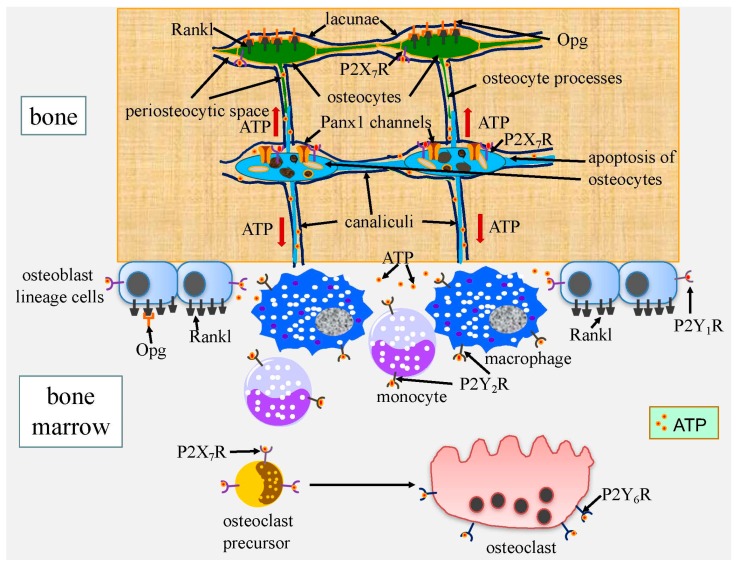
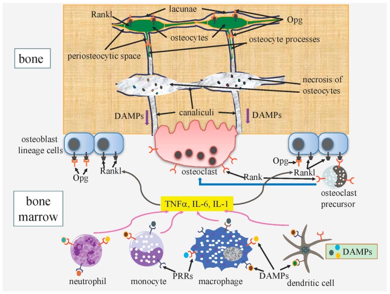
Cell death in skeletal component cells, including chondrocytes, osteoblasts, and osteocytes, plays roles in skeletal development, maintenance, and repair as well as in the pathogenesis of osteoarthritis and osteoporosis. Chondrocyte proliferation, differentiation, and apoptosis are important steps for endochondral ossification. Although the inactivation of P53 and RB is involved in the pathogenesis of osteosarcomas, the deletion of p53 and inactivation of Rb are insufficient to enhance chondrocyte proliferation, indicating the presence of multiple inhibitory mechanisms against sarcomagenesis in chondrocytes. The inflammatory processes induced by mechanical injury and chondrocyte death through the release of danger-associated molecular patterns (DAMPs) are involved in the pathogenesis of posttraumatic osteoarthritis. The overexpression of BCLXL increases bone volume with a normal structure and maintains bone during aging by inhibiting osteoblast apoptosis. p53 inhibits osteoblast proliferation and enhances osteoblast apoptosis, thereby reducing bone formation, but also exerts positive effects on osteoblast differentiation through the Akt–FoxOs pathway. Apoptotic osteocytes release ATP, which induces the receptor activator of nuclear factor κ-B ligand (Rankl) expression and osteoclastogenesis, from pannexin 1 channels. Osteocyte death ultimately results in necrosis; DAMPs are released to the bone surface and promote the production of proinflammatory cytokines, which induce Rankl expression, and osteoclastogenesis is further enhanced.
Overview
The claims and statistics of this paper haven’t been checked yet.
Run the check — free, no account needed. It reads the open-access text and checks the sources it cites.
Integrity signals
Registry
Cites
Statistics
Figures
Verification
Claims the paper makes
The claims and statistics of this paper haven’t been checked yet.
Run the check — free, no account needed. It reads the open-access text and checks the sources it cites.
Claims other papers make about this one
Each is checked against this paper’s own text: does it say what the citing paper says it does?
The cited source says what the claim attributes to it.
In the citing paper
“Necrotic osteocytes release damage-associated molecular pattern (DAMP) proteins after the cell membrane is disrupted.61”
From Bone remodeling: an operational process ensuring survival and bone mechanical competence (2022) · cites this paper as [61]
In the citing paper
“In vivo, osteocyte apoptosis is fundamental for initiating bone remodeling,61 independent of the stimulus causing osteocyte death such as estrogen loss,62 fatigue,63 or unloading.64”
From Bone remodeling: an operational process ensuring survival and bone mechanical competence (2022) · cites this paper as [61]
Self-check
Percentages, sums and reported differences re-computed from the paper’s own numbers.
The self-check needs the paper’s full text.
Citations
Cites
Stöckl, S., Göttl, C., Grifka, J. K., & Grässel, S. (2013) · Cellular Physiology and Biochemistry · doi:10.1159/000350089 · Cited by 4,670
Osteoarthritis: A disease of the joint as an organ
Loeser, R. F., Goldring, S. R., Scanzello, C. R., & Goldring, M. B. (2012) · Arthritis & Rheumatism · doi:10.1002/art.34453 · Cited by 3,234 · cited 1×
“The signaling pathway through PRRs induces synovitis, transduces inflammation, increases the expression of matrix metalloproteinases (MMPs) and aggrecanases, and induces oxidative stress and cell death [55,56].”
BackgroundHigh-mobility group box 1 protein (HMGB1): nuclear weapon in the immune arsenal
Lotze, M. T., & Tracey, K. J. (2005) · Nature reviews. Immunology · doi:10.1038/nri1594 · Cited by 2,490
Regulation of Rate of Cartilage Differentiation by Indian Hedgehog and PTH-Related Protein
Vortkamp, A., Lee, K., Lanske, B., Segre, G. V., Kronenberg, H. M., & Tabin, C. J. (1996) · Science · doi:10.1126/science.273.5275.613 · Cited by 1,999
Evidence for osteocyte regulation of bone homeostasis through RANKL expression
Nakashima, T., Hayashi, M., Fukunaga, T., Kurata, K., Oh-hora, M., Feng, J. Q., Bonewald, L. F., Kodama, T., Wutz, A., Wagner, E. F., et al. (2011) · Nature Medicine · doi:10.1038/nm.2452 · Cited by 1,778 · cited 1×
“The osteocyte-specific deletion of Rankl results in the inhibition of bone resorption, indicating that osteocytes are a major source of Rankl [121,122].”
BackgroundWeinstein, R. S., Jilka, R. L., Parfitt, A. M., & Manolagas, S. C. (1998) · Journal of Clinical Investigation · doi:10.1172/jci2799 · Cited by 1,753
St‐Jacques, B., Hammerschmidt, M., & McMahon, A. P. (1999) · Genes & Development · doi:10.1101/gad.13.16.2072 · Cited by 1,742 · cited 1×
“Ihh enhances chondrocyte proliferation [3].”
Park, J. S., Svetkauskaite, D., He, Q., Kim, J., Strassheim, D., Ishizaka, A., & Abraham, E. H. (2004) · Journal of Biological Chemistry · doi:10.1074/jbc.m306793200 · Cited by 1,582
p53 mutant mice that display early ageing-associated phenotypes
Tyner, S. D., Venkatachalam, S., Choi, J., Jones, S., Ghebranious, N., Igelmann, H., Lu, X., Soron, G., Cooper, B., Brayton, C., et al. (2002) · Nature · doi:10.1038/415045a · Cited by 1,459 · cited 1×
“Mice with activated mutations in p53 exhibit osteopenia [86].”
BackgroundCyclin D as a therapeutic target in cancer
Musgrove, E. A., Caldon, C. E., Barraclough, J., Stone, A., & Sutherland, R. L. (2011) · Nature Reviews Cancer · doi:10.1038/nrc3090 · Cited by 1,370 · cited 2×
“This leads to the transcriptional activation of E2f-responsive genes that are essential for DNA synthesis, including cyclins E and A [16].”
BackgroundMatrix-embedded cells control osteoclast formation
Xiong, J., Onal, M., Jilka, R. L., Weinstein, R. S., Manolagas, S. C., & O'Brien, C. A. (2011) · Nature Medicine · doi:10.1038/nm.2448 · Cited by 1,304 · cited 1×
“The osteocyte-specific deletion of Rankl results in the inhibition of bone resorption, indicating that osteocytes are a major source of Rankl [121,122].”
BackgroundNongenotropic, Sex-Nonspecific Signaling through the Estrogen or Androgen Receptors
Kousteni, S., Bellido, T., Plotkin, L. I., O’Brien, C. A., Bodenner, D., Li, H., Han, K., Gregorio, G. B. D., Katzenellenbogen, J. A., Katzenellenbogen, B. S., et al. (2001) · Cell · doi:10.1016/s0092-8674(01)00268-9 · Cited by 1,163
Akt, FoxO and regulation of apoptosis
Zhang, X., Tang, N., Hadden, T. J., & Rishi, A. K. (2011) · Biochimica et Biophysica Acta (BBA) - Molecular Cell Research · doi:10.1016/j.bbamcr.2011.03.010 · Cited by 1,149 · cited 3×
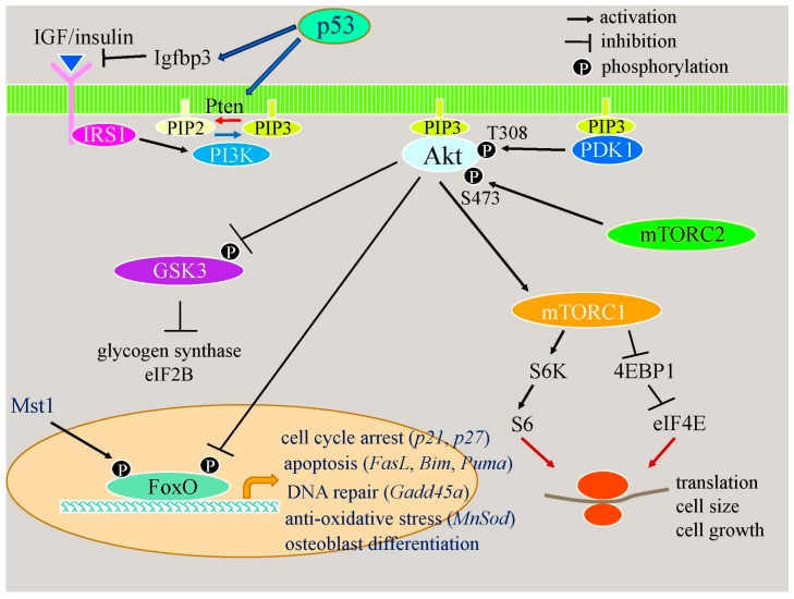
“Sci. 2016, 17, 2045 7 of 16 function of p53 in bone formation is to inhibit the proliferation of and enhance apoptosis in osteoblasts, leading to reduced bone formation. p53 also has a positive effect on osteoblast differentiation. p53 and FoxOs have similar functions, including inhibition of the cell cycle, DNA repai…”
BackgroundInfluentialPannexin 1 channels mediate ‘find-me’ signal release and membrane permeability during apoptosis
Chekeni, F. B., Elliott, M. R., Sandilos, J. K., Walk, S. F., Kinchen, J. M., Lazarowski, E. R., Armstrong, A. J., Penuela, S., Laird, D. W., Salvesen, G. S., et al. (2010) · Nature · doi:10.1038/nature09413 · Cited by 1,120 · cited 2×
“a “find me” signal necessary for macropha e recruitment [96].”
BackgroundIncreased bone formation by prevention of osteoblast apoptosis with parathyroid hormone
Jilka, R. L., Weinstein, R. S., Bellido, T., Roberson, P. K., PARFITT, A. M., & Manolagas, S. C. (1999) · Journal of Clinical Investigation · doi:10.1172/jci6610 · Cited by 1,042
Cyclin D1 provides a link between development and oncogenesis in the retina and breast
Sicinski, P., Donaher, J. L., Parker, S. B., Li, T., Fazeli, A., Gardner, H., Haslam, S. Z., Bronson, R. T., Elledge, S. J., & Weinberg, R. A. (1995) · Cell · doi:10.1016/0092-8674(95)90034-9 · Cited by 1,021
Mammary hyperplasia and carcinoma in MMTV-cyclin D1 transgenic mice
Wang, T. C., Cardiff, R. D., Zukerberg, L. R., Lees, E. M., Arnold, A., & Schmidt, E. V. (1994) · Nature · doi:10.1038/369669a0 · Cited by 964
Necrotic death as a cell fatePreprint
Zong, W., & Thompson, C. B. (2006) · Genes & Development · doi:10.1101/gad.1376506 · Cited by 952 · cited 1×
“Necrosis leads to the rupture of the cytoplasmic membrane, with most of the intracellular content being released into the extracellular environment [111].”
BackgroundPrevention of osteocyte and osteoblast apoptosis by bisphosphonates and calcitonin
Plotkin, L. I., Weinstein, R. S., Parfitt, A. M., Roberson, P. K., Manolagas, S. C., & Bellido, T. (1999) · Journal of Clinical Investigation · doi:10.1172/jci6800 · Cited by 890
Targeted ablation of osteocytes induces osteoporosis with defective mechanotransduction.
Tatsumi, S., Ishii, K., Amizuka, N., Li, M., Kobayashi, T., Kohno, K., Ito, M., Takeshita, S., & Ikeda, K. (2007) · Cell Metabolism · doi:10.1016/j.cmet.2007.05.001 · Cited by 848 · cited 2×
“Osteocyte ablation by diphtheria toxin, the deletion of Gja1 (connexin 43), which constitutes gap junction channels and hemichannels, and osteocyte-specific transgenic mice expressing mutant Gja1, which blocks gap junction channels and hemichannels, cause osteocyte apoptosis, leading to secondary necrosis, enhanced bo…”
BackgroundRegulation of bone development and extracellular matrix protein genes by RUNX2
Komori, T. (2009) · Cell and Tissue Research · doi:10.1007/s00441-009-0832-8 · Cited by 806 · cited 1×
“A number of processes, including chondrocyte hypertrophy, inflammatory processes, and chondrocyte death, are involved in the pathogenesis of OA [9,30,31].”
BackgroundSelf-citationO’Brien, C. A., Jia, D., Plotkin, L. I., Bellido, T., Powers, C. C., Stewart, S. A., Manolagas, S. C., & Weinstein, R. S. (2003) · Endocrinology · doi:10.1210/en.2003-0990 · Cited by 771
FoxO transcription factors; Regulation by AKT and 14-3-3 proteins
Tzivion, G., Dobson, M., & Ramakrishnan, G. (2011) · Biochimica et Biophysica Acta (BBA) - Molecular Cell Research · doi:10.1016/j.bbamcr.2011.06.002 · Cited by 757 · cited 1×
“FoxO proteins are inactivated through the phosphorylation of Thr24, S256, and S319 in FoxO1 and that of Thr32, S253, and S315 in FoxO3 by Akt, and are activated through the phosphorylation of S212 in FoxO1 and S207 in FoxO3a by Mst1, and Akt and Mst1 are activated by phosphorylation [85] (Figure 2).”
BackgroundKousteni, S. (2001) · Cell · doi:10.1016/s0092-8674(02)08100-x · Cited by 757
Hypertrophic chondrocytes can become osteoblasts and osteocytes in endochondral bone formation
Yang, L., Tsang, K. Y., Tang, H. C., Chan, D. T.-M., & Cheah, K. S. E. (2014) · Proceedings of the National Academy of Sciences · doi:10.1073/pnas.1302703111 · Cited by 755 · cited 1×
“Terminal hypertrophic chondrocytes gradually die by apoptosis or become osteoblasts, and the terminal hypertrophic chondrocyte layer is replaced with bone [10,11].”
BackgroundVerborgt, O., Gibson, G. J., & Schaffler, M. B. (2000) · Journal of Bone and Mineral Research · doi:10.1359/jbmr.2000.15.1.60 · Cited by 731 · cited 1×
“The microdamage of bone has been associated with osteocyte apoptosis and targeted bone resorption [105,106].”
BackgroundMice lacking cyclin D1 are small and show defects in eye and mammary gland development.
Fantl, V, Stamp, G., Andrews, A., Rosewell, I., & Dickson, C. (1995) · Genes & Development · doi:10.1101/gad.9.19.2364 · Cited by 691 · cited 1×
“In the growth plate, Cyclin D1 is strongly expressed in the proliferating layer [15], and Cyclin D1-deficient mice show dwarfism with a diminished proliferating layer in the growth plate [13,15,17,18].”
BackgroundEmerging regulators of the inflammatory process in osteoarthritis
Liu‐Bryan, R., & Terkeltaub, R. A. (2014) · Nature Reviews Rheumatology · doi:10.1038/nrrheum.2014.162 · Cited by 680 · cited 5×
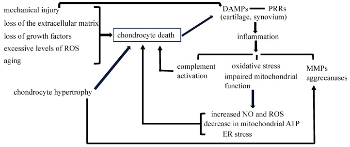
“Mechanical injury increases danger-associated molecular patterns (DAMPs), including released (HMGB1) protein, purine metabolites, heat-shock proteins, and uric acid, which initiate noninfectious inflammatory responses through various pattern recognition receptors (PRRs) expressed in OA cartilage and the synovium, incl…”
BackgroundInfluentialLong, F., Zhang, X. M., Karp, S. J., Yang, Y., & McMahon, A. P. (2001) · Development · doi:10.1242/dev.128.24.5099 · Cited by 654
The autophagosomal–lysosomal compartment in programmed cell death
Bursch, W. (2001) · Cell Death and Differentiation · doi:10.1038/sj.cdd.4400852 · Cited by 620 · cited 1×
“, apoptosis, autophagic cell death, and necrosis, under physiological and pathological conditions [108].”
BackgroundCarter, M. E., & Brunet, A. (2007) · Current Biology · doi:10.1016/j.cub.2007.01.008 · Cited by 588
Yoshida, C. A., Yamamoto, H., Fujita, T., Furuichi, T., Ito, K., Inoue, K., Yamana, K., Zanma, A., Takada, K., Ito, Y., et al. (2004) · Genes & Development · doi:10.1101/gad.1174704 · Cited by 588 · cited 1×
“Runx2 induces the expression of Ihh, which is also strongly expressed in prehypertrophic chondrocytes [1,2].”
BackgroundSelf-citationZhou, X., von der Mark, K., Henry, S. P., Norton, W., Adams, H. P., & Crombrugghe, B. D. (2014) · PLoS Genetics · doi:10.1371/journal.pgen.1004820 · Cited by 585 · cited 1×
“Terminal hypertrophic chondrocytes gradually die by apoptosis or become osteoblasts, and the terminal hypertrophic chondrocyte layer is replaced with bone [10,11].”
BackgroundIdentification of a central role for complement in osteoarthritis
Wang, Q., Rozelle, A. L., Lepus, C. M., Scanzello, C. R., Song, J. J., Larsen, D. M., Crish, J. F., Bebek, G., Ritter, S. Y., Lindström, T. M., et al. (2011) · Nature Medicine · doi:10.1038/nm.2543 · Cited by 576
The role of mitochondria in osteoarthritis
Blanco, F. J., Rego‐Pérez, I., & Ruíz‐Romero, C. (2011) · Nature Reviews Rheumatology · doi:10.1038/nrrheum.2010.213 · Cited by 549
Kosinski, M. R., Zhao, S. Z., Dedhiya, S. D., Osterhaus, J. T., & Ware, J. E. (2000) · Arthritis & Rheumatism · doi:10.1002/1529-0131(200007)43:7 · Cited by 502
Linkage of chondrocyte apoptosis and cartilage degradation in human osteoarthritis
Hashimoto, S., Ochs, R., Komiya, S., & Lotz, M. (1998) · Arthritis & Rheumatism · doi:10.1002/1529-0131(199809)41:9 · Cited by 500
Messmer, D., Yang, H., Telusma, G., Knoll, F., Li, J., Messmer, B., Tracey, K. J., & Chiorazzi, N. (2004) · The Journal of Immunology · doi:10.4049/jimmunol.173.1.307 · Cited by 447
Hurford, R. K., Cobrinik, D., Lee, M. H., & Dyson, N. (1997) · Genes & Development · doi:10.1101/gad.11.11.1447 · Cited by 431
Shared role of the pRB-related p130 and p107 proteins in limb development.Preprint
Cobrinik, D. E., Lee, M. H., Hannon, G. J., Mulligan, G., Bronson, R. T., Dyson, N. J., Harlow, E. E., Beach, D., Weinberg, R. A., & Jacks, T. (1996) · Genes & Development · doi:10.1101/gad.10.13.1633 · Cited by 426 · cited 2×
“Rb family proteins play an important role in chondrocyte proliferation and differentiation [19,20].”
BackgroundThe Death of Osteocytes via Apoptosis Accompanies Estrogen Withdrawal in Human Bone
Tomkinson, A., REEVE, J., Shaw, R. W., & Noble, B. (1997) · The Journal of Clinical Endocrinology & Metabolism · doi:10.1210/jcem.82.9.4200 · Cited by 417
The biochemistry and function of pannexin channels
Peñuela, S., Gehi, R., & Laird, D. W. (2012) · Biochimica et Biophysica Acta (BBA) - Biomembranes · doi:10.1016/j.bbamem.2012.01.017 · Cited by 400 · cited 1×
“There are three members in the pannexin family: pannexin 1 (Panx1) and pannexin 3 (Panx3) are expressed in bone cells, with Panx1 already being extensively examined [95].”
BackgroundYang, Y., Topol, L. Z., Lee, H., & Wu, J. (2003) · Development · doi:10.1242/dev.00324 · Cited by 398 · cited 1×
“In the growth plate, Cyclin D1 is strongly expressed in the proliferating layer [15], and Cyclin D1-deficient mice show dwarfism with a diminished proliferating layer in the growth plate [13,15,17,18].”
BackgroundPelletier, J., Jovanović, D., Fernandes, J., Manning, P. T., Connor, J. R., Currie, M. G., Battista, J. A. D., & Martel‐Pelletier, J. (1998) · Arthritis & Rheumatism · doi:10.1002/1529-0131(199807)41:7 · Cited by 344
Tissue specific regulation of VEGF expression during bone development requires Cbfa1/Runx2
Zelzer, E., Glotzer, D. J., Hartmann, C., Thomas, D., Fukai, N., Söker, S., & Olsen, B. R. (2001) · Mechanisms of Development · doi:10.1016/s0925-4773(01)00428-2 · Cited by 344
Osteocyte Apoptosis Controls Activation of Intracortical Resorption in Response to Bone Fatigue
Cardoso, L., Herman, B. C., Verborgt, O., Laudier, D., Majeska, R. J., & Schaffler, M. B. (2009) · Journal of Bone and Mineral Research · doi:10.1359/jbmr.081210 · Cited by 340
P2X7 receptor-Pannexin1 complex: pharmacology and signaling
Iglesias, R., Locovei, S., Roque, A., Alberto, A. P., Dahl, G., Spray, D. C., & Scemes, E. (2008) · American Journal of Physiology-Cell Physiology · doi:10.1152/ajpcell.00228.2008 · Cited by 331
Kühn, K., DʼLima, D. D., Hashimoto, S., & Lotz, M. (2003) · Osteoarthritis and Cartilage · doi:10.1016/j.joca.2003.09.015 · Cited by 317 · cited 1×
“Apoptosis, autophagic cell death, and necrosis have been detected in OA cartilage [32].”
BackgroundAmbrogini, E., Almeida, M., Martín-Millán, M., Paik, J.-H., DePinho, R. A., Li, H., Goellner, J. J., Weinstein, R. S., Jilka, R. L., O’Brien, C. A., et al. (2010) · Cell Metabolism · doi:10.1016/j.cmet.2009.12.009 · Cited by 313
Human chondrocyte apoptosis in response to mechanical injury
DʼLima, D. D., Hashimoto, S., CHEN, P. C., Colwell, C., & Lotz, M. (2001) · Osteoarthritis and Cartilage · doi:10.1053/joca.2001.0468 · Cited by 305
Apoptotic chondrocyte death in human osteoarthritis.
Kim, H. A., Lee, Y. J., Seong, S. C., Choe, K. W., & Song, Y. W. (2000) · PubMed · Cited by 287
Osteoblast differentiation and skeletal development are regulated by Mdm2–p53 signaling
Lengner, C. J., Steinman, H. A., Gagnon, J. A., Smith, T. W., Henderson, J. E., Kream, B. E., Stein, G. S., Lian, J. B., & Jones, S. N. (2006) · The Journal of Cell Biology · doi:10.1083/jcb.200508130 · Cited by 270 · cited 1×
“6-kb Col1a1 promoter in Cre transgenic mice resulted in reduced bone formation, and the differentiation of primary osteoblasts was inhibited in vitro [87].”
BackgroundRAGE and TLRs: Relatives, friends or neighbours?
Ibrahim, Z. A., Armour, C. L., Phipps, S., & Sukkar, M. B. (2013) · Molecular Immunology · doi:10.1016/j.molimm.2013.07.008 · Cited by 263
Bivi, N., Condon, K. W., Allen, M. R., Farlow, N., Passeri, G., Brun, L. R., Rhee, Y., Bellido, T., & Plotkin, L. I. (2012) · Journal of Bone and Mineral Research · doi:10.1002/jbmr.548 · Cited by 261 · cited 1×
“Osteocyte ablation by diphtheria toxin, the deletion of Gja1 (connexin 43), which constitutes gap junction channels and hemichannels, and osteocyte-specific transgenic mice expressing mutant Gja1, which blocks gap junction channels and hemichannels, cause osteocyte apoptosis, leading to secondary necrosis, enhanced bo…”
BackgroundWang, X., Kua, H.-Y., Hu, Y., Guo, K., Zeng, Q., Wu, Q., Ng, H., Karsenty, G., Crombrugghe, B. D., Yeh, J. K., et al. (2005) · The Journal of Cell Biology · doi:10.1083/jcb.200507106 · Cited by 254 · cited 2×
“The introduction of p53 into wild-type and p53−/− osteoblasts inhibits osteoblast differentiation [90].”
BackgroundResultChondrocyte death by apoptosis is associated with cartilage matrix degradation
Thomas, C. M., Fuller, C. J., Whittles, C. E., & Sharif, M. (2006) · Osteoarthritis and Cartilage · doi:10.1016/j.joca.2006.06.012 · Cited by 248
Estrogen Prevents Glucocorticoid-Induced Apoptosis in Osteoblasts in Vivo and in Vitro1
Gohel, A., McCarthy, M.-B., & Gronowicz, G. (1999) · Endocrinology · doi:10.1210/endo.140.11.7135 · Cited by 244
Osteocyte apoptosis and control of bone resorption following ovariectomy in mice
Emerton, K. B., Hu, B., Woo, A. A., Sinofsky, A., Hernandez, C., Majeska, R. J., Jepsen, K. J., & Schaffler, M. B. (2010) · Bone · doi:10.1016/j.bone.2009.11.006 · Cited by 220
Chondroptosis: A variant of apoptotic cell death in chondrocytes?
Roach, H. I., Aigner, T. G., & Kouri, J. B. (2004) · APOPTOSIS · doi:10.1023/b:appt.0000025803.17498.26 · Cited by 219 · cited 1×
“Therefore, the mixed morphology of apoptosis, autophagic cell death, and necrosis has been reported in OA cartilage [34].”
BackgroundOsteocyte function, osteocyte death and bone fracture resistance
Noble, B., & REEVE, J. (2000) · Molecular and Cellular Endocrinology · doi:10.1016/s0303-7207(99)00174-4 · Cited by 211
SIRT1 regulation of apoptosis of human chondrocytes
Takayama, K., Ishida, K., Matsushita, T., Fujita, N., Hayashi, S., Sasaki, K., Tei, K., Kubo, S., Matsumoto, T., Fujioka, H., et al. (2009) · Arthritis & Rheumatism · doi:10.1002/art.24864 · Cited by 210
Connexins, pannexins, innexins: novel roles of “hemi-channels”
Scemes, E., Spray, D. C., & Meda, P. (2008) · Pflügers Archiv - European Journal of Physiology · doi:10.1007/s00424-008-0591-5 · Cited by 203 · cited 1×
“Two junctional proteins—connexins and pannexins—form membrane channels permeable to large molecules in vertebrates [94].”
BackgroundSchelbergen, R., Blom, A., van den Bosch, M. V. D., Slöetjes, A., Abdollahi-Roodsaz, S., Schreurs, B. W., Mort, J., Vogl, T., Roth, J., van den Berg, W., et al. (2012) · Arthritis & Rheumatism · doi:10.1002/art.33495 · Cited by 195 · cited 1×
“The signaling pathway through PRRs induces synovitis, transduces inflammation, increases the expression of matrix metalloproteinases (MMPs) and aggrecanases, and induces oxidative stress and cell death [55,56].”
BackgroundStructure and Development of the Skeleton
Marks, S. C., & Odgren, P. R. (2002) · Elsevier eBooks · doi:10.1016/b978-012098652-1.50103-7 · Cited by 194 · cited 2×
“Both communication systems are extended to osteoblasts on the bone surface [93].”
BackgroundChondrocyte apoptosis: a cause or consequence of osteoarthritis?
Zamli, Z., & Sharif, M. (2011) · International Journal of Rheumatic Diseases · doi:10.1111/j.1756-185x.2011.01618.x · Cited by 178 · cited 1×
“A number of processes, including chondrocyte hypertrophy, inflammatory processes, and chondrocyte death, are involved in the pathogenesis of OA [9,30,31].”
BackgroundSharif, M., Whitehouse, A., Sharman, P., Perry, M., & Adams, M. (2004) · Arthritis & Rheumatism · doi:10.1002/art.20020 · Cited by 173
Control of RANKL gene expression
O’Brien, C. A. (2009) · Bone · doi:10.1016/j.bone.2009.08.050 · Cited by 170
Caspase inhibitors reduce severity of cartilage lesions in experimental osteoarthritis
DʼLima, D. D., Hermida, J. C., Hashimoto, S., Colwell, C. W., & Lotz, M. (2006) · Arthritis & Rheumatism · doi:10.1002/art.21874 · Cited by 169 · cited 1×
“The intra-articular administration of caspase inhibitors has been shown to inhibit chondrocyte death and cartilage degradation in OA rabbit models [46,47].”
BackgroundAccelerated, aging‐dependent development of osteoarthritis in α1 integrin–deficient mice
Zemmyo, M., Meharra, E. J., Kühn, K., Creighton‐Achermann, L., & Lotz, M. (2003) · Arthritis & Rheumatism · doi:10.1002/art.11246 · Cited by 159 · cited 2×
“proteins to various intracellular cytoskeletal proteins [53].”
BackgroundHow severe must repetitive loading be to kill chondrocytes in articular cartilage?
Clements, K. M., Bee, Z. C., Crossingham, G. V., Adams, M. A., & Sharif, M. (2001) · Osteoarthritis and Cartilage · doi:10.1053/joca.2000.0417 · Cited by 152
Bone microdamage and cell apoptosis
Noble, B. (2003) · European Cells and Materials · doi:10.22203/ecm.v006a05 · Cited by 152 · cited 1×
“The microdamage of bone has been associated with osteocyte apoptosis and targeted bone resorption [105,106].”
BackgroundKennedy, O. D., Laudier, D. M., Majeska, R. J., Sun, H. B., & Schaffler, M. B. (2014) · Bone · doi:10.1016/j.bone.2014.03.049 · Cited by 151
Foxo1, a Novel Regulator of Osteoblast Differentiation and Skeletogenesis
Teixeira, C. C., Liu, Y., Thant, L. M., Pang, J., Palmer, G. D., & Alikhani, M. (2010) · Journal of Biological Chemistry · doi:10.1074/jbc.m109.079962 · Cited by 150
Hirschmann-Jax, C., Foster, A. E., Wulf, G. G., Goodell, M. A., & Brenner, M. K. (2004) · Cell Cycle · doi:10.4161/cc.4.2.1406 · Cited by 147
Cell signals transduced by complement
Bohana‐Kashtan, O. (2004) · Molecular Immunology · doi:10.1016/j.molimm.2004.04.007 · Cited by 145 · cited 1×
“Complement may be activated by DAMPs, and activated complement components may induce chondrocyte death [62,63].”
BackgroundTGFβ and PTHrP Control Chondrocyte Proliferation by Activating Cyclin D1 Expression
Beier, F., Ali, Z., Mok, D., Taylor, A. C., Leask, T., Albanese, C., Pestell, R. G., & Luvalle, P. A. (2001) · Molecular Biology of the Cell · doi:10.1091/mbc.12.12.3852 · Cited by 137 · cited 1×
“In the growth plate, Cyclin D1 is strongly expressed in the proliferating layer [15], and Cyclin D1-deficient mice show dwarfism with a diminished proliferating layer in the growth plate [13,15,17,18].”
BackgroundGagarina, V., Gabay, O., Dvir‐Ginzberg, M., Lee, E. J., Brady, J. K., Quon, M. J., & Hall, D. J. (2010) · Arthritis & Rheumatism · doi:10.1002/art.27369 · Cited by 137
Effect of nitric oxide on mitochondrial respiratory activity of human articular chondrocytes
Maneiro, E., López-Armada, M. J., de Andres, M. C., Caramés, B., Martín, M. A., Bonilla, A., del Hoyo, P., Galdo, F., Arenas, J., & Blanco, F. J. (2005) · Annals of the Rheumatic Diseases · doi:10.1136/ard.2004.022152 · Cited by 134
Glucocorticoid Signaling and Bone Biology
Komori, T. (2016) · Hormone and Metabolic Research · doi:10.1055/s-0042-110571 · Cited by 131 · cited 2×
“Excess GCs increase the levels of the pro-apoptotic factors, Bim and Bak; decrease that of the pro-survival factor, BclXL; up-regulate the expression of the p53 protein; increase ROS levels; inhibit Akt; and enhance ER stress [35].”
BackgroundSelf-citationUehara, Y., Hirose, J., Yamabe, S., Okamoto, N., Okada, T., Oyadomari, S., & Mizuta, H. (2014) · Osteoarthritis and Cartilage · doi:10.1016/j.joca.2014.04.025 · Cited by 124 · cited 1×
“Furthermore, oxidative stress and mitochondrial dysfunction cause endoplasmic reticulum (ER) stress, and the unsuccessful resolution of ER stress by the unfolded protein response (UPR) promotes chondrocyte apoptosis via Chop [64] (Figure 1).”
BackgroundKurz, B., Lemke, A. K., Kehn, M., Domm, C., Patwari, P., Frank, E. H., Grodzinsky, A. J., & Schünke, M. (2004) · Arthritis & Rheumatism · doi:10.1002/art.11438 · Cited by 123
Functions of the osteocyte network in the regulation of bone mass
Komori, T. (2013) · Cell and Tissue Research · doi:10.1007/s00441-012-1546-x · Cited by 113 · cited 13×
“Regulation of the Release of Osteocyte-Derived Opg to the Bone Surface May Be a Major Role for Rankl on Osteocytes Osteocytes strongly express Rankl and Opg, which is a secreted decoy receptor of Rankl [120–122].”
BackgroundInfluentialSelf-citationP2Y6 Nucleotide Receptors Activate NF-κB and Increase Survival of Osteoclasts
Korčok, J., Raimundo, L., Du, X., Sims, S. M., & Dixon, S. J. (2005) · Journal of Biological Chemistry · doi:10.1074/jbc.m410764200 · Cited by 112
Apoptosis of Chondrocytes in Transgenic Mice Lacking Collagen II
Yang, C., Li, S.-W., Helminen, H. J., Khillan, J. S., Bao, Y., & Prockop, D. J. (1997) · Experimental Cell Research · doi:10.1006/excr.1997.3692 · Cited by 112
Zhu, L., Zhu, L., Xie, E., & Chang, L. (1995) · Molecular and Cellular Biology · doi:10.1128/mcb.15.7.3552 · Cited by 112
Connexin 43 Channels Are Essential for Normal Bone Structure and Osteocyte Viability
Xu, H., Gu, S., Riquelme, M. A., Burra, S., Callaway, D. A., Cheng, H., Guda, T., Schmitz, J. E., Fajardo, R. J., Werner, S. L., et al. (2014) · Journal of Bone and Mineral Research · doi:10.1002/jbmr.2374 · Cited by 108 · cited 1×
“Osteocyte ablation by diphtheria toxin, the deletion of Gja1 (connexin 43), which constitutes gap junction channels and hemichannels, and osteocyte-specific transgenic mice expressing mutant Gja1, which blocks gap junction channels and hemichannels, cause osteocyte apoptosis, leading to secondary necrosis, enhanced bo…”
BackgroundIncreased apoptotic chondrocytes in articular cartilage from adult heterozygous SirT1 mice
Gabay, O., Oppenhiemer, H., Meir, H., Zaal, K. J. M., Sanchez, C., & Dvir‐Ginzberg, M. (2012) · Annals of the Rheumatic Diseases · doi:10.1136/ard.2011.200504 · Cited by 107
Carlo, M. D., & Loeser, R. F. (2008) · Current Rheumatology Reports · doi:10.1007/s11926-008-0007-8 · Cited by 106 · cited 4×
“Chondrocyte Death during Mechanical Injury May Contribute to the Development of OA Chondrocyte death may be induced by mechanical injury, the loss of the extracellular matrix, loss of growth factors, or excessive levels of reactive oxygen species (ROS) [49] (Figure 1).”
BackgroundInfluentialMoriishi, T., Fukuyama, R., Ito, M., Miyazaki, T., Maeno, T., Kawai, Y., Komori, H., & Komori, T. (2012) · PLoS ONE · doi:10.1371/journal.pone.0040143 · Cited by 105 · cited 2×
“In BCl2 transgenic mice, osteocyte death does not induce bone resorption because the extracellular communication system is also disrupted [120].”
BackgroundSelf-citationBuckley, K. M., Hipskind, R. A., Gartland, A., Bowler, W. B., & Gallagher, J. A. (2002) · Bone · doi:10.1016/s8756-3282(02)00877-3 · Cited by 98
Zhang, M., Mani, S. B., He, Y., Hall, A. M., Xu, L., Li, Y., Zurakowski, D., Jay, G. D., & Warman, M. L. (2016) · Journal of Clinical Investigation · doi:10.1172/jci83676 · Cited by 95 · cited 2×
“However, the significance of the DAMPs released from necrotic chondrocytes still needs to be investigated because the ablation of chondrocytes by diphtheria toxin prior to the operation for surgical OA reduces cartilage damage [48].”
BackgroundResultLi, T.-F., Dong, Y., Ionescu, A. M., Rosier, R. N., Zuscik, M. J., Schwarz, E. M., O’Keefe, R. J., & Drissi, H. (2004) · Experimental Cell Research · doi:10.1016/j.yexcr.2004.05.025 · Cited by 94
Cheung, W. Y., Fritton, J. C., Morgan, S. A., Seref-Ferlengez, Z., Basta-Pljakic, J., Thi, M. M., Suadicani, S. O., Spray, D. C., Majeska, R. J., & Schaffler, M. B. (2016) · Journal of Bone and Mineral Research · doi:10.1002/jbmr.2740 · Cited by 89 · cited 3×
“Since bone resorption after osteocyte apoptosis is attenuated in Panx1−/− mice [110], the release of ATP from Panx1 channels appears to be a prerequisite for bone resorption.”
BackgroundInfluentialFOXO1 modulates osteoblast differentiation
Siqueira, M. F., Flowers, S. J., Bhattacharya, R., Faibish, D., Behl, Y., Kotton, D. N., Gerstenfeld, L. C., Morán, E., & Graves, D. T. (2011) · Bone · doi:10.1016/j.bone.2011.01.019 · Cited by 88
Chondrocyte death during murine osteoarthritis
Mistry, D., Oue, Y., Chambers, M. G., Kayser, M. V., & Mason, R. M. (2003) · Osteoarthritis and Cartilage · doi:10.1016/j.joca.2003.10.006 · Cited by 87
Calbó, J., Parreño, M., Sotillo, E., Yong, T., Mazo, A., Garriga, J., & Graña, X. (2002) · Journal of Biological Chemistry · doi:10.1074/jbc.m209181200 · Cited by 87
Iwamoto, M., Kitagaki, J., Tamamura, Y., Gentili, C., Koyama, E., Enomoto, H., Komori, T., Pacifici, M., & Enomoto‐Iwamoto, M. (2002) · Osteoarthritis and Cartilage · doi:10.1053/joca.2002.0860 · Cited by 86
Self-citationJilka, R. L., O’Brien, C. A., Roberson, P. K., Bonewald, L. F., Weinstein, R. S., & Manolagas, S. C. (2013) · Journal of Bone and Mineral Research · doi:10.1002/jbmr.2007 · Cited by 85 · cited 1×
“The deletion of Bak and Bax in osteoblasts also increases the mineral density and trabecular bone volume of femurs, whereas the cortical bone volume of femurs and cancellous bone volume of vertebrae are not increased and cortical bone shows severe porosity in aged mice [77].”
BackgroundGarnero, P., Gineyts, É., Schaffer, A. V., Seaman, J. J., & Delmas, P. D. (1998) · Arthritis & Rheumatism · doi:10.1002/1529-0131(199802)41:2 · Cited by 79
Stanislaus, D., Yang, X., Liang, J., Wolfe, J. M. T., Cain, R. L., Onyia, J. E., Falla, N., Marder, P., Bidwell, J. P., Queener, S. W., et al. (2000) · Bone · doi:10.1016/s8756-3282(00)00309-4 · Cited by 72
Mueller, A., Odze, R. D., Jenkins, T. D., Shahsesfaei, A., Nakagawa, H., Inomoto, T., & Rustgi, A. K. (1997) · PubMed · Cited by 70
Allen, R. T., Robertson, C. M., Harwood, F. L., Sasho, T., Williams, S. K., Pomerleau, A. C., & Amiel, D. (2004) · Osteoarthritis and Cartilage · doi:10.1016/j.joca.2004.08.003 · Cited by 68 · cited 1×
“Furthermore, a significant decrease has been reported in chondrocyte numbers in articular cartilage with aging [39,44,45].”
BackgroundRossi, F., MacLean, H. E., Yuan, W., Francis, R. O., Semenova, E., Lin, C. S., Kronenberg, H. M., & Cobrinik, D. E. (2002) · Developmental Biology · doi:10.1006/dbio.2002.0691 · Cited by 68 · cited 2×
“Rb family proteins play an important role in chondrocyte proliferation and differentiation [19,20].”
BackgroundMoriishi, T., Maruyama, Z., Fukuyama, R., Ito, M., Miyazaki, T., Kitaura, H., Ohnishi, H., Furuichi, T., Kawai, Y., Masuyama, R., et al. (2011) · PLoS ONE · doi:10.1371/journal.pone.0027487 · Cited by 68 · cited 7×
“Osteoblastogenesis in BCL2-overexpressing primary osteoblasts in vitro is also controversial, and was previously reported to be enhanced and inhibited [81].”
BackgroundInfluentialSelf-citationEstrogen Prevents Glucocorticoid-Induced Apoptosis in Osteoblasts in Vivo and in Vitro
Gohel, A. (1999) · Endocrinology · doi:10.1210/en.140.11.5339 · Cited by 68
Nagase, Y., Iwasawa, M., Akiyama, T., Kadono, Y., Nakamura, M., Oshima, Y., Yasui, T., Matsumoto, T., Hirose, J., Nakamura, H., et al. (2009) · Journal of Biological Chemistry · doi:10.1074/jbc.m109.016915 · Cited by 59 · cited 2×
“In vitro osteoblast cultures also showed that the differentiation of Bcl2−/− osteoblasts was similar to or impaired more than that of wild-type osteoblasts [78,79].”
BackgroundResultChondrocyte cell death mediated by reactive oxygen species-dependent activation of PKC-betaI.
Delcarlo, M., & Loeser, R. (2006) · American Journal of Physiology - Cell Physiology · doi:10.1152/ajpcell.00214.2005 · Cited by 56
Role of Bcl2 in Osteoclastogenesis and PTH Anabolic Actions in Bone
Yamashita, J., Datta, N. S., Chun, Y. P., Yang, D.-Y., Carey, A. A., Kreider, J. M., Goldstein, S. A., & McCauley, L. K. (2007) · Journal of Bone and Mineral Research · doi:10.1359/jbmr.071211 · Cited by 55 · cited 2×
“In vitro osteoblast cultures also showed that the differentiation of Bcl2−/− osteoblasts was similar to or impaired more than that of wild-type osteoblasts [78,79].”
BackgroundResultLemaire, I. B., Falzoni, S., Zhang, B., Pellegatti, P., & Virgilio, F. D. (2011) · The Journal of Immunology · doi:10.4049/jimmunol.1002780 · Cited by 53
Sakai, A., Sakata, T., Tanaka, S., Okazaki, R., Kunugita, N., Norimura, T., & Nakamura, T. (2002) · Journal of Bone and Mineral Research · doi:10.1359/jbmr.2002.17.1.119 · Cited by 48
Bcl2 Deficiency Activates FoxO through Akt Inactivation and Accelerates Osteoblast Differentiation
Moriishi, T., Kawai, Y., Komori, H., Rokutanda, S., Eguchi, Y., Tsujimoto, Y., Asahina, I., & Komori, T. (2014) · PLoS ONE · doi:10.1371/journal.pone.0086629 · Cited by 47 · cited 9×
“The inhibition of osteoblast proliferation in Bcl2−/− mice is, at least in part, due to the up-regulation of p53 and FoxOs [80].”
BackgroundResultInfluentialSelf-citationBCL2 inhibits cell adhesion, spreading, and motility by enhancing actin polymerization
Ke, H., Parron, V. I., Reece, J. M., Zhang, J. Y., Akiyama, S. K., & French, J. E. (2010) · Cell Research · doi:10.1038/cr.2010.21 · Cited by 45 · cited 1×
“3-kb Col1a1 promoter because the osteocyte processes and canaliculi are severely reduced, possibly due to the formation of a complex of BCL2, actin, and gelsolin, which reduces gelsolin-severing activity in order to increase actin polymerization [81,119].”
BackgroundImpaired Vascular Invasion of Cbfa1-Deficient Cartilage Engrafted in the Spleen
Himeno, M., Enomoto, H., Liu, W., Ishizeki, K., Nomura, S., Kitamura, Y., & Komori, T. (2002) · Journal of Bone and Mineral Research · doi:10.1359/jbmr.2002.17.7.1297 · Cited by 44
Self-citationATP P2 receptors and regulation of bone effector cells.
Gallagher, J. A. (2004) · PubMed · Cited by 41
Burkhart, D. L., Wirt, S. E., Zmoos, A., Kareta, M. S., & Sage, J. (2010) · PLoS Genetics · doi:10.1371/journal.pgen.1001003 · Cited by 38
Bohensky, J., Shapiro, I. M., Leshinsky, S., Watanabe, H., & Srinivas, V. (2007) · Journal of Cellular Physiology · doi:10.1002/jcp.21117 · Cited by 36 · cited 1×
“Similar to osteocytes, autophagy also protects chondrocytes from apoptosis, and its failure leads to apoptosis [35,36].”
BackgroundImai, K., & Senoo, H. (1998) · The Anatomical Record · doi:10.1002/(sici)1097-0185(199804)250:4 · Cited by 34
Beneficial effects of intra-articular caspase inhibition therapy following osteochondral injury
Dang, A. C., Warren, A. P., & Kim, H. T. (2006) · Osteoarthritis and Cartilage · doi:10.1016/j.joca.2005.12.010 · Cited by 33 · cited 1×
“The intra-articular administration of caspase inhibitors has been shown to inhibit chondrocyte death and cartilage degradation in OA rabbit models [46,47].”
BackgroundDevelopment of the chick olfactory nerve
Vermeer, P. D., & Silverman, A. (1999) · Developmental Dynamics · doi:10.1002/(sici)1097-0177(199904)214:4 · Cited by 33
Self-citationMoriishi, T., Fukuyama, R., Miyazaki, T., Furuichi, T., Ito, M., & Komori, T. (2016) · Journal of Bone and Mineral Research · doi:10.1002/jbmr.2808 · Cited by 33 · cited 2×
“The bone structure is similar to, but stronger than that in wild-type mice and the increased bone mass is maintained during aging in both sexes [76].”
BackgroundSelf-citationRevisiting the “Cdk-Centric” View of the Mammalian Cell Cycle
Malumbres, M. (2004) · Cell Cycle · doi:10.4161/cc.4.2.1410 · Cited by 32 · cited 1×
“Cyclin D activates Cdk4 and Cdk6 in the G1 phase, cyclin E activates Cdk2 in the G1/S phase, cyclin A activates Cdk2 and Cdk1 in the S phase, and cyclin B activates Cdk1 in the M phase [12].”
BackgroundIto, K., Maruyama, Z., Sakai, A., Izumi, S., Moriishi, T., Yoshida, C. A., Miyazaki, T., Komori, H., Takada, K., Kawaguchi, H., et al. (2013) · Oncogene · doi:10.1038/onc.2013.130 · Cited by 30 · cited 6×
“Rb is highly phosphorylated, while unphosphorylated p107 levels are elevated in Cdk6/Cyclin D1 double transgenic mice [21].”
BackgroundInfluentialSelf-citationHerzog, C. R., Noh, S. J., Lantry, L. E., Guan, K.-L., & You, M. (1999) · Molecular Carcinogenesis · doi:10.1002/(sici)1098-2744(199906)25:2 · Cited by 26
Cyclic Loading Can Denature Type II Collagen in Articular Cartilage
Clements, K. M., Hollander, A. P., Sharif, M., & Adams, M. A. (2004) · Connective Tissue Research · doi:10.1080/03008200490514121 · Cited by 21
Regulation of Rb family proteins by Cdk6/Ccnd1 in growth plates
Komori, T. (2013) · Cell Cycle · doi:10.4161/cc.25515 · Cited by 14 · cited 1×
“Under physiological conditions, unphosphorylated p107 associates with repressing E2fs, whereas unphosphorylated p107 also associates with activating E2fs when p107 is up-regulated, resulting in the dysregulation of E2f target gene expression, as reported in Cdk6/Cyclin D1 double transgenic mice [28,29].”
BackgroundSelf-citationLoss of pRB and p107 disrupts cartilage development and promotes enchondroma formation
Landman, A. S., Danielian, P. S., & Lees, J. A. (2012) · Oncogene · doi:10.1038/onc.2012.496 · Cited by 13 · cited 1×
“in p107−/−p130−/− or p107−/−Rb−/− mice and chondrocyte proliferation also increases without elevations in chondrocyte apoptosis [19,20,27].”
Trabecular bone mass and bone formation are preserved after limb immobilisation in p53 null mice
Okazaki, R., Sakai, A., Ootsuyama, A., Sakata, T., Nakamura, T., & Norimura, T. (2004) · Annals of the Rheumatic Diseases · doi:10.1136/ard.2003.011643 · Cited by 8
Cited by
Sorted by citations
Molecular mechanosensors in osteocytes
Qin, L., Liu, W., Cao, H., & Xiao, G. (2020) · Bone Research · doi:10.1038/s41413-020-0099-y · Cited by 484
“These apoptotic osteocytes further induced Rankl expression and osteoclastogenesis, which resulted in increased bone resorption and bone mass loss.(176) Interestingly, in experimental animals, treatment with an apoptosis inhibitor (the pancaspase inhibitor QVD-OPh) rescued osteocyte cell loss and bone mass reduction d…”
BackgroundBone remodeling: an operational process ensuring survival and bone mechanical competence
Bolamperti, S., Villa, I., & Rubinacci, A. (2022) · Bone Research · doi:10.1038/s41413-022-00219-8 · Cited by 482
“Necrotic osteocytes release damage-associated molecular pattern (DAMP) proteins after the cell membrane is disrupted.(61) DAMP proteins flow through the canalicular network and reach the bone/marrow interface, where they bind to pattern recognition receptors (PRRs) on bone marrow cells.”
BackgroundThe Regulation of Bone Metabolism and Disorders by Wnt Signaling
Maeda, K., Kobayashi, Y., Koide, M., Uehara, S., Okamoto, M., Ishihara, A., Kayama, T., Saito, M., & Marumo, K. (2019) · International Journal of Molecular Sciences · doi:10.3390/ijms20225525 · Cited by 360
“OA is a degenerative disease associated with articular cartilage damage, osteophyte formation, and hardening of the subchondral bone [152,153].”
BackgroundRegulation and Role of Transcription Factors in Osteogenesis
Chan, W. C. W., Tan, Z., To, M. K.-T., & Chan, D. T.-M. (2021) · International Journal of Molecular Sciences · doi:10.3390/ijms22115445 · Cited by 276
“p53 and FoxOs have similar functions in inhibiting cell cycles and inducing cell death in osteoblasts [141].”
BackgroundOsteocyte necrosis triggers osteoclast-mediated bone loss through macrophage-inducible C-type lectin
Andreev, D., Liu, M., Weidner, D., Kachler, K., Faas, M., Grüneboom, A., Schlötzer-Schrehardt, U., Muñoz, L. E., Steffen, U., Grötsch, B., et al. (2020) · Journal of Clinical Investigation · doi:10.1172/jci134214 · Cited by 188
“However, because phagocytes cannot reach and engulf apoptotic osteocytes in their isolated lacunae, these cells automatically undergo secondary necrosis, losing their membrane integrity and releasing damage-associated molecular patterns (DAMPs) into their environment (10).”
Background
Metadata
- Authors
- Komori, T.
- Affiliations
- Nagasaki University (JP)
- Corresponding author
- Komori, Toshihisa
- ORCID
- Komori, Toshihisa
- Venue
- International Journal of Molecular Sciences
- Publisher
- MDPI AG
- Published
- 2016-12-06
- Volume
- 17
- Issue
- 12
- Pages
- 2045
- ISSN
- 1422-0067 (ISSN-L)
- PMC
- PMC5187845
- OpenAlex
- W2560057190
- Cited by
- 176
- References
- 128
- Citations by year
- 2026: 12 · 2025: 16 · 2024: 17 · 2023: 36 · 2022: 24 · 2021: 19 · 2020: 21 · 2019: 9 · 2018: 14 · 2017: 8
- FWCI
- 0.93
- Type
- journal article
- Language
- en
- Open access
- gold
- doi.org
- Licence
- Locations
- International Journal of Molecular Sciences (publishedVersion, cc-by)
- PubMed Central (publishedVersion, cc-by)
- PubMed (publishedVersion)
- Funders
- Japanese Ministry of Education, Culture, Sports, Science and Technology, Japan (26221310)
- Topics
- Life Sciences › Biochemistry, Genetics and Molecular Biology › Molecular Biology › Bone Metabolism and Diseases
- Life Sciences › Biochemistry, Genetics and Molecular Biology › Molecular Biology › Cell death mechanisms and regulation
- Health Sciences › Medicine › Rheumatology › Osteoarthritis Treatment and Mechanisms
- Keywords
- cell death, chondrocytes, osteoblasts, osteocytes, apoptosis, osteoarthritis, osteoporosis, endochondral ossification, p53, retinoblastoma protein, osteosarcoma, post-traumatic osteoarthritis, damage-associated molecular patterns, Bcl-xL, RANKL, osteoclastogenesis, pannexin-1 channels, ATP release, skeletal development, bone preservation
- MeSH terms
- Alarmins, Animals, Humans, Osteoblasts, Osteocytes, Apoptosis, Chondrocytes
About these checks
Registry, retraction, Cites and statistics results are computed, not judged, with one exception: a printed reference that no index lists is read once by a model before it counts as a citation error. Claims are extracted by a model and kept only when their quote appears verbatim in the text; each cited source is then checked, with a model’s help, for whether it says what the claim relies on. Those verdicts can be wrong — read the claim’s quote and the cited source before relying on one.
Statistics and arithmetic are recomputed, not judged.
Confidence bands are shown only for fields calibrated on our annotated test set. None is calibrated yet, so no band is shown.
What counts as a claim ›How we measure confidence ›How we check this ›
Are you an author, or do you see a problem with this page? Request a review
