

The DOI matches its Crossref record; 48 of the 48 references it cites were found in a registry or an index.
Anti-Inflammatory Effect of Titrated Extract of Centella asiatica in Phthalic Anhydride-Induced Allergic Dermatitis Animal Model
Open accessAbstract
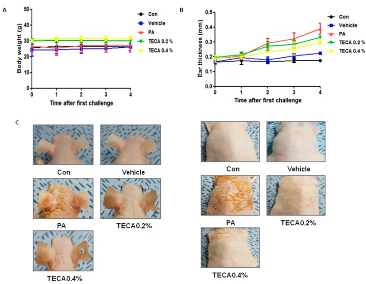
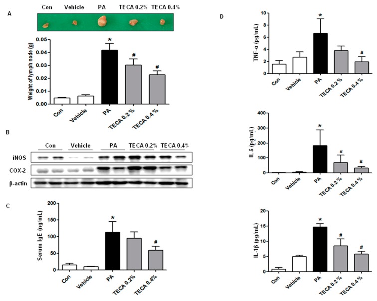
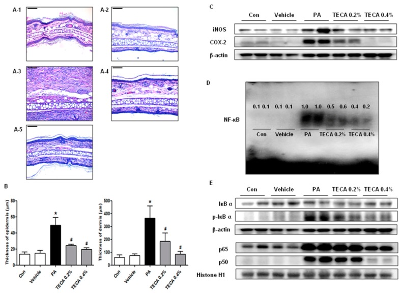
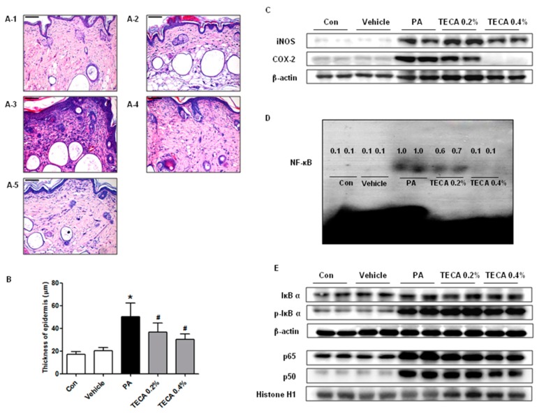
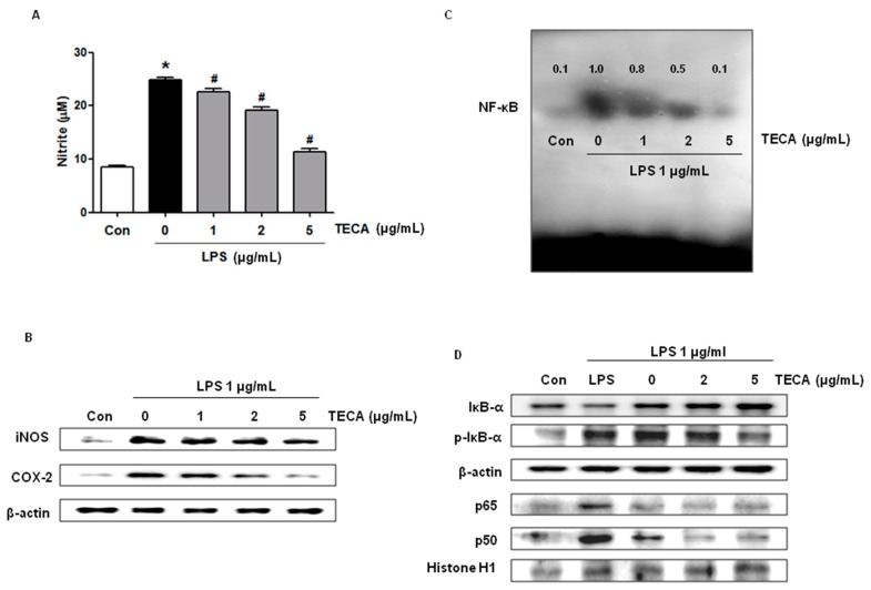
has potent antioxidant and anti-inflammatory properties. However, its anti-dermatitic effect has not yet been reported. In this study, we investigated the anti-dermatitic effects of titrated extract of Centella asiatica (TECA) in a phthalic anhydride (PA)-induced atopic dermatitis (AD) animal model as well as in vitro model. An AD-like lesion was induced by the topical application of five percent PA to the dorsal skin or ear of Hos:HR-1 mouse. After AD induction, 100 μL of 0.2% and 0.4% of TECA (40 μg or 80 μg/cm²) was spread on the dorsum of the ear or back skin three times a week for four weeks. We evaluated dermatitis severity, histopathological changes and changes in protein expression by Western blotting for inducible nitric oxide synthase (iNOS), cyclooxygenase-2 (COX-2), and NF-κB activity, which were determined by electromobility shift assay (EMSA). We also measured TNF-α, IL-1β, IL-6, and IgE concentration in the blood of AD mice by enzyme-linked immunosorbent assay (ELISA). TECA treatment attenuated the development of PA-induced atopic dermatitis. Histological analysis showed that TECA inhibited hyperkeratosis, mast cells and infiltration of inflammatory cells. TECA treatment inhibited expression of iNOS and COX-2, and NF-κB activity as well as the release of TNF-α, IL-1β, IL-6, and IgE. In addition, TECA (1, 2, 5 μg/mL) potently inhibited Lipopolysaccharide (LPS) (1 μg/mL)-induced NO production, expression of iNOS and COX-2, and NF-κB DNA binding activities in RAW264.7 macrophage cells. Our data demonstrated that TECA could be a promising agent for AD by inhibition of NF-κB signaling.
Overview
The claims and statistics of this paper haven’t been checked yet.
Run the check — free, no account needed. It reads the open-access text and checks the sources it cites.
Integrity signals
Registry
Cites
Statistics
Figures
Verification
Claims the paper makes
The claims and statistics of this paper haven’t been checked yet.
Run the check — free, no account needed. It reads the open-access text and checks the sources it cites.
Claims other papers make about this one
Each is checked against this paper’s own text: does it say what the citing paper says it does?
The source backs part of the claim, not all of it — or backs it for a different population, condition or comparison.
In the citing paper
“Studies utilizing phytosomal formulations of C. asiatica (0.2–0.4%) in models of atopic dermatitis have shown substantial decreases in inflammatory markers such as immunoglobulin E (IgE), cyclooxygenase-2 (COX-2), inducible nitric oxide synthase (iNOS), tumor necrosis factor alpha (TNF-α), and mast cell counts through modulation of the NF-κB signaling pathway (Park et al. 2017).”
From Centella asiatica (L.) Urb. in skin health and cosmeceuticals: mechanisms, clinical evidence, and advanced delivery systems (2025) · cites this paper as Park et al. 2017
Experimental — accuracy not yet measuredThe source backs part of the claim, not all of it — or backs it for a different population, condition or comparison.
In the citing paper
“as demonstrated by the inhibition of NF-κB, COX-2, iNOS, and IgE in dermatitis models (Park et al. 2017).”
From Centella asiatica (L.) Urb. in skin health and cosmeceuticals: mechanisms, clinical evidence, and advanced delivery systems (2025) · cites this paper as Park et al. 2017
Experimental — accuracy not yet measuredThe source backs part of the claim, not all of it — or backs it for a different population, condition or comparison.
In the citing paper
“Phytosome complexes showed added anti-inflammatory effects in atopic and contact dermatitis models, emphasizing the importance of delivery vehicle–bioactive interactions.”
From Centella asiatica (L.) Urb. in skin health and cosmeceuticals: mechanisms, clinical evidence, and advanced delivery systems (2025) · cites this paper as Park et al. 2017
Experimental — accuracy not yet measuredThe check wasn’t confident either way, so it gives no verdict — never disputed.
In the citing paper
“Phytosomes, complexing polar bioactives such as madecassoside with phospholipids, offer enhanced anti-inflammatory action and better skin permeation”
From Centella asiatica (L.) Urb. in skin health and cosmeceuticals: mechanisms, clinical evidence, and advanced delivery systems (2025) · cites this paper as Park et al. 2017
Experimental — accuracy not yet measured
Self-check
Percentages, sums and reported differences re-computed from the paper’s own numbers.
The self-check needs the paper’s full text.
Citations
Cites
Nuclear Factor-κB — A Pivotal Transcription Factor in Chronic Inflammatory Diseases
Barnes, P. J., & Karin, M. (1997) · New England Journal of Medicine · doi:10.1056/nejm199704103361506 · Cited by 4,622
Atopic dermatitis: New insights and opportunities for therapeutic intervention
Leung, D. Y. M. (2000) · Journal of Allergy and Clinical Immunology · doi:10.1067/mai.2000.106484 · Cited by 717
Cytokines and chemokines orchestrate atopic skin inflammation
Homey, B., Steinhoff, M. S., Ruzicka, T., & Leung, D. Y. M. (2006) · Journal of Allergy and Clinical Immunology · doi:10.1016/j.jaci.2006.03.047 · Cited by 584
NF-κB in the regulation of epithelial homeostasis and inflammation
Wullaert, A., Bonnet, M., & Pasparakis, M. (2010) · Cell Research · doi:10.1038/cr.2010.175 · Cited by 538
Barnes, P. J. (1997) · The International Journal of Biochemistry & Cell Biology · doi:10.1016/s1357-2725(96)00159-8 · Cited by 498
IgE, Mast Cells, and Eosinophils in Atopic Dermatitis
Liu, F., Goodarzi, H., & Chen, H. (2011) · Clinical Reviews in Allergy & Immunology · doi:10.1007/s12016-011-8252-4 · Cited by 474
Triterpene Composition and Bioactivities of Centella asiatica
Hashim, P., Sidek, H., Helan, M. H. M., Sabery, A., Palanisamy, U. D., & Ilham, M. (2011) · Molecules · doi:10.3390/molecules16021310 · Cited by 266
Centella asiatica in Dermatology: An Overview
Bylka, W., Znajdek-Awiżeń, P., Studzińska-Sroka, E., Dańczak‐Pazdrowska, A., & Brzezińska, M. (2014) · Phytotherapy Research · doi:10.1002/ptr.5110 · Cited by 231
Centella asiatica in cosmetology
Bylka, W., Znajdek-Awiżeń, P., Studzińska-Sroka, E., & Brzezińska, M. (2013) · Advances in Dermatology and Allergology · doi:10.5114/pdia.2013.33378 · Cited by 229
(1999) · PubMed · Cited by 171
Cited by
Sorted by citations
Items 101–101 of 101
Potensi Kombinasi Pegagan dan Dadap Serep dalam Menurunkan Risiko Nonalcoholic Fatty Liver Disease
Kaniawati, M., Sulaeman, A., Susilawati, E., Selifiana, N., Kasihtheisya, N., Syahyudin, W., & Parasitia, O. (2026) · JFIOnline · doi:10.35617/jfionline.v18i2.768
Metadata
- Authors
- Park, J., Choi, J., Son, D., Park, E., Song, M., Hellström, M., & Hong, J.
- Affiliations
- Chungbuk National University (KR)
- The Catholic University of Korea Daejeon St. Mary's Hospital (KR)
- University of Gothenburg (SE)
- Corresponding author
- Hong, Jin
- Venue
- International Journal of Molecular Sciences
- Publisher
- MDPI AG
- Published
- 2017-03-30
- Volume
- 18
- Issue
- 4
- Pages
- 738
- ISSN
- 1422-0067 (ISSN-L)
- PMC
- PMC5412323
- OpenAlex
- W2600159981
- Cited by
- 97
- References
- 48
- Citations by year
- 2026: 9 · 2025: 10 · 2024: 14 · 2023: 10 · 2022: 11 · 2021: 16 · 2020: 13 · 2019: 7 · 2018: 5 · 2017: 2
- FWCI
- 8.41
- Type
- journal article
- Language
- en
- Open access
- gold
- doi.org
- Licence
- Locations
- International Journal of Molecular Sciences (publishedVersion, cc-by)
- Europe PMC (PubMed Central) (publishedVersion, cc-by)
- PubMed Central (publishedVersion, cc-by)
- PubMed (publishedVersion)
- Funders
- National Research Foundation
- Ministry of Trade, Industry and Energy (1415139249, MOTIE, 1415139249)
- Ministry of Science, ICT and Future Planning (MRC.2008-0062275, 2008-0062275)
- National Research Foundation of Korea (MRC 2008‐0062275, 2008‐0062275)
- Medical Research Council (2008-0062275)
- Topics
- Health Sciences › Medicine › Dermatology › Dermatology and Skin Diseases
- Health Sciences › Medicine › Complementary and alternative medicine › Medicinal Plants and Neuroprotection
- Health Sciences › Medicine › Dermatology › Contact Dermatitis and Allergies
- Keywords
- Centella asiatica, atopic dermatitis, anti-inflammatory activity, NF-κB signaling, inducible nitric oxide synthase, cyclooxygenase-2, mast cells, hyperkeratosis, cytokines, IgE, lipopolysaccharide-induced nitric oxide, macrophages, topical herbal extract
- MeSH terms
- Animals, Anti-Inflammatory Agents, Cell Line, Interleukins, Macrophages, Phthalic Anhydrides, Plant Extracts, Skin, Triterpenes, Tumor Necrosis Factor-alpha, NF-kappa B, Dermatitis, Allergic Contact, Centella, Mice, Cyclooxygenase 2, Nitric Oxide Synthase Type II
About these checks
Registry, retraction, Cites and statistics results are computed, not judged, with one exception: a printed reference that no index lists is read once by a model before it counts as a citation error. Claims are extracted by a model and kept only when their quote appears verbatim in the text; each cited source is then checked, with a model’s help, for whether it says what the claim relies on. Those verdicts can be wrong — read the claim’s quote and the cited source before relying on one.
Statistics and arithmetic are recomputed, not judged.
Confidence bands are shown only for fields calibrated on our annotated test set. None is calibrated yet, so no band is shown.
What counts as a claim ›How we measure confidence ›How we check this ›
Are you an author, or do you see a problem with this page? Request a review
