

The DOI matches its Crossref record; 4 of the 4 references it cites were found in a registry or an index.
ERK Inhibition Increases RANKL-Induced Osteoclast Differentiation in RAW 264.7 Cells by Stimulating AMPK Activation and RANK Expression and Inhibiting Anti-Osteoclastogenic Factor Expression
Abstract
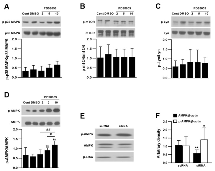
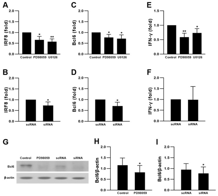
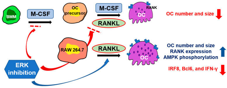
Bone absorption is necessary for the maintenance of bone homeostasis. An osteoclast (OC) is a monocyte–macrophage lineage cell that absorbs bone tissue. Extracellular signal-regulated kinases (ERKs) are known to play important roles in regulating OC growth and differentiation. In this study, we examined specific downstream signal pathways affected by ERK inhibition during OC differentiation. Our results showed that the ERK inhibitors PD98059 and U0126 increased receptor activator of NF-κB ligand (RANKL)-induced OC differentiation in RAW 264.7 cells, implying a negative role in OC differentiation. This is supported by the effect of ERK2-specific small interfering RNA on increasing OC differentiation. In contrast to our findings regarding the RAW 264.7 cells, the ERK inhibitors attenuated the differentiation of bone marrow-derived cells into OCs. The ERK inhibitors significantly increased the phosphorylation of adenosine 5′-monophosphate-activated protein kinase (AMPK) but not the activation of p38 MAPK, Lyn, and mTOR. In addition, while the ERK inhibition increased the expression of the RANKL receptor RANK, it decreased the expression of negative mediators of OC differentiation, such as interferon regulatory factor-8, B-cell lymphoma 6, and interferon-γ. These dichotomous effects of ERK inhibition suggest that while ERKs may play positive roles in bone marrow-derived cells, ERKs may also play negative regulatory roles in RAW 264.7 cells. These data provide important information for drug development utilizing ERK inhibitors in OC-related disease treatment.
Overview
The claims and statistics of this paper haven’t been checked yet.
Run the check — free, no account needed. It reads the open-access text and checks the sources it cites.
Integrity signals
Registry
Cites
Statistics
Figures
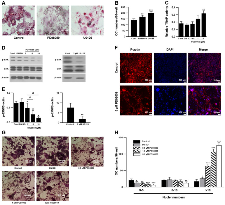
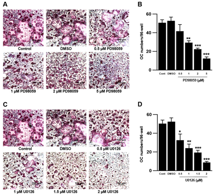
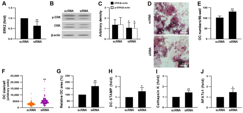
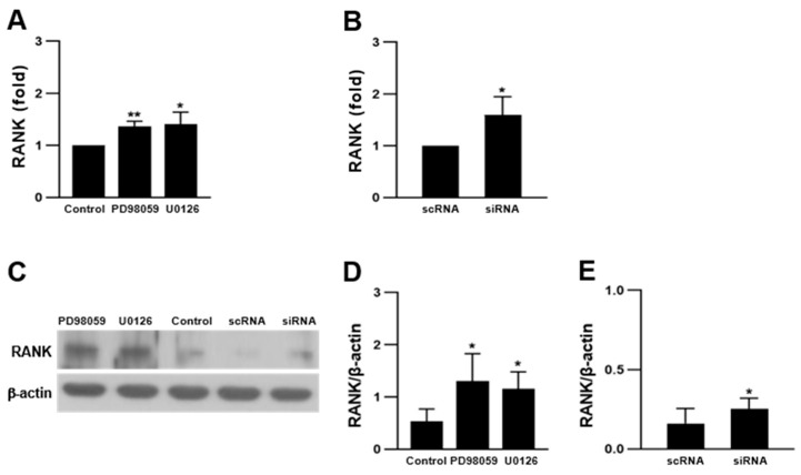
Verification
Claims the paper makes
The claims and statistics of this paper haven’t been checked yet.
Run the check — free, no account needed. It reads the open-access text and checks the sources it cites.
Claims other papers make about this one
Each is checked against this paper’s own text: does it say what the citing paper says it does?
In the citing paper
“Conversely, the role of ERK in regulating osteoclast differentiation appears to be context- and stage-dependent [32].”
From Anti-Osteoporotic Potential of Water Extract of Anethum graveolens L. Seeds (2023) · cites this paper as [32]
Self-check
Percentages, sums and reported differences re-computed from the paper’s own numbers.
The self-check needs the paper’s full text.
Citations
Cites
Hsu, H., Lacey, D. L., Dunstan, C. R., Solovyev, I., Colombero, A., Timms, E., Tan, H.-L., Elliott, G., Kelley, M. J., Sarosi, I., et al. (1999) · Proceedings of the National Academy of Sciences · doi:10.1073/pnas.96.7.3540 · Cited by 1,636
T-Cell mediated regulation of osteoclastogenesis by signalling cross-talk between RANKL and IFN-γ.
Takayanagi, H., Ogasawara, K., Hida, S., Chiba, T., Murata, S., Sato, K., Takaoka, A., Yokochi, T., Oda, H., Tanaka, K., et al. (2000) · Nature · doi:10.1038/35046102 · Cited by 1,358
Matsumoto, M., Kogawa, M., Wada, S., Takayanagi, H., Tsujimoto, M., Katayama, S., Hisatake, K., & Nogi, Y. (2004) · Journal of Biological Chemistry · doi:10.1074/jbc.m408795200 · Cited by 436
Sims, N. A., & Martin, T. J. (2015) · Frontiers in Endocrinology · doi:10.3389/fendo.2015.00041 · Cited by 184
Research works printed but not linked: 54
- Seeman, 2006, Bone quality-the material and structural basis of bone strength and fragility, N. Engl. J. Med., 354, 2250, 10.1056/NEJMra053077 · DOI
- Chen, 2018, Osteoblast-osteoclast interactions, Connect. Tissue Res., 59, 99, 10.1080/03008207.2017.1290085 · DOI
- Zhu, S., Ehnert, S., Roub, M., Häussling, V., Aspera, R.H., Chen, T., and Nussler, A.K. (2018). From the clinical problem to the basic research: Co-culture models of osteoblasts and osteoclasts. Int. J. Mol. Sci., 19. · DOI
- Bellido, 2014, Osteocyte-driven bone remodeling, Calcif. Tissue Int., 94, 25, 10.1007/s00223-013-9774-y · DOI · Paper page
- Schaffler, 2014, Osteocytes: Master orchestrators of bone, Calcif. Tissue Int., 94, 5, 10.1007/s00223-013-9790-y · DOI · Paper page
- Park, 2017, Current understanding of RANK signaling in osteoclast differentiation and maturation, Mol. Cells, 40, 706, 10.14348/molcells.2017.0225 · DOI · Paper page
- Teitelbaum, 2000, Bone resorption by osteoclasts, Science, 289, 1504, 10.1126/science.289.5484.1504 · DOI · Paper page
- Quinn, 1998, A combination of osteoclast differentiation factor and macrophage-colony stimulating factor is sufficient for both human and mouse osteoclast formation in vitro, Endocrinology, 139, 4424, 10.1210/endo.139.10.6331 · DOI · Paper page
- Boyle, 2003, Osteoclast differentiation and activation, Nature, 423, 337, 10.1038/nature01658 · DOI · Paper page
- Feng, 2013, Osteoclasts: New insights, Bone Res., 1, 11, 10.4248/BR201301003 · DOI
- Teitelbaum, 2003, Genetic regulation of osteoclast development and function, Nat. Rev. Genet., 4, 638, 10.1038/nrg1122 · DOI
- Ross, 2006, M-CSF, c-fms, and signaling in osteoclasts and their precursors, Ann. N. Y. Acad. Sci., 1068, 110, 10.1196/annals.1346.014 · DOI
- Lee, K., Seo, I., Choi, M.H., and Jeong, D. (2018). Roles of mitogen-activated protein kinases in osteoclast biology. Int. J. Mol. Sci., 19. · DOI
- Wada, 2006, RANKL-RANK signaling in osteoclastogenesis and bone disease, Trends Mol. Med., 12, 17, 10.1016/j.molmed.2005.11.007 · DOI
- Hotokezaka, 2002, U0126 and PD98059, specific inhibitors of MEK, accelerate differentiation of RAW264.7 cells into osteoclast-like cells, J. Biol. Chem., 277, 47366, 10.1074/jbc.M208284200 · DOI
- He, Y., Staser, K., Rhodes, S.D., Liu, Y., Wu, X., Park, S.-J., Yuan, J., Yang, X., Li, X., and Jiang., L. (2011). ERK1 positively regulates osteoclast differentiation and bone resorptive activity. PLoS ONE, 6. · DOI
- Ihn, 2015, The 1,2,3-triazole derivative KP-A021 suppresses osteoclast differentiation and function by inhibiting RANKL-mediated MEK-ERK signaling pathway, Exp. Biol. Med., 240, 1690, 10.1177/1535370215576310 · DOI
- Li, 2015, Inhibition of RANKL induced osteoclastogenesis through the suppression of the ERK signaling pathway by astragaloside IV and attenuation of titanium-particle-induced osteolysis, Int. J. Mol. Med., 36, 1335, 10.3892/ijmm.2015.2330 · DOI
- Kim, 2017, The inactivation of ERK1/2, p38 and NF-κB is involved in the down-regulation of osteoclastogenesis and function by A2B adenosine receptor stimulation, Mol. Cells, 40, 752, 10.14348/molcells.2017.0098 · DOI
- Zhao, 2015, Osteoprotegerin induces podosome disassembly in osteoclasts through calcium, ERK, and p38 MAPK signaling pathways, Cytokine, 71, 199, 10.1016/j.cyto.2014.10.007 · DOI
- Agidigbi, 2020, Inhibition of MEK/ERK upregulates GSH production and increases RANKL-induced osteoclast differentiation in RAW 264.7 cells, Free Radic. Res., 54, 894, 10.1080/10715762.2020.1742896 · DOI
- Cai, 2018, High glucose downregulates the effects of autophagy on osteoclastogenesis via the AMPK/mTOR/ULK1 pathway, Biochem. Biophys. Res. Commun., 503, 428, 10.1016/j.bbrc.2018.04.052 · DOI
- Lee, 2010, AMP kinase acts as a negative regulator of RANKL in the differentiation of osteoclasts, Bone, 47, 926, 10.1016/j.bone.2010.08.001 · DOI
- Oh, 2016, Cytosolic malate dehydrogenase regulates RANKL-mediated osteoclastogenesis via AMPK/c-Fos/NFATc1 signaling, Biochem. Biophys. Res. Commun., 475, 125, 10.1016/j.bbrc.2016.05.055 · DOI
- Quinn, 2010, Germline deletion of AMP-activated protein kinase beta subunits reduces bone mass without altering osteoclast differentiation or function, FASEB J., 24, 275, 10.1096/fj.09-137158 · DOI
- Jeyabalan, 2012, AMP-activated protein kinase pathway and bone metabolism, J. Endocrinol., 212, 277, 10.1530/JOE-11-0306 · DOI
- Kang, 2013, Genetic deletion of catalytic subunits of AMP-activated protein kinase increases osteoclasts and reduces bone mass in young adult mice, J. Biol. Chem., 288, 12187, 10.1074/jbc.M112.430389 · DOI
- Tong, 2020, Suppression of AMP-activated protein kinase reverses osteoprotegerin-induced inhibition of osteoclast differentiation by reducing autophagy, Cell Prolif., 53, e12714, 10.1111/cpr.12714 · DOI
- Guo, H., Ding, D., Wang, L., Yan, J., Ma, L., and Jin, Q. (2021). Metformin attenuates osteoclast-mediated abnormal subchondral bone remodeling and alleviates osteoarthritis via AMPK/NF-κB/ERK signaling pathway. PLoS ONE, 16. · DOI
- Li, 2018, Glycyrrhizin suppresses RANKL-induced osteoclastogenesis and oxidative stress through inhibiting NF-κB and MAPK and activating AMPK/Nrf2, Calcif. Tissue Int., 103, 324, 10.1007/s00223-018-0425-1 · DOI
- Zhao, 2009, Interferon regulatory factor-8 regulates bone metabolism by suppressing osteoclastogenesis, Nat. Med., 15, 1066, 10.1038/nm.2007 · DOI · Paper page
- Miyauchi, 2010, The Blimp1–Bcl6 axis is critical to regulate osteoclast differentiation and bone homeostasis, J. Exp. Med., 207, 751, 10.1084/jem.20091957 · DOI
- Ihn, 2017, Afatinib ameliorates osteoclast differentiation and function through downregulation of RANK signaling pathways, BMB Rep., 50, 150, 10.5483/BMBRep.2017.50.3.223 · DOI
- Takahashi, 1986, Recombinant human interferon-gamma inhibits formation of human osteoclast-like cells, J. Immunol., 137, 3544, 10.4049/jimmunol.137.11.3544 · DOI
- Fox, 2000, Interferon gamma directly inhibits TRANCE-induced osteoclastogenesis, Biochem. Biophys. Res. Commun., 276, 868, 10.1006/bbrc.2000.3577 · DOI
- Madyastha, 2000, IFN-gamma enhances osteoclast generation in cultures of peripheral blood from osteopetrotic patients and normalizes superoxide production, J. Interferon Cytokine Res., 20, 645, 10.1089/107999000414826 · DOI
- Gao, 2007, IFN-gamma stimulates osteoclast formation and bone loss in vivo via antigen-driven T cell activation IFN-gamma stimulates osteoclast formation and bone loss in vivo via antigen-driven T cell activation, J. Clin. Investig., 117, 122, 10.1172/JCI30074 · DOI
- Koyama, 2007, ZD1839 induces p15INK4b and causes G1 arrest by inhibiting the mitogen-activated protein kinase/extracellular signal-regulated kinase pathway, Mol. Cancer Ther., 6, 1579, 10.1158/1535-7163.MCT-06-0814 · DOI
- Stewart, 2008, PD98059 enhances C2 myoblast differentiation through p38 MAPK activation: A novel role for PD98059, J. Endocrinol., 198, 243, 10.1677/JOE-08-0151 · DOI
- Busca, R., Christen, R., Lovern, M., Clifford, A.M., Yue, J.X., Goss, G.G., Pouysségur, J., and Lenormand, P. (2015). ERK1 and ERK2 present functional redundancy in tetrapods despite higher evolution rate of ERK1. BMC Evol. Biol., 15. · DOI
- Ang, 2015, Functional redundancy of ERK1 and ERK2 MAP kinases during development, Cell Rep., 12, 913, 10.1016/j.celrep.2015.07.011 · DOI
- Oh, 2015, Obatoclax regulates the proliferation and fusion of osteoclast precursors through the inhibition of ERK activation by RANKL, Mol. Cells, 38, 279, 10.14348/molcells.2015.2340 · DOI
- Atkins, 2006, RANK expression as a cell surface marker of human osteoclast precursors in peripheral blood, bone marrow, and giant cell tumors of bone, J. Bone Miner. Res., 21, 1339, 10.1359/jbmr.060604 · DOI
- Horne, 2005, The role(s) of Src kinase and Cbl proteins in the regulation of osteoclast differentiation and function, Immunol. Rev., 208, 106, 10.1111/j.0105-2896.2005.00335.x · DOI
- Kim, 2009, The Src family kinase, Lyn, suppresses osteoclastogenesis in vitro and in vivo, Proc. Natl. Acad. Sci. USA, 106, 2325, 10.1073/pnas.0806963106 · DOI
- Zhao, 2011, Negative regulation of osteoclastogenesis and bone resorption by cytokines and transcriptional repressors, Arthritis Res. Ther., 13, 234, 10.1186/ar3379 · DOI · Paper page
- Rahman, 2015, Proliferation-coupled osteoclast differentiation by RANKL: Cell density as a determinant of osteoclast formation, Bone, 81, 392, 10.1016/j.bone.2015.08.008 · DOI
- Ng, 2018, Comparative characterization of osteoclasts derived from murine bone marrow macrophages and RAW 264.7 cells using quantitative proteomics, JBMR Plus, 2, 328, 10.1002/jbm4.10058 · DOI
- Li, S., Yang, B., Teguh, D., Zhou, L., Xu, J., and Rong, L. (2016). Amyloid b peptide enhances RANKL-induced osteoclast activation through NF-κB, ERK, and calcium oscillation signaling. Int. J. Mol. Sci., 17. · DOI
- Steinberg, 2009, AMPK in health and disease, Physiol. Rev., 89, 1025, 10.1152/physrev.00011.2008 · DOI
- Shah, 2010, AMP-activated protein kinase (AMPK) activation regulates in vitro bone formation and bone mass, Bone, 47, 309, 10.1016/j.bone.2010.04.596 · DOI
- Yuan, 2020, The MAPK and AMPK signalings: Interplay and implication in targeted cancer therapy, J. Hematol. Oncol., 13, 113, 10.1186/s13045-020-00949-4 · DOI
- Kang, 2016, NADPH oxidase gp91phox contributes to RANKL-induced osteoclast differentiation by upregulating NFTAc1, Sci. Rep., 6, 38014, 10.1038/srep38014 · DOI
- Kim, 2001, Rac2 is an essential regulator of neutrophil nicotinamide adenine dinucleotide phosphate oxidase activation in response to specific signaling pathways, J. Immunol., 166, 1223, 10.4049/jimmunol.166.2.1223 · DOI · Paper page
Cited by
Anti-Osteoporotic Potential of Water Extract of Anethum graveolens L. Seeds
Jang, S.-A., Lee, S.-J., Hwang, Y.-H., & Ha, H. (2023) · Nutrients · doi:10.3390/nu15194302 · Cited by 2
Metadata
- Authors
- Choi, E.-B., Agidigbi, T. S., Kang, I.-S., & Kim, C.
- Venue
- International Journal of Molecular Sciences
- Publisher
- MDPI AG
- Published
- 2022-11-04
- Volume
- 23
- Issue
- 21
- Pages
- 13512
- ISSN
- 1422-0067
- PMC
- PMC9656104
- Cited by
- 22
- References
- 4
- Language
- en
- Funders
- National Research Foundation of Korea (2020R1F1A1068065, 2022R1I1A1A01055015)
About these checks
Registry, retraction, Cites and statistics results are computed, not judged, with one exception: a printed reference that no index lists is read once by a model before it counts as a citation error. Claims are extracted by a model and kept only when their quote appears verbatim in the text; each cited source is then checked, with a model’s help, for whether it says what the claim relies on. Those verdicts can be wrong — read the claim’s quote and the cited source before relying on one.
Statistics and arithmetic are recomputed, not judged.
Confidence bands are shown only for fields calibrated on our annotated test set. None is calibrated yet, so no band is shown.
What counts as a claim ›How we measure confidence ›How we check this ›
Are you an author, or do you see a problem with this page? Request a review
How to cite
Cite CoChat for the checks on this page, and the paper for its own findings.
- This page
- CoChat. (2026). Citation check: ERK Inhibition Increases RANKL-Induced Osteoclast Differentiation in RAW 264.7 Cells by Stimulating AMPK Activation and RANK Expression and Inhibiting Anti-Osteoclastogenic Factor Expression. https://app.cochat.ai/papers/10.3390/ijms232113512
- The paper
- Choi, E.-B., Agidigbi, T. S., Kang, I.-S., & Kim, C. (2022). ERK Inhibition Increases RANKL-Induced Osteoclast Differentiation in RAW 264.7 Cells by Stimulating AMPK Activation and RANK Expression and Inhibiting Anti-Osteoclastogenic Factor Expression. International Journal of Molecular Sciences. https://doi.org/10.3390/ijms232113512
Data sources
Bibliographic data: Semantic Scholar, OpenAlex, Crossref.
Contains information from the Semantic Scholar Academic Graph Datasets, made available under the ODC Attribution License (ODC-BY 1.0).
Semantic Scholar’s data is described in: Kinney, R., et al. (2023). The Semantic Scholar Open Data Platform. arXiv:2301.10140.
