

The DOI matches its Crossref record; 7 of the 7 references it cites were found in a registry or an index.
Toxicity of Magnetic Nanoparticles in Medicine: Contributing Factors and Modern Assessment Methods
Abstract
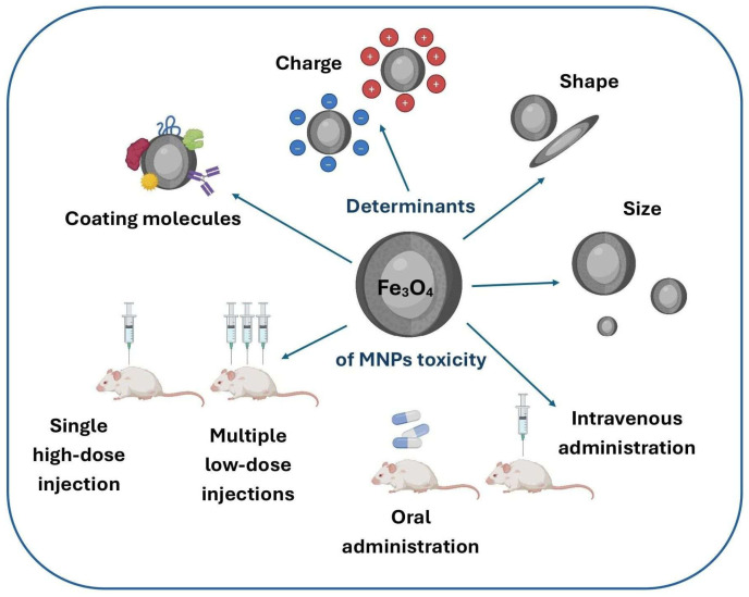
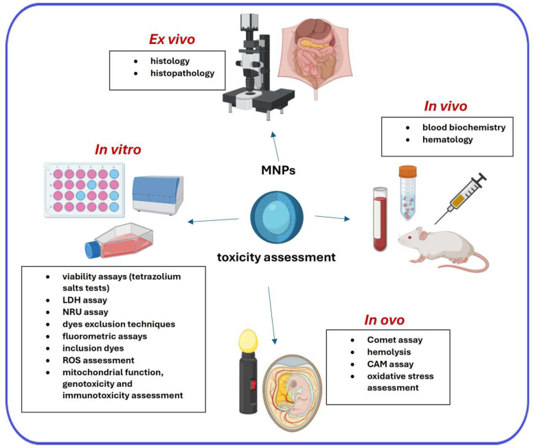
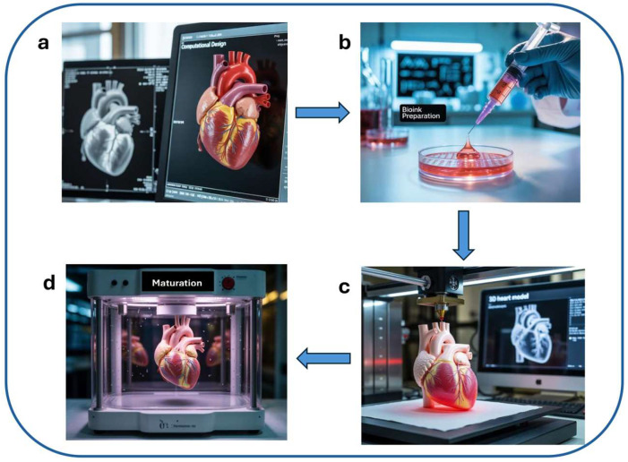
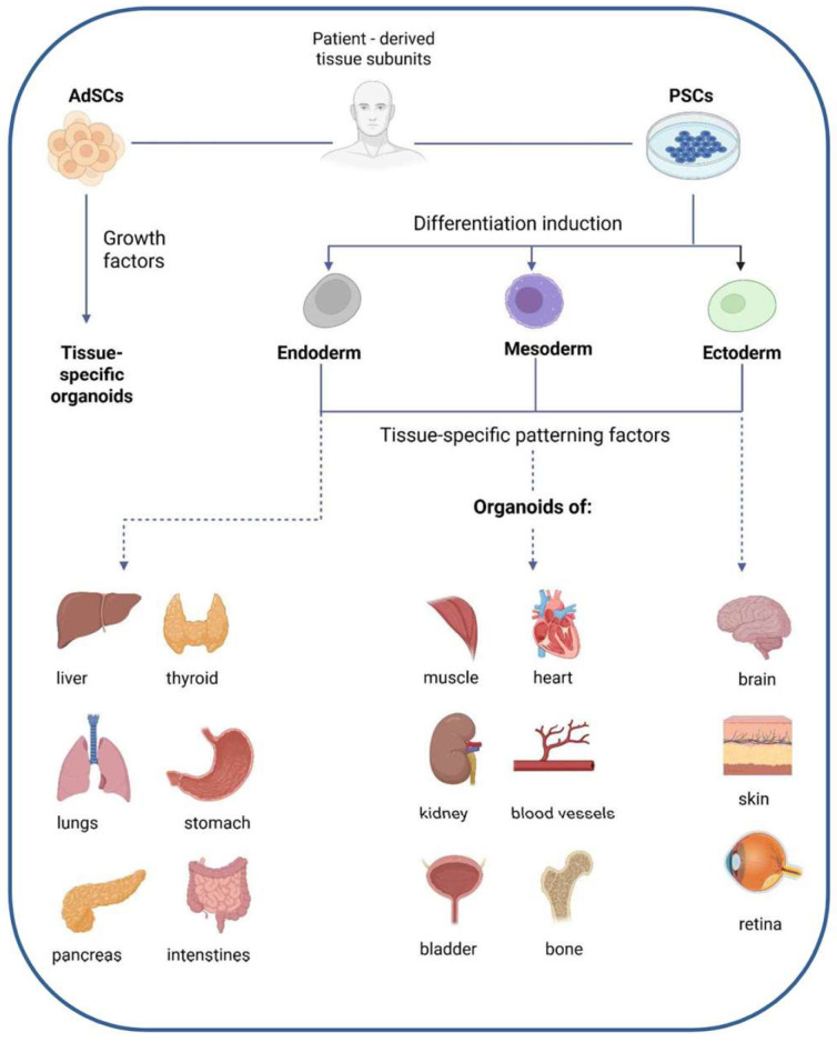
With the rapid evolution of nanotechnology, magnetic iron oxide nanoparticles (MNPs)—primarily Fe3O4 and γ-Fe2O3—have gained prominence in biomedicine. Their extensive specific surface area, tunable surface functionalities, and intrinsic magnetic characteristics render them highly versatile for diverse clinical applications, including tumor visualization through Magnetic Resonance Imaging (MRI), radiolabeling, targeted radiotherapy, hyperthermia, gene transfer, drug delivery, Magnetic Particle Imaging (MPI), magnetic blood filtration and theranostic strategies. Nevertheless, ensuring the biocompatibility and non-toxicity of these nanostructures remains a fundamental prerequisite for their medical implementation. Hence, it is essential to continuously refine our understanding of MNP-related toxicity and pursue comprehensive research on this front. This article consolidates up-to-date insights into the evaluation of MNPs’ toxicological profiles, emphasizing the influence of physicochemical properties such as morphology, surface modifications, and electrostatic characteristics, along with operational factors like dosage and administration routes. Traditional toxicity testing strategies, including in vitro assays as first-line screening tools, together with standard ex vivo and in vivo models, are discussed. Special attention is given to the emerging role of New Approach Methodologies (NAMs), such as organoid formation, 3D bioprinting, in ovo chicken embryo assays, and image cytometry. These techniques offer ethical, human-relevant, and informative alternatives to animal testing, supporting more predictive and translationally relevant toxicity assessment of MNPs. Taken together, the integration of conventional assays with innovative NAMs, alongside careful consideration of physicochemical and operational factors, is essential to translate the laboratory promise of MNPs into safe and clinically effective nanomedicines.
Overview
The claims and statistics of this paper haven’t been checked yet.
Run the check — free, no account needed. It reads the open-access text and checks the sources it cites.
Integrity signals
Registry
Cites
Statistics
Figures
Verification
Claims the paper makes
The claims and statistics of this paper haven’t been checked yet.
Run the check — free, no account needed. It reads the open-access text and checks the sources it cites.
Claims other papers make about this one
Each is checked against this paper’s own text: does it say what the citing paper says it does?
In the citing paper
“Comprehensive toxicological evaluations are necessary to ensure safety over extended periods, especially for repeated dosing regimens [69, 98].”
From Advances in green-synthesized magnetic nanoparticles for targeted cancer therapy: mechanisms, applications, and future perspectives (2026) · cites this paper as [98]
In the citing paper
“While MNPs are often considered biocompatible, their long-term accumulation and degradation products (e.g., free iron ions) may induce oxidative stress or interfere with normal cellular processes.”
From Advances in green-synthesized magnetic nanoparticles for targeted cancer therapy: mechanisms, applications, and future perspectives (2026) · cites this paper as [98]
In the citing paper
“Toxicity and clearance remain partially unresolved.”
From Advances in green-synthesized magnetic nanoparticles for targeted cancer therapy: mechanisms, applications, and future perspectives (2026) · cites this paper as [98]
Self-check
Percentages, sums and reported differences re-computed from the paper’s own numbers.
The self-check needs the paper’s full text.
Citations
Cites
Organoids as an in vitro model of human development and disease
Fatehullah, A., Tan, S. H., & Barker, N. (2016) · Nature Cell Biology · doi:10.1038/ncb3312 · Cited by 1,508
Identification of erythroferrone as an erythroid regulator of iron metabolism
Kautz, L., Jung, G., Valore, E. V., Rivella, S., Nemeth, E., & Ganz, T. (2014) · Nature Genetics · doi:10.1038/ng.2996 · Cited by 1,127
In vivo delivery, pharmacokinetics, biodistribution and toxicity of iron oxide nanoparticles
Arami, H., Khandhar, A., Liggitt, D., & Krishnan, K. M. (2015) · Chemical Society Reviews · doi:10.1039/c5cs00541h · Cited by 830
Iron Homeostasis and Ferroptosis in Human Diseases: Mechanisms and Therapeutic Prospects
Ru, Q., Li, Y., Chen, L., Wu, Y., Min, J., & Wang, F. (2024) · Signal Transduction and Targeted Therapy · doi:10.1038/s41392-024-01969-z · Cited by 724
Guidelines for the use of flow cytometry and cell sorting in immunological studies (third edition)
Cossarizza, A., Chang, H., Radbruch, A., Abrignani, S., Addo, R., Akdis, M., Andrä, I., Andreata, F., Annunziato, F., Arranz, E., et al. (2021) · European Journal of Immunology · doi:10.1002/eji.202170126 · Cited by 531 · cited 1×
“Imaging flow cytometry (IFC) offers single-cell, high-content morphology and localization data, yet it may be confounded by nanoparticle autofluorescence, quenching, spectral overlap/compensation, and scattering artifacts without rigorous controls and appropriate reference materials [162,176].”
NCOA4-Mediated Ferritinophagy: A Potential Link to Neurodegeneration
Quiles del Rey, M., & Mancias, J. D. (2019) · Frontiers in Neuroscience · doi:10.3389/fnins.2019.00238 · Cited by 227
Remya, N. S., Syama, S., Sabareeswaran, A., & Mohanan, P. V. (2016) · International Journal of Pharmaceutics · doi:10.1016/j.ijpharm.2016.06.119 · Cited by 97 · cited 2×
“Common assessments include the analysis of RBC and WBC morphology following nanoparticle exposure, along with the determination of the erythrocyte sedimentation rate (ESR) [85,87].”
Research works printed but not linked: 169
- Stanicki, 2023, An update on the applications and characteristics of magnetic iron oxide nanoparticles for drug delivery, Expert Opin. Drug Deliv., 19, 321, 10.1080/17425247.2022.2047020 · DOI
- Avval, 2020, Introduction of magnetic and supermagnetic nanoparticles in new approach of targeting drug delivery and cancer therapy application, Drug Metab. Rev., 52, 157, 10.1080/03602532.2019.1697282 · DOI
- Wang, 2023, Advances in smart delivery of magnetic field-targeted drugs in cardiovascular diseases, Drug Deliv., 30, 2256495, 10.1080/10717544.2023.2256495 · DOI
- Ulbrich, 2016, Targeted drug delivery with polymers and magnetic nanoparticles: Covalent and noncovalent approaches, release control, and clinical studies, Chem. Rev., 116, 5338, 10.1021/acs.chemrev.5b00589 · DOI
- Bao, 2018, Magnetic iron oxide nanoparticles as T1 c ontrast agents for magnetic resonance imaging, J. Mater. Chem. C, 6, 1280, 10.1039/C7TC05854C · DOI
- Palzer, J., Eckstein, L., Slabu, I., Reisen, O., Neumann, U.P., and Roeth, A.A. (2021). Iron oxide nanoparticle-based hyperthermia as a treatment option in various gastrointestinal malignancies. Nanomaterials, 11. · DOI
- Rahman, 2023, Magnetic resonance imaging and iron-oxide nanoparticles in the era of personalized medicine, Nanotheranostics, 7, 424, 10.7150/ntno.86467 · DOI · Paper page
- Mahmoudi, 2018, Magnetic hyperthermia therapy for the treatment of glioblastoma: A review of the therapy’s history, efficacy and application in humans, Int. J. Hyperth., 34, 1316, 10.1080/02656736.2018.1430867 · DOI
- Mu, 2018, siRNA delivery with stem cell membrane-coated magnetic nanoparticles for imaging-guided photothermal therapy and gene therapy, ACS Biomater. Sci. Eng., 4, 3895, 10.1021/acsbiomaterials.8b00858 · DOI
- Russell, 2021, Impact of superparamagnetic iron oxide nanoparticles on in vitro and in vivo radiosensitisation of cancer cells, Radiat. Oncol., 16, 104, 10.1186/s13014-021-01829-y · DOI
- Sizikov, A.A., Nikitin, P.I., and Nikitin, M.P. (2021). Magnetofection in vivo by nanomagnetic carriers systemically administered into the bloodstream. Pharmaceutics, 13. · DOI
- Pellico, 2021, Radiolabelling of nanomaterials for medical imaging and therapy, Chem. Soc. Rev., 50, 3355, 10.1039/D0CS00384K · DOI
- Rodrigues, 2019, Antimicrobial magnetic nanoparticle-based therapies for controlling infectious diseases, Int. J. Pharm., 555, 356, 10.1016/j.ijpharm.2018.11.043 · DOI
- Velazquez-Albino, A.C., Imhoff, E.D., and Rinaldi-Ramos, C.M. (2025). Advances in engineering nanoparticles for magnetic particle imaging (MPI). Sci. Adv., 11. · DOI
- Li, Z., Xue, L., Wang, P., Ren, X., Zhang, Y., Wang, C., and Sun, J. (2023). Biological scaffolds assembled with magnetic nanoparticles for bone tissue engineering: A review. Materials, 16. · DOI
- Carvalho, T.S.S., Torres, P.M.C., Belo, J.H., Mano, J., and Olhero, S.M. (2023). Bioactive magnetic materials in bone tissue engineering: A review of recent findings in CaP-based particles and 3D-printed scaffolds. Adv. NanoBiomed Res., 3. · DOI
- Bauso, L.V., La Fauci, V., Longo, C., and Calabrese, G. (2024). Bone tissue engineering and nanotechnology: A promising combination for bone regeneration. Biology, 13. · DOI
- Waalders, N., Van Lier, D., Gerretsen, J., Moran, L., Stegmann, K.A., Twigger, W., Blanco-Andujar, C., Frodsham, G., Kox, M., and Pickkers, P. (2024). Preclinical and first-in-human safety studies on a novel magnetism-based haemofiltration method. Sci. Rep., 14. · DOI
- Wallace, 2016, The regulation of iron absorption and homeostasis, Clin. Biochem. Rev., 37, 51
- Meng, 2022, Lysosomal iron recycling in mouse macrophages is dependent upon both LcytB and Steap3 reductases, Blood Adv., 6, 1692, 10.1182/bloodadvances.2021005609 · DOI
- Yambire, K.F., Rostosky, C., Watanabe, T., Pacheu-Grau, D., Torres-Odio, S., Sanchez-Guerrero, A., Sendorovich, O., Meyron-Holtz, E.G., Milosevic, J., and Frahm, J. (2019). Impaired lysosomal acidification triggers iron deficiency and inflammation in vivo. eLife, 8. · DOI
- Arosio, P., Cairo, G., and Bou-Abdallah, F. (2024). A brief history of ferritin, an ancient and versatile protein. Int. J. Mol. Sci., 26. · DOI
- 2010, The iron redox and hydrolysis chemistry of the ferritins, Biochim. Biophys. Acta Gen. Subj., 1800, 719, 10.1016/j.bbagen.2010.03.021 · DOI
- Obi, C.D., Bhuiyan, T., Dailey, H.A., and Medlock, A.E. (2022). Ferrochelatase: Mapping the intersection of iron and porphyrin metabolism in the mitochondria. Front. Cell Dev. Biol., 10. · DOI
- Maio, 2020, Mammalian iron–sulfur cluster biogenesis: Recent insights into the roles of frataxin, acyl carrier protein and ATPase-mediated transfer to recipient proteins, Curr. Opin. Chem. Biol., 55, 34, 10.1016/j.cbpa.2019.11.014 · DOI
- Pearson, S.A., and Cowan, J.A. (2021). Evolution of the human mitochondrial ABCB7 2Fe–2S4 cluster exporter and the molecular mechanism of an E433K disease-causing mutation. Arch. Biochem. Biophys., 697. · DOI
- Li, 2022, Structures of Atm1 provide insight into [2Fe-2S] cluster export from mitochondria, Nat. Commun., 13, 4339, 10.1038/s41467-022-32006-8 · DOI
- White, 2013, HRG1 is essential for heme transport from the phagolysosome of macrophages during erythrophagocytosis, Cell Metab., 17, 261, 10.1016/j.cmet.2013.01.005 · DOI
- Helman, 2023, The biology of mammalian multi-copper ferroxidases, BioMetals, 36, 263, 10.1007/s10534-022-00370-z · DOI
- Wang, 2023, Ferritinophagy: Research advance and clinical significance in cancers, Cell Death Discov., 9, 463, 10.1038/s41420-023-01753-y · DOI
- Colucci, S., Altamura, S., Marques, O., Müdder, K., Agarvas, A.R., Hentze, M.W., and Muckenthaler, M.U. (2022). Iron-dependent BMP6 regulation in liver sinusoidal endothelial cells is instructed by hepatocyte-derived secretory signals. HemaSphere, 6. · DOI
- Camaschella, 2020, Iron metabolism and iron disorders revisited in the hepcidin era, Haematologica, 105, 260, 10.3324/haematol.2019.232124 · DOI · Paper page
- 2018, Magnetic iron oxide nanoparticles as drug carriers: Clinical relevance, Nanomedicine, 13, 953, 10.2217/nnm-2017-0336 · DOI
- Weissleder, 1989, Superparamagnetic iron oxide: Pharmacokinetics and toxicity, Am. J. Roentgenol., 152, 167, 10.2214/ajr.152.1.167 · DOI
- Bulte, 2004, Iron oxide MR contrast agents for molecular and cellular imaging, NMR Biomed., 17, 484, 10.1002/nbm.924 · DOI · Paper page
- McCullough, 2013, Ferumoxytol in clinical practice: Implications for MRI, J. Magn. Reson. Imaging, 37, 1476, 10.1002/jmri.23879 · DOI
- Ulrich, 2011, Efficacy and safety of intratumoral thermotherapy using magnetic iron-oxide nanoparticles combined with external beam radiotherapy on patients with recurrent glioblastoma multiforme, J. Neurooncol., 103, 317, 10.1007/s11060-010-0389-0 · DOI
- Johannsen, 2007, Thermotherapy of prostate cancer using magnetic nanoparticles: Feasibility, imaging, and three-dimensional temperature distribution, Eur. Urol., 52, 1653, 10.1016/j.eururo.2006.11.023 · DOI
- Gonzalez, 2022, Current hurdles to the translation of nanomedicines from bench to the clinic, Drug Deliv. Transl. Res., 12, 500, 10.1007/s13346-021-01024-2 · DOI
- Mai, 2017, Magnetic nanoparticles: Reactive oxygen species generation and potential therapeutic applications, J. Nanopart Res., 19, 253, 10.1007/s11051-017-3943-2 · DOI
- Abakumov, M.A., Semkina, A.S., Skorikov, A.S., Vishnevskiy, D.A., Ivanova, A.V., Mironova, E., Davydova, G.A., Majouga, A.G., and Chekhonin, V.P. (2018). Toxicity of iron oxide nanoparticles: Size and coating effects. J. Biochem. Mol. Toxicol., 32. · DOI
- Machnicka, 2023, In vivo biodistribution and clearance of magnetic iron oxide nanoparticles for medical applications, Int. J. Nanomed., 18, 4067, 10.2147/IJN.S415063 · DOI
- Frantellizzi, 2020, New frontiers in molecular imaging with superparamagnetic iron oxide nanoparticles (SPIONs): Efficacy, toxicity, and future applications, Nucl. Med. Mol. Imaging, 54, 65, 10.1007/s13139-020-00635-w · DOI
- Yang, 2015, Size dependent biodistribution and toxicokinetics of iron oxide magnetic nanoparticles in mice, Nanoscale, 7, 625, 10.1039/C4NR05061D · DOI
- Li, J., Yuan, Z., Liu, H., Feng, J., and Chen, Z. (2019). Size-dependent tissue-specific biological effects of core–shell structured Fe3O4@SiO2–NH2 nanoparticles. J. Nanobiotechnol., 17. · DOI
- Wu, 2022, Ultrasmall iron oxide nanoparticles cause significant toxicity by specifically inducing acute oxidative stress to multiple organs, Part. Fibre Toxicol., 19, 24, 10.1186/s12989-022-00465-y · DOI
- Xie, 2016, Size-dependent cytotoxicity of Fe3O4 nanoparticles induced by biphasic regulation of oxidative stress in different human hepatoma cells, Int. J. Nanomed., 11, 3557, 10.2147/IJN.S105575 · DOI
- Kaygisiz, 2017, Genotoxic evaluation of different sizes of iron oxide nanoparticles and ionic form by SMART, Allium and comet assay, Toxicol. Ind. Health, 33, 802, 10.1177/0748233717722907 · DOI
- Lee, 2014, Rod-shaped iron oxide nanoparticles are more toxic than sphere-shaped nanoparticles to murine macrophage cells, Environ. Toxicol. Chem., 33, 2759, 10.1002/etc.2735 · DOI
- Hu, 2016, External magnetic field-induced targeted delivery of highly sensitive iron oxide nanocubes for MRI of myocardial infarction, Small, 12, 4707, 10.1002/smll.201600263 · DOI
- Tonson, 2018, Effects of nanoprobe morphology on cellular binding and inflammatory responses: Hyaluronan-conjugated magnetic nanoworms for magnetic resonance imaging of atherosclerotic plaques, ACS Appl. Mater. Interfaces, 10, 11495, 10.1021/acsami.7b19708 · DOI
- Mahmoudi, 2009, Cell toxicity of superparamagnetic iron oxide nanoparticles, J. Colloid Interface Sci., 336, 510, 10.1016/j.jcis.2009.04.046 · DOI
- Bourquin, J., Milosevic, A., Hauser, D., Lehner, R., Blank, F., Petri-Fink, A., and Rothen-Rutishauser, B. (2018). Biodistribution, clearance, and long-term fate of clinically relevant nanomaterials. Adv. Mater., 30. · DOI
- Turrina, C., Klassen, A., Milani, D., Rojas-González, D.M., Ledinski, G., Auer, D., Sartori, B., Cvirn, G., Mela, P., and Berensmeier, S. (2023). Superparamagnetic iron oxide nanoparticles for their application in the human body: Influence of the surface. Heliyon, 9. · DOI
- Rabel, 2019, Simulation of the long-term fate of superparamagnetic iron oxide-based nanoparticles using simulated biological fluids, Nanomedicine, 14, 1681, 10.2217/nnm-2018-0382 · DOI
- Ajdary, 2021, Potential toxicity of nanoparticles on the reproductive system in animal models: A review, J. Reprod. Immunol., 148, 103384, 10.1016/j.jri.2021.103384 · DOI
- Zavisova, 2019, Effect of magnetic nanoparticles coating on cell proliferation and uptake, J. Magn. Magn. Mater., 472, 66, 10.1016/j.jmmm.2018.09.116 · DOI
- Alemdar, 2019, Comparison of ionic polymers in the targeted drug delivery applications as the coating materials on the Fe3O4 nanoparticles, Mater. Sci. Eng. C, 103, 109838, 10.1016/j.msec.2019.109838 · DOI
- Zheng, 2018, Effects of starch-coating of magnetite nanoparticles on cellular uptake, toxicity and gene expression profiles in adult zebrafish, Sci. Total Environ., 622–623, 930, 10.1016/j.scitotenv.2017.12.018 · DOI
- Liu, Y., Xia, Q., Liu, Y., Zhang, S., Cheng, F., Zhong, Z., Wang, L., Li, H., and Xiao, K. (2014). Genotoxicity assessment of magnetic iron oxide nanoparticles with different particle sizes and surface coatings. Nanotechnology, 25. · DOI
- Lazaratos, M., Karathanou, K., Mainas, E., Chatzigoulas, A., Pippa, N., Demetzos, C., and Cournia, Z. (2020). Coating of magnetic nanoparticles affects their interactions with model cell membranes. Biochim. Biophys. Acta Gen. Subj., 1864. · DOI
- Yang, 2013, Difference between toxicities of iron oxide magnetic nanoparticles with various surface-functional groups against human normal fibroblasts and fibrosarcoma cells, Materials, 6, 4689, 10.3390/ma6104689 · DOI
- Blank, 2011, Biomedical nanoparticles modulate specific CD4+ T cell stimulation by inhibition of antigen processing in dendritic cells, Nanotoxicology, 5, 606, 10.3109/17435390.2010.541293 · DOI · Paper page
- Bhattacharjee, 2010, Role of surface charge and oxidative stress in cytotoxicity of organic monolayer-coated silicon nanoparticles towards macrophage NR8383 cells, Part. Fibre Toxicol., 7, 25, 10.1186/1743-8977-7-25 · DOI
- Gokduman, 2018, Dose-, treatment- and time-dependent toxicity of superparamagnetic iron oxide nanoparticles on primary rat hepatocytes, Nanomedicine, 13, 1267, 10.2217/nnm-2017-0387 · DOI
- Gong, M., Yang, H., Zhang, S., Yang, Y., Zhang, D., Qi, Y., and Zou, L. (2015). Superparamagnetic core/shell GoldMag nanoparticles: Size-, concentration- and time-dependent cellular nanotoxicity on human umbilical vein endothelial cells and the suitable conditions for magnetic resonance imaging. J. Nanobiotechnol., 13. · DOI
- Nguyen, 2020, Cytotoxicity, cellular uptake and apoptotic responses in human coronary artery endothelial cells exposed to ultrasmall superparamagnetic iron oxide nanoparticles, J. Appl. Toxicol., 40, 918, 10.1002/jat.3953 · DOI
- Lomphithak, T., Helvacioglu, S., Armenia, I., Keshavan, S., Ovejero, J.G., Baldi, G., Ravagli, C., Grazú, V., and Fadeel, B. (2023). High-dose exposure to polymer-coated iron oxide nanoparticles elicits autophagy-dependent ferroptosis in susceptible cancer cells. Nanomaterials, 13. · DOI
- Sadeghi, 2015, Toxic effects of the Fe2O3 nanoparticles on the liver and lung tissue, Bratisl. Med. J., 116, 373, 10.4149/BLL_2015_071 · DOI
- Aydogan, 2020, Nanotoxicity: A challenge for future medicine, Turk. J. Med. Sci., 50, 1180, 10.3906/sag-1912-209 · DOI
- Fakhardo, 2019, Toxicity patterns of clinically relevant metal oxide nanoparticles, ACS Appl. Bio Mater., 2, 4427, 10.1021/acsabm.9b00615 · DOI
- Amraeai, 2013, Evaluation of iron oxide nanoparticles effects on tissue and enzymes of thyroid in rats, Int. Res. J. Biol. Sci., 2, 67
- Nemmar, 2016, Ultrasmall superparamagnetic iron oxide nanoparticles acutely promote thrombosis and cardiac oxidative stress and DNA damage in mice, Part. Fibre Toxicol., 13, 22, 10.1186/s12989-016-0132-x · DOI
- Zhao, 2012, A frustrating problem: Accelerated blood clearance of PEGylated solid lipid nanoparticles following subcutaneous injection in rats, Eur. J. Pharm. Biopharm., 81, 506, 10.1016/j.ejpb.2012.04.023 · DOI
- Feng, Q., Liu, Y., Huang, J., Chen, K., Huang, J., and Xiao, K. (2018). Uptake, distribution, clearance, and toxicity of iron oxide nanoparticles with different sizes and coatings. Sci. Rep., 8. · DOI
- Amin, 2014, Cytotoxicity of magnetic nanoparticles on normal and malignant human skin cells, Nano LIFE, 4, 1440002, 10.1142/S1793984414400029 · DOI
- Yu, 2012, Dextran and polymer polyethylene glycol (PEG) coating reduce both 5 and 30 nm iron oxide nanoparticle cytotoxicity in 2D and 3D cell culture, Int. J. Mol. Sci., 13, 5554, 10.3390/ijms13055554 · DOI · Paper page
- Jurewicz, 2020, Evaluation of magnetite nanoparticle-based toxicity on embryo–larvae stages of zebrafish (Danio rerio), ACS Appl. Nano Mater., 3, 1621, 10.1021/acsanm.9b02330 · DOI
- Kamiloglu, 2020, Guidelines for cell viability assays, Food Front., 1, 332, 10.1002/fft2.44 · DOI
- Shukla, 2015, In vitro toxicity assessment of chitosan oligosaccharide coated iron oxide nanoparticles, Toxicol. Rep., 2, 27, 10.1016/j.toxrep.2014.11.002 · DOI
- Agotegaray, 2016, Influence of chitosan coating on magnetic nanoparticles in endothelial cells and acute tissue biodistribution, J. Biomater. Sci. Polym. Ed., 27, 1069, 10.1080/09205063.2016.1170417 · DOI
- Uggeri, 2004, Calcein-AM is a detector of intracellular oxidative activity, Histochem. Cell Biol., 122, 499, 10.1007/s00418-004-0712-y · DOI
- Caro, 2019, Comprehensive toxicity assessment of PEGylated magnetic nanoparticles for in vivo applications, Colloids Surf. B Biointerfaces, 177, 253, 10.1016/j.colsurfb.2019.01.051 · DOI
- Nosrati, 2019, Glutathione (GSH) peptide conjugated magnetic nanoparticles as blood–brain barrier shuttle for MRI-monitored brain delivery of paclitaxel, ACS Biomater. Sci. Eng., 5, 1677, 10.1021/acsbiomaterials.8b01420 · DOI
- Chandekar, 2021, One-spot fabrication and in vivo toxicity evaluation of core–shell magnetic nanoparticles, Mater. Sci. Eng. C, 122, 111898, 10.1016/j.msec.2021.111898 · DOI
- Obireddy, 2022, ROS-generating amine-functionalized magnetic nanoparticles coupled with carboxymethyl chitosan for pH-responsive release of doxorubicin, Int. J. Nanomed., 17, 589, 10.2147/IJN.S338897 · DOI
- Do, X.H., Nguyen, T.D., Le, T.T.H., To, T.T., Bui, T.V.K., Pham, N.H., Lam, K., Hoang, T.M.N., and Ha, P.T. (2023). High biocompatibility, MRI enhancement, and dual chemo- and thermal-therapy of curcumin-encapsulated alginate/Fe3O4 nanoparticles. Pharmaceutics, 15. · DOI
- Jiang, Y., Xu, X., Lu, J., Yin, C., Li, G., Bai, L., Zhang, T., Mo, J., Wang, X., and Shi, Q. (2023). Development of ε-poly(L-lysine) carbon dots-modified magnetic nanoparticles and their applications as novel antibacterial agents. Front. Chem., 11. · DOI
- Berridge, 2005, Tetrazolium dyes as tools in cell biology: New insights into their cellular reduction, Biotechnol. Annu. Rev., 11, 127, 10.1016/S1387-2656(05)11004-7 · DOI · Paper page
- Kumar, 2018, Analysis of cell viability by the lactate dehydrogenase assay, Cold Spring Harb. Protoc., 2018, 469
- Repetto, 2008, Neutral red uptake assay for the estimation of cell viability/cytotoxicity, Nat. Protoc., 3, 1125, 10.1038/nprot.2008.75 · DOI
- Rozhina, 2021, Biocompatibility of magnetic nanoparticles coating with polycations using A549 cells, J. Biotechnol., 325, 25, 10.1016/j.jbiotec.2020.12.003 · DOI
- Gillissen, 2016, The modified FACS calcein AM retention assay: A high throughput flow cytometer based method to measure cytotoxicity, J. Immunol. Methods, 434, 16, 10.1016/j.jim.2016.04.002 · DOI
- Mieloch, A.A., Żurawek, M., Giersig, M., Rozwadowska, N., and Rybka, J.D. (2020). Bioevaluation of superparamagnetic iron oxide nanoparticles (SPIONs) functionalized with dihexadecyl phosphate (DHP). Sci. Rep., 10. · DOI
- Weydert, 2010, Measurement of superoxide dismutase, catalase and glutathione peroxidase in cultured cells and tissue, Nat. Protoc., 5, 51, 10.1038/nprot.2009.197 · DOI
- Csonka, 2015, Measurement of NO in biological samples, Br. J. Pharmacol., 172, 1620, 10.1111/bph.12832 · DOI
- Cheng, 2018, Detection of mitochondria-generated reactive oxygen species in cells using multiple probes and methods: Potentials, pitfalls, and the future, J. Biol. Chem., 293, 10363, 10.1074/jbc.RA118.003044 · DOI
- Nakkala, 2018, Green synthesized silver nanoparticles: Catalytic dye degradation, in vitro anticancer activity and in vivo toxicity in rats, Mater. Sci. Eng. C, 91, 372, 10.1016/j.msec.2018.05.048 · DOI
- Liao, 2009, The comet assay: A sensitive method for detecting DNA damage in individual cells, Methods, 48, 46, 10.1016/j.ymeth.2009.02.016 · DOI
- Nasirzadeh, 2021, 8-Hydroxy-2′-deoxyguanosine (8-OHdG) as a biomarker of oxidative DNA damage induced by occupational exposure to nanomaterials: A systematic review, Nanotoxicology, 15, 850, 10.1080/17435390.2021.1936254 · DOI
- Siddiqui, M.A., Wahab, R., Saquib, Q., Ahmad, J., Farshori, N.N., Al-Sheddi, E.S., Al-Oqail, M.M., Al-Massarani, S.M., and Al-Khedhairy, A.A. (2023). Iron oxide nanoparticles induced cytotoxicity, oxidative stress, cell cycle arrest, and DNA damage in human umbilical vein endothelial cells. J. Trace Elem Med. Biol., 76. · DOI
- Fontaine, M., Bartolami, E., Prono, M., Béal, D., Blosi, M., Costa, A.L., Ravagli, C., Baldi, G., Sprio, S., and Tampieri, A. (2023). Nanomaterial genotoxicity evaluation using the high-throughput p53-binding protein 1 (53BP1) assay. PLoS ONE, 18. · DOI
- Sharma, 2016, Terminal deoxynucleotidyl transferase dUTP nick end labeling (TUNEL) assay using bench top flow cytometer for evaluation of sperm DNA fragmentation in fertility laboratories: Protocol, reference values, and quality control, J. Assist. Reprod. Genet., 33, 291, 10.1007/s10815-015-0635-7 · DOI
- Patsula, 2019, Toxicity evaluation of monodisperse PEGylated magnetic nanoparticles for nanomedicine, Nanotoxicology, 13, 510, 10.1080/17435390.2018.1555624 · DOI
- Mor, G., and Alvero, A.B. (2008). Caspase activity assays. Apoptosis and Cancer, Humana Press. · DOI
- Hofer, S., Hofstätter, N., Punz, B., Hasenkopf, I., Johnson, L., and Himly, M. (2022). Immunotoxicity of nanomaterials in health and disease: Current challenges and emerging approaches for identifying immune modifiers in susceptible populations. Wiley Interdiscip. Rev. Nanomed. Nanobiotechnol., 14. · DOI
- 2005, Inhibition of Na+/K+-ATPase and Mg2+-ATPase by metal ions and prevention and recovery of inhibited activities by chelators, J. Enzyme Inhib. Med. Chem., 20, 469, 10.1080/14756360500213280 · DOI
- 2019, Assessment of Na+/K+-ATPase, Mg2+-ATPase, Ca2+-ATPase, and total-ATPase activities in gills of freshwater mussels exposed to penconazole, Commagene J. Biol., 3, 125
- Anuje, 2021, Synthesis, characterization, and cytotoxicity evaluation of polyethylene glycol-coated iron oxide nanoparticles for radiotherapy application, J. Med. Phys., 46, 154, 10.4103/jmp.JMP_102_20 · DOI
- Prodan, 2013, Iron oxide magnetic nanoparticles: Characterization and toxicity evaluation by in vitro and in vivo assays, J. Nanomater., 2013, 587021, 10.1155/2013/587021 · DOI
- Jarockyte, G., Daugelaite, E., Stasys, M., Statkute, U., Poderys, V., Tseng, T.C., Hsu, S.-H., Karabanovas, V., and Rotomskis, R. (2016). Accumulation and toxicity of superparamagnetic iron oxide nanoparticles in cells and experimental animals. Int. J. Mol. Sci., 17. · DOI
- Malvindi, M.A., De Matteis, V., Galeone, A., Brunetti, V., Anyfantis, G.C., Athanassiou, A., Cingolani, R., and Pompa, P.P. (2014). Toxicity assessment of silica coated iron oxide nanoparticles and biocompatibility improvement by surface engineering. PLoS ONE, 9. · DOI
- Torres, 2011, Cell death induced by the application of alternating magnetic fields to nanoparticle-loaded dendritic cells, Nanotechnology, 22, 205101, 10.1088/0957-4484/22/20/205101 · DOI
- Costa, 2016, In vitro cytotoxicity of superparamagnetic iron oxide nanoparticles on neuronal and glial cells: Evaluation of nanoparticle interference with viability tests, J. Appl. Toxicol., 36, 361, 10.1002/jat.3213 · DOI
- Hoskins, 2012, Dilemmas in the reliable estimation of the in vitro cell viability in magnetic nanoparticle engineering: Which tests and what protocols?, Nanoscale Res. Lett., 7, 77, 10.1186/1556-276X-7-77 · DOI
- Han, 2011, Validation of an LDH assay for assessing nanoparticle toxicity, Toxicology, 287, 99, 10.1016/j.tox.2011.06.011 · DOI
- Reddy, 2017, Biomarkers of oxidative stress for in vivo assessment of toxicological effects of iron oxide nanoparticles, Saudi J. Biol. Sci., 24, 1172, 10.1016/j.sjbs.2015.09.029 · DOI
- Pongrac, 2016, Oxidative stress response in neural stem cells exposed to different superparamagnetic iron oxide nanoparticles, Int. J. Nanomed., 11, 1701
- Radu, 2015, Magnetite nanoparticles induced adaptive mechanisms counteract cell death in human pulmonary fibroblasts, Toxicol. Vitr., 29, 1492, 10.1016/j.tiv.2015.06.002 · DOI
- Gaharwar, 2017, Iron oxide nanoparticles induced cytotoxicity, oxidative stress and DNA damage in lymphocytes, J. Appl. Toxicol., 37, 1232, 10.1002/jat.3485 · DOI
- Seo, 2017, Investigation of the genetic toxicity by dextran-coated superparamagnetic iron oxide nanoparticles in HepG2 cells using the comet assay and cytokinesis-block micronucleus assay, Toxicol. Environ. Health Sci., 9, 23, 10.1007/s13530-017-0299-z · DOI
- Jacewicz, D., Siedlecka-Kroplewska, K., Drzeżdżon, J., Piotrowska, A., Wyrzykowski, D., Tesmar, A., Żamojć, K., and Chmurzyński, L. (2017). Method for detection of hydrogen peroxide in HT22 cells. Sci. Rep., 7. · DOI
- Erofeev, A., Gorelkin, P., Garanina, A., Alova, A., Efremova, M., Vorobyeva, N., Edwards, C., Korchev, Y., and Majouga, A. (2018). Novel method for rapid toxicity screening of magnetic nanoparticles. Sci. Rep., 8. · DOI
- Cellai, F., Munnia, A., Viti, J., Doumett, S., Ravagli, C., Ceni, E., Mello, T., Polvani, S., Giese, R.W., and Baldi, G. (2017). Magnetic hyperthermia and oxidative damage to DNA of human hepatocarcinoma cells. Int. J. Mol. Sci., 18. · DOI
- Woo, S., Kim, S., Kim, H., Cheon, Y.W., Yoon, S., Oh, J.H., and Park, J. (2021). Charge-modulated synthesis of highly stable iron oxide nanoparticles for in vitro and in vivo toxicity evaluation. Nanomaterials, 11. · DOI
- Radosinska, J., Jasenovec, T., Radosinska, D., Balis, P., Puzserova, A., Skratek, M., Manka, J., and Bernatova, I. (2021). Ultra-small superparamagnetic iron-oxide nanoparticles exert different effects on erythrocytes in normotensive and hypertensive rats. Biomedicines, 9. · DOI
- Ruíz-Baltazar, Á.D.J., Reyes-López, S.Y., Méndez-Lozano, N., and Juárez-Moreno, K. (2024). Evaluation of superparamagnetic Fe3O4-Ag decorated nanoparticles: Cytotoxicity studies in human fibroblasts (HFF-1) and breast cancer cells (MCF-7). Appl. Sci., 14. · DOI
- Paulini, F., Marangon, A.R.M., Azevedo, C.L., Brito, J.L.M., Lemos, M.S., Sousa, M.H., Veiga-Sonza, F.H., Sonza, P.E.N., Lucci, C.M., and Azevedo, R.B. (2022). In vivo evaluation of DMSA-coated magnetic nanoparticle toxicity and biodistribution in rats: A long-term follow-up. Nanomaterials, 12. · DOI
- Rogoveanu, 2022, Liver histopathological changes related to intraperitoneal administration of salicylic acid/Fe3O4 nanoparticles to C57BL/6 mice, Curr. Health Sci. J., 48, 146
- Sewell, F., Alexander-White, C., Brescia, S., Currie, R.A., Roberts, R., Roper, C., Vickers, C., Westmoreland, C., and Kimber, I. (2024). New approach methodologies (NAMs): Identifying and overcoming hurdles to accelerated adoption. Toxicol. Res., 13. · DOI
- Usmani, S.M., Bremer-Hoffmann, S., Cheyns, K., Cubadda, F., Dumit, V.I., Escher, S.E., Fessard, V., Gutleb, A.C., Léger, T., and Liu, Y.-C. (2024). Review of new approach methodologies for application in risk assessment of nanoparticles in the food and feed sector: Status and challenges. EFSA Support. Publ., 21. · DOI
- Yang, 2024, Applications of 3D organoids in toxicological studies: A comprehensive analysis based on bibliometrics and advances in toxicological mechanisms, Arch. Toxicol., 98, 2309, 10.1007/s00204-024-03777-4 · DOI
- Gerbolés, A.G., Galetti, M., Rossi, S., Lo Muzio, F.P., Pinelli, S., Delmonte, N., Malvezzi, C.C., Macaluso, C., Miragoli, M., and Foresti, R. (2023). Three-dimensional bioprinting of organoid-based scaffolds (OBST) for long-term nanoparticle toxicology investigation. Int. J. Mol. Sci., 24. · DOI
- Groll, J., Boland, T., Blunk, T., Burdick, J.A., Cho, D.W., Dalton, P.D., Derby, B., Forgacs, G., Li, Q., and Mironov, V.A. (2016). Biofabrication: Reappraising the definition of an evolving field. Biofabrication, 8. · DOI · Paper page
- Li, 2020, Inkjet bioprinting of biomaterials, Chem. Rev., 120, 10793, 10.1021/acs.chemrev.0c00008 · DOI
- Mandrycky, 2016, 3D bioprinting for engineering complex tissues, Biotechnol. Adv., 34, 422, 10.1016/j.biotechadv.2015.12.011 · DOI
- Li, 2016, Recent advances in bioprinting techniques: Approaches, applications and future prospects, J. Transl. Med., 14, 271, 10.1186/s12967-016-1028-0 · DOI
- Daly, 2021, Bioprinting for the biologist, Cell, 184, 18, 10.1016/j.cell.2020.12.002 · DOI
- Theus, A.S., Ning, L., Kabboul, G., Hwang, B., Tomov, M.L., LaRock, C.N., Bauser-Heaton, H., Mahmoudi, M., and Serpooshan, V. (2022). 3D bioprinting of nanoparticle-laden hydrogel scaffolds with enhanced antibacterial and imaging properties. iScience, 25. · DOI
- Ning, 2024, Targeted rapamycin delivery via magnetic nanoparticles to address stenosis in a 3D bioprinted in vitro model of pulmonary veins, Adv. Sci., 11, 2400476, 10.1002/advs.202400476 · DOI
- Palzer, 2021, Magnetic fluid hyperthermia as treatment option for pancreatic cancer cells and pancreatic cancer organoids, Int. J. Nanomed., 16, 2965, 10.2147/IJN.S288379 · DOI
- Sun, A., Hayat, H., Liu, S., Tull, E., Bishop, J.O., Dwan, B.F., Gudi, M., Talebloo, N., Dizon, J.R., and Li, W. (2021). 3D in vivo magnetic particle imaging of human stem cell-derived islet organoid transplantation using a machine learning algorithm. Front. Cell Dev. Biol., 9. · DOI
- Yazdani, M. (2014). In Vitro, In Ovo and In Vivo Models for Cytotoxicity, Oxidative Stress, Neurotoxicity and DNA Damage. [Ph.D. Thesis, Oslo University Hospital]. Available online: https://www.researchgate.net/publication/342241588.
- Ghimire, 2022, Use of chicken embryo model in toxicity studies of endocrine-disrupting chemicals and nanoparticles, Chem. Res. Toxicol., 35, 550, 10.1021/acs.chemrestox.1c00399 · DOI
- Sarnella, A., Ferrara, Y., Terlizzi, C., Albanese, S., Monti, S., Licenziato, L., and Mancini, M. (2024). The chicken embryo: An old but promising model for in vivo preclinical research. Biomedicines, 12. · DOI
- Ribatti, 2008, Chapter 5: Chick embryo chorioallantoic membrane as a useful tool to study angiogenesis, Int. Rev. Cell Mol. Biol., 266, 181, 10.1016/S1937-6448(08)01405-6 · DOI
- Ribatti, 2014, The chick embryo chorioallantoic membrane as a model for tumor biology, Exp. Cell Res., 328, 314, 10.1016/j.yexcr.2014.06.010 · DOI
- Auerbach, 2000, Angiogenesis assays: Problems and pitfalls, Cancer Metastasis Rev., 19, 167, 10.1023/A:1026574416001 · DOI
- Tufan, 2005, The chick embryo chorioallantoic membrane as a model system for the study of tumor angiogenesis, invasion and development of anti-angiogenic agents, Curr. Cancer Drug Targets, 5, 249, 10.2174/1568009054064624 · DOI
- Patel, 2019, Toxicity evaluation of magnetic iron oxide nanoparticles reveals neuronal loss in chicken embryo, Drug Chem. Toxicol., 42, 1, 10.1080/01480545.2017.1413110 · DOI
- Akbari, 2022, Morphological assessment of iron oxide nanoparticles effects on the chicken embryo: Teratogenic aspect, ASJ, 19, 89
- Scheschenja, M., Jedelská, J., Juchems, E., Weinmann, M., Pagenstecher, A., Helmprobst, F., Buchholz, M., Tatura, M., Schaefer, J., and Bakowsky, U. (2025). Chick chorioallantoic membrane model as a preclinical platform for cryoablation studies. Eur. Radiol. Exp., 9. · DOI
- Tkachenko, 2022, Flow cytometry in nanotoxicology: Brief overview, Inter Collegas, 8, 278, 10.35339/ic.8.4.278-289 · DOI
- Monteiro-Riviere, N.A., and Tran, C.L. (2014). Advanced methodologies and techniques for assessing nanomaterial toxicity. Nanotoxicology, CRC Press. · DOI
- Gupta, 2019, Deep learning in image cytometry: A review, Cytom. A, 95, 366, 10.1002/cyto.a.23701 · DOI
- Rees, 2022, Imaging flow cytometry, Nat. Rev. Methods Primers, 2, 67, 10.1038/s43586-022-00167-x · DOI
- Eustaquio, T., and Leary, J.F. (2012). Single-cell nanotoxicity assays of superparamagnetic iron oxide nanoparticles. Methods in Molecular Biology, Humana Press. · DOI
- Eustaquio, 2016, Nanobarcoded superparamagnetic iron oxide nanoparticles for nanomedicine: Quantitative studies of cell–nanoparticle interactions by scanning image cytometry, Cytom. A, 89, 207, 10.1002/cyto.a.22699 · DOI
- Friedrich, 2015, Flow cytometry for intracellular SPION quantification: Specificity and sensitivity in comparison with spectroscopic methods, Int. J. Nanomed., 10, 4185, 10.2147/IJN.S82714 · DOI
- Polasky, C., Studt, T., Steuer, A.K., Loyal, K., Lüdtke-Buzug, K., Bruchhage, K.L., and Pries, R. (2022). Impact of superparamagnetic iron oxide nanoparticles on THP-1 monocytes and monocyte-derived macrophages. Front. Mol. Biosci., 9. · DOI
- Jiang, 2021, Ferroptosis: Mechanisms, biology and role in disease, Nat. Rev. Mol. Cell Biol., 22, 266, 10.1038/s41580-020-00324-8 · DOI
- Kuno, S., Fujita, H., Tanaka, Y., Ogra, Y., and Iwai, K. (2022). Iron-induced NCOA4 condensation regulates ferritin fate and iron homeostasis. Embo Rep., 23. · DOI
- Ucar, 2022, Magnetic nanoparticles-induced neurotoxicity and oxidative stress in brain of rainbow trout: Mitigation by ulexite through modulation of antioxidant, anti-inflammatory, and antiapoptotic activities, Sci. Total Environ., 838, 155718, 10.1016/j.scitotenv.2022.155718 · DOI
- Bardestani, A., Ebrahimpour, S., Esmaeili, A., and Esmaeili, A. (2021). Quercetin attenuates neurotoxicity induced by iron oxide nanoparticles. J. Nanobiotechnology, 19. · DOI
- Bouwmeester, M.C., Bernal, P.N., Oosterhoff, L.A., Van Wolferen, M.E., Lehmann, V., Vermaas, M., Bucholz, M.-B., Peiffer, Q.C., Malda, J., and van der Laan, L.J.W. (2021). Bioprinting of human liver-derived epithelial organoids for toxicity studies. Macromol. Biosci., 21. · DOI
- Krewski, 2010, Toxicity testing in the 21st century: A vision and a strategy, J. Toxicol. Environ. Health B Crit. Rev., 13, 51, 10.1080/10937404.2010.483176 · DOI
- Hartung, 2021, The state of the scientific revolution in toxicology, ALTEX, 38, 3, 10.14573/altex.2101051 · DOI
- DeLoid, 2017, Preparation, characterization, and in vitro dosimetry of dispersed, engineered nanomaterials, Nat. Protoc., 12, 355, 10.1038/nprot.2016.172 · DOI
- Nelissen, I., Haase, A., Anguissola, S., Rocks, L., Jacobs, A., Willems, H., Riebeling, C., Luch, A., Piret, J.-P., and Toussaint, O. (2020). Improving quality in nanoparticle-induced cytotoxicity testing by a tiered inter-laboratory comparison study. Nanomaterials, 10. · DOI
Cited by
Sorted by citations
El-khawaga, A. M., Youssef, O. I., El-dydamoni, O. A., Ragab, W. M., & Mohammed, R. A. E. (2026) · European Journal of Medical Research · doi:10.1186/s40001-025-03804-9 · Cited by 14
Metadata
- Authors
- Nowak-Jary, J., & Machnicka, B.
- Venue
- International Journal of Molecular Sciences
- Publisher
- MDPI AG
- Published
- 2025-09-03
- Volume
- 26
- Issue
- 17
- Pages
- 8586
- ISSN
- 1422-0067
- PMC
- PMC12428864
- Cited by
- 25
- References
- 7
- Language
- en
About these checks
Registry, retraction, Cites and statistics results are computed, not judged, with one exception: a printed reference that no index lists is read once by a model before it counts as a citation error. Claims are extracted by a model and kept only when their quote appears verbatim in the text; each cited source is then checked, with a model’s help, for whether it says what the claim relies on. Those verdicts can be wrong — read the claim’s quote and the cited source before relying on one.
Statistics and arithmetic are recomputed, not judged.
Confidence bands are shown only for fields calibrated on our annotated test set. None is calibrated yet, so no band is shown.
What counts as a claim ›How we measure confidence ›How we check this ›
Are you an author, or do you see a problem with this page? Request a review
How to cite
Cite CoChat for the checks on this page, and the paper for its own findings.
- This page
- CoChat. (2026). Citation check: Toxicity of Magnetic Nanoparticles in Medicine: Contributing Factors and Modern Assessment Methods. https://app.cochat.ai/papers/10.3390/ijms26178586
- The paper
- Nowak-Jary, J., & Machnicka, B. (2025). Toxicity of Magnetic Nanoparticles in Medicine: Contributing Factors and Modern Assessment Methods. International Journal of Molecular Sciences. https://doi.org/10.3390/ijms26178586
