

The DOI matches its Crossref record; 3 of the 3 references it cites were found in a registry or an index.
Chitosan nanoparticle encapsulation of antibacterial essential oils.
Abstract
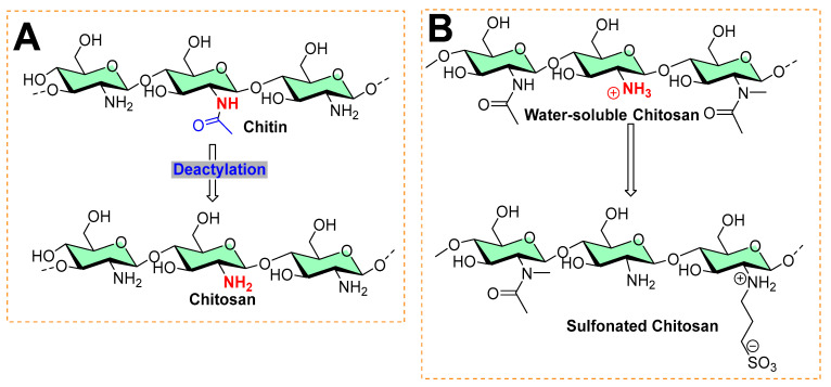
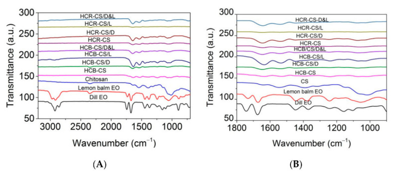
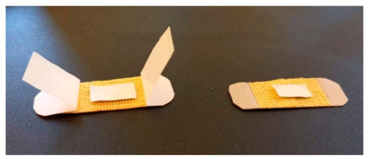
Chitosan is the most suitable encapsulation polymer because of its natural abundance, biodegradability, and surface functional groups in the form of free NH2 groups. The presence of NH2 groups allows for the facile grafting of functionalized molecules onto the chitosan surface, resulting in multifunctional materialistic applications. Quaternization of chitosan’s free amino is one of the typical chemical modifications commonly achieved under acidic conditions. This quaternization improves its ionic character, making it ready for ionic–ionic surface modification. Although the cationic nature of chitosan alone exhibits antibacterial activity because of its interaction with negatively-charged bacterial membranes, the nanoscale size of chitosan further amplifies its antibiofilm activity. Additionally, the researcher used chitosan nanoparticles as polymeric materials to encapsulate antibiofilm agents (such as antibiotics and natural phytochemicals), serving as an excellent strategy to combat biofilm-based secondary infections. This paper provided a summary of available carbohydrate-based biopolymers as antibiofilm materials. Furthermore, the paper focuses on chitosan nanoparticle-based encapsulation of basil essential oil (Ocimum basilicum), mandarin essential oil (Citrus reticulata), Carum copticum essential oil (“Ajwain”), dill plant seed essential oil (Anethum graveolens), peppermint oil (Mentha piperita), green tea oil (Camellia sinensis), cardamom essential oil, clove essential oil (Eugenia caryophyllata), cumin seed essential oil (Cuminum cyminum), lemongrass essential oil (Cymbopogon commutatus), summer savory essential oil (Satureja hortensis), thyme essential oil, cinnamomum essential oil (Cinnamomum zeylanicum), and nettle essential oil (Urtica dioica). Additionally, chitosan nanoparticles are used for the encapsulation of the major essential components carvacrol and cinnamaldehyde, the encapsulation of an oil-in-water nanoemulsion of eucalyptus oil (Eucalyptus globulus), the encapsulation of a mandarin essential oil nanoemulsion, and the electrospinning nanofiber of collagen hydrolysate–chitosan with lemon balm (Melissa officinalis) and dill (Anethum graveolens) essential oil.
Overview
The claims and statistics of this paper haven’t been checked yet.
Run the check — free, no account needed. It reads the open-access text and checks the sources it cites.
Integrity signals
Registry
Cites
Statistics
Figures
Verification
Claims the paper makes
The claims and statistics of this paper haven’t been checked yet.
Run the check — free, no account needed. It reads the open-access text and checks the sources it cites.
Claims other papers make about this one
Each is checked against this paper’s own text: does it say what the citing paper says it does?
In the citing paper
“Chitosan Nanoparticles serve as an excellent strategy for enhancing the biological activities of several natural compounds as well as the used antibiotics via encapsulating the anti-biofilm agents (Negi and Kesari, 2022).”
From Induction role of chitosan nanoparticles to Anethum graveolens extract against food-borne bacteria, oxidant, and diabetic activities in vitro (2023) · cites this paper as Negi and Kesari, 2022
Self-check
Percentages, sums and reported differences re-computed from the paper’s own numbers.
The self-check needs the paper’s full text.
Citations
Cites
Prokaryotes: The unseen majority
Whitman, W. B., Coleman, D. C., & Wiebe, W. J. (1998) · Proceedings of the National Academy of Sciences · doi:10.1073/pnas.95.12.6578 · Cited by 4,871 · cited 1×
“The biofilms comprise about 90% of microbial biomass of bacteria in the world [8], often producing secondary or nosocomial infections in patients with delineating diseases or histories of chronic disorders.”
BackgroundZakrzewska, A., Boorsma, A., Brul, S., Hellingwerf, K. J., & Klis, F. M. (2005) · Eukaryotic Cell · doi:10.1128/ec.4.4.703-715.2005 · Cited by 161
Jun, J.-Y., Jung, M.-J., Jeong, I.-H., Yamazaki, K., Kawai, Y., & Kim, B.-M. (2018) · Marine Drugs · doi:10.3390/md16090301 · Cited by 160
Research works printed but not linked: 166
- Manyi-Loh, C., Mamphweli, S., Meyer, E., and Okoh, A. (2018). Antibiotic use in agriculture and its consequential resistance in environmental sources: Potential public health implications. Molecules, 23. · DOI · Paper page
- Darouiche, 2004, Treatment of infections associated with surgical implants, N. Engl. J. Med., 350, 1422, 10.1056/NEJMra035415 · DOI
- Neoh, 2017, Surface modification strategies for combating catheter-related complications: Recent advances and challenges, J. Mater. Chem. B, 5, 2045, 10.1039/C6TB03280J · DOI
- Kimura, S.Y., and Nakamura, Y. (2020). Poor Quality Pharmaceuticals in Global Public Health, Springer. · DOI
- Khan, 2020, Is enhancing the professionalism of healthcare providers critical to tackling antimicrobial resistance in low-and middle-income countries?, Hum. Resour. Health, 18, 10, 10.1186/s12960-020-0452-7 · DOI
- Edwards, S.E., Morel, C.M., Busse, R., and Harbarth, S. (2018). Combatting antibiotic resistance together: How can we enlist the help of industry?. Antibiotics, 7. · DOI
- Morel, 2020, Industry incentives and antibiotic resistance: An introduction to the antibiotic susceptibility bonus, J. Antibiot., 73, 421, 10.1038/s41429-020-0300-y · DOI
- Alali, 2020, Community hospital stethoscope cleaning practices and contamination rates, Am. J. Infect. Control., 48, 1365, 10.1016/j.ajic.2020.04.019 · DOI
- Donlan, 2002, Biofilms: Microbial life on surfaces, Emerg. Infect. Dis., 8, 881, 10.3201/eid0809.020063 · DOI · Paper page
- Lima, 2021, Antimicrobial coatings based on chitosan to prevent implant-associated infections: A systematic review, Iscience, 24, 103480, 10.1016/j.isci.2021.103480 · DOI
- Heim, 2014, Myeloid-derived suppressor cells contribute to Staphylococcus aureus orthopedic biofilm infection, J. Immunol., 192, 3778, 10.4049/jimmunol.1303408 · DOI
- Sutherland, 2001, Biofilm exopolysaccharides: A strong and sticky framework, Microbiology, 147, 3, 10.1099/00221287-147-1-3 · DOI
- Pangestuti, 2011, Neuroprotective effects of marine algae, Mar. Drugs, 9, 803, 10.3390/md9050803 · DOI
- Wang, 2012, Surface properties of polyurethanes modified by bioactive polysaccharide-based polyelectrolyte multilayers, Colloids Surf. B Biointerfaces, 100, 77, 10.1016/j.colsurfb.2012.05.030 · DOI
- Vishwakarma, 2019, Evaluating the antibacterial and antibiofilm potential of sulphated polysaccharides extracted from green algae Chlamydomonas reinhardtii, J. Appl. Microbiol., 127, 1004, 10.1111/jam.14364 · DOI
- Fiorica, 2013, A new hyaluronic acid pH sensitive derivative obtained by ATRP for potential oral administration of proteins, Int. J. Pharm., 457, 150, 10.1016/j.ijpharm.2013.09.005 · DOI
- Palumbo, 2012, In situ forming hydrogels of new amino hyaluronic acid/benzoyl-cysteine derivatives as potential scaffolds for cartilage regeneration, Soft Matter, 8, 4918, 10.1039/c2sm07310b · DOI
- Palumbo, 2015, A polycarboxylic/amino functionalized hyaluronic acid derivative for the production of pH sensible hydrogels in the prevention of bacterial adhesion on biomedical surfaces, Int. J. Pharm., 478, 70, 10.1016/j.ijpharm.2014.11.015 · DOI
- Pallavicini, 2017, Silver nanoparticles synthesized and coated with pectin: An ideal compromise for anti-bacterial and anti-biofilm action combined with wound-healing properties, J. Colloid Interface Sci., 498, 271, 10.1016/j.jcis.2017.03.062 · DOI
- Bertoglio, F., de Vita, L., D’Agostino, A., Fernandez, Y.D., Falqui, A., Casu, A., Merli, D., Milanese, C., Rossi, S., and Taglietti, A. (2020). Increased antibacterial and antibiofilm properties of silver nanoparticles using silver fluoride as precursor. Molecules, 25. · DOI
- Khelissa, 2021, Microencapsulation of benzalkonium chloride enhanced its antibacterial and antibiofilm activities against Listeria monocytogenes and Escherichia coli, J. Appl. Microbiol., 131, 1136, 10.1111/jam.15010 · DOI
- Khelissa, 2021, Anti-biofilm activity of dodecyltrimethylammonium chloride microcapsules against Salmonella enterica serovar Enteritidis and Staphylococcus aureus, Biofouling, 37, 49, 10.1080/08927014.2021.1873958 · DOI
- Paramasivan, 2014, The use of chitosan–dextran gel shows anti-inflammatory, antibiofilm, and antiproliferative properties in fibroblast cell culture, Am. J. Rhinol. Allergy, 28, 361, 10.2500/ajra.2014.28.4069 · DOI
- Li, 2021, Acid-induced self-catalyzing platform based on dextran-coated copper peroxide nanoaggregates for biofilm treatment, ACS Appl. Mater. Interfaces, 13, 29269, 10.1021/acsami.1c03409 · DOI
- Hoque, 2017, Direct synthesis of dextran-based antibacterial hydrogels for extended release of biocides and eradication of topical biofilms, ACS Appl. Mater. Interfaces, 9, 15975, 10.1021/acsami.7b03208 · DOI
- Naha, 2019, Dextran-coated iron oxide nanoparticles as biomimetic catalysts for localized and pH-activated biofilm disruption, ACS Nano, 13, 4960, 10.1021/acsnano.8b08702 · DOI
- Goel, 2019, Microwave assisted κ-carrageenan capped silver nanocomposites for eradication of bacterial biofilms, Carbohydr. Polym., 206, 854, 10.1016/j.carbpol.2018.11.033 · DOI
- Hamblin, 2016, Chitin and chitosan: Production and application of versatile biomedical nanomaterials, Int. J. Adv. Res., 4, 411
- Periayah, 2016, Chitosan: A promising marine polysaccharide for biomedical research, Pharmacogn. Rev., 10, 39, 10.4103/0973-7847.176545 · DOI
- Raafat, 2009, Chitosan and its antimicrobial potential–a critical literature survey, Microb. Biotechnol., 2, 186, 10.1111/j.1751-7915.2008.00080.x · DOI
- Ray, S.S. (2013). Environmentally Friendly Polymer Nanocomposites: Types, Processing and Properties, Elsevier. · DOI
- Je, 2006, Chitosan derivatives killed bacteria by disrupting the outer and inner membrane, J. Agric. Food Chem., 54, 6629, 10.1021/jf061310p · DOI
- Helander, 2001, Chitosan disrupts the barrier properties of the outer membrane of Gram-negative bacteria, Int. J. Food Microbiol., 71, 235, 10.1016/S0168-1605(01)00609-2 · DOI
- Khan, 2020, Chitosan and their derivatives: Antibiofilm drugs against pathogenic bacteria, Colloids Surf. B Biointerfaces, 185, 110627, 10.1016/j.colsurfb.2019.110627 · DOI
- Costa, 2013, Study of the effects of chitosan upon Streptococcus mutans adherence and biofilm formation, Anaerobe, 20, 27, 10.1016/j.anaerobe.2013.02.002 · DOI
- Costa, 2017, Investigation of chitosan’s antibacterial activity against vancomycin resistant microorganisms and their biofilms, Carbohydr. Polym., 174, 369, 10.1016/j.carbpol.2017.06.087 · DOI
- Tan, 2016, Inhibition of mixed fungal and bacterial biofilms on silicone by carboxymethyl chitosan, Colloids Surf. B Biointerfaces, 148, 193, 10.1016/j.colsurfb.2016.08.061 · DOI
- Reighard, 2015, Disruption and eradication of P. aeruginosa biofilms using nitric oxide-releasing chitosan oligosaccharides, Biofouling, 31, 775, 10.1080/08927014.2015.1107548 · DOI · Paper page
- Peng, 2011, Quaternized chitosan inhibits icaA transcription and biofilm formation by Staphylococcus on a titanium surface, Antimicrob. Agents Chemother., 55, 860, 10.1128/AAC.01005-10 · DOI
- Jiang, 2014, Quaternized chitosans bind onto preexisting biofilms and eradicate pre-attached microorganisms, J. Mater. Chem. B, 2, 8518, 10.1039/C4TB01131G · DOI
- Sahariah, 2018, Quaternary ammoniumyl chitosan derivatives for eradication of staphylococcus aureus biofilms, Biomacromolecules, 19, 3649, 10.1021/acs.biomac.8b00718 · DOI
- Qin, 2020, Cationic chitosan derivatives as potential antifungals: A review of structural optimization and applications, Carbohydr. Polym., 236, 116002, 10.1016/j.carbpol.2020.116002 · DOI
- Carlson, 2008, Anti-biofilm properties of chitosan-coated surfaces, J. Biomater. Sci. Polym. Ed., 19, 1035, 10.1163/156856208784909372 · DOI
- Negi, 2018, Inverse docking based screening and identification of protein targets for Cassiarin alkaloids against Plasmodium falciparum, Saudi Pharm. J., 26, 546, 10.1016/j.jsps.2018.01.017 · DOI
- Benet, 2016, BDDCS, the Rule of 5 and drugability, Adv. Drug Deliv. Rev., 101, 89, 10.1016/j.addr.2016.05.007 · DOI
- Zhu, 2021, Encapsulation strategies to enhance the antibacterial properties of essential oils in food system, Food Control, 123, 107856, 10.1016/j.foodcont.2020.107856 · DOI
- Annunziata, 2011, Nanoencapsulation of essential oils to enhance their antimicrobial activity in foods, LWT Food Sci. Technol., 44, 1908, 10.1016/j.lwt.2011.03.003 · DOI
- Bazargani, 2016, Antibiofilm activity of essential oils and plant extracts against Staphylococcus aureus and Escherichia coli biofilms, Food Control, 61, 156, 10.1016/j.foodcont.2015.09.036 · DOI
- Saviuc, 2015, Essential oils with microbicidal and antibiofilm activity, Curr. Pharm. Biotechnol., 16, 137, 10.2174/138920101602150112151549 · DOI
- Liu, 2020, Fabrication and characterization of chitosan nanoemulsions loading thymol or thyme essential oil for the preservation of refrigerated pork, Int. J. Biol. Macromol., 162, 1509, 10.1016/j.ijbiomac.2020.07.207 · DOI
- Khan, 2019, Antibiofilm and antivirulence properties of chitosan-polypyrrole nanocomposites to Pseudomonas aeruginosa, Microb. Pathog., 128, 363, 10.1016/j.micpath.2019.01.033 · DOI
- Auger, 2006, Autoinducer 2 affects biofilm formation by Bacillus cereus, Appl. Environ. Microbiol., 72, 937, 10.1128/AEM.72.1.937-941.2006 · DOI
- Qayyum, 2016, Nanoparticles vs. biofilms: A battle against another paradigm of antibiotic resistance, MedChemComm, 7, 1479, 10.1039/C6MD00124F · DOI
- Grenha, 2012, Chitosan nanoparticles: A survey of preparation methods, J. Drug Target., 20, 291, 10.3109/1061186X.2011.654121 · DOI
- Erbacher, 1998, Chitosan-based vector/DNA complexes for gene delivery: Biophysical characteristics and transfection ability, Pharm. Res., 15, 1332, 10.1023/A:1011981000671 · DOI
- Hu, 2002, Synthesis and characterization of chitosan–poly (acrylic acid) nanoparticles, Biomaterials, 23, 3193, 10.1016/S0142-9612(02)00071-6 · DOI
- Du, 2004, Novel polyelectrolyte carboxymethyl konjac glucomannan–chitosan nanoparticles for drug delivery, Macromol. Rapid Commun., 25, 954, 10.1002/marc.200300314 · DOI
- Sarmento, 2006, Development and comparison of different nanoparticulate polyelectrolyte complexes as insulin carriers, Int. J. Pept. Res. Ther., 12, 131, 10.1007/s10989-005-9010-3 · DOI
- Sarmento, 2006, Development and characterization of new insulin containing polysaccharide nanoparticles, Colloids Surf. B Biointerfaces, 53, 193, 10.1016/j.colsurfb.2006.09.012 · DOI
- Lin, 2007, Preparation and characterization of nanoparticles shelled with chitosan for oral insulin delivery, Biomacromolecules, 8, 146, 10.1021/bm0607776 · DOI
- Bayat, 2008, Preparation and characterization of insulin nanoparticles using chitosan and its quaternized derivatives, Nanomed. Nanotechnol. Biol. Med., 4, 115, 10.1016/j.nano.2008.01.003 · DOI
- Avadi, 2010, Preparation and characterization of insulin nanoparticles using chitosan and Arabic gum with ionic gelation method, Nanomed. Nanotechnol. Biol. Med., 6, 58, 10.1016/j.nano.2009.04.007 · DOI
- Grenha, 2010, Development of new chitosan/carrageenan nanoparticles for drug delivery applications, J. Biomed. Mater. Res. Part A, 92, 1265, 10.1002/jbm.a.32466 · DOI
- Kaihara, 2011, In situ synthesis of polysaccharide nanoparticles via polyion complex of carboxymethyl cellulose and chitosan, Colloids Surf. B Biointerfaces, 85, 343, 10.1016/j.colsurfb.2011.03.008 · DOI
- Lin, 2008, Multi-ion-crosslinked nanoparticles with pH-responsive characteristics for oral delivery of protein drugs, J. Control. Release, 132, 141, 10.1016/j.jconrel.2008.08.020 · DOI
- Alonso, 2009, New generation of hybrid poly/oligosaccharide nanoparticles as carriers for the nasal delivery of macromolecules, Biomacromolecules, 10, 243, 10.1021/bm800975j · DOI
- Yeh, 2011, Novel protein-loaded chondroitin sulfate–chitosan nanoparticles: Preparation and characterization, Acta Biomater., 7, 3804, 10.1016/j.actbio.2011.06.026 · DOI
- Calvo, 1997, Novel hydrophilic chitosan-polyethylene oxide nanoparticles as protein carriers, J. Appl. Polym. Sci., 63, 125, 10.1002/(SICI)1097-4628(19970103)63:1<125::AID-APP13>3.0.CO;2-4 · DOI
- Alonso, 2001, Chitosan nanoparticles: A new vehicle for the improvement of the delivery of drugs to the ocular surface. Application to cyclosporin A, Int. J. Pharm., 224, 159, 10.1016/S0378-5173(01)00760-8 · DOI
- Ma, 2002, Formulation pH modulates the interaction of insulin with chitosan nanoparticles, J. Pharm. Sci., 91, 1396, 10.1002/jps.10149 · DOI
- Wang, 2008, Preparation of estradiol chitosan nanoparticles for improving nasal absorption and brain targeting, Eur. J. Pharm. Biopharm., 70, 735, 10.1016/j.ejpb.2008.07.005 · DOI
- Fan, 2012, Formation mechanism of monodisperse, low molecular weight chitosan nanoparticles by ionic gelation technique, Colloids Surf. B Biointerfaces, 90, 21, 10.1016/j.colsurfb.2011.09.042 · DOI
- Ohya, 1994, Release behavior of 5-fluorouracil from chitosan-gel nanospheres immobilizing 5-fluorouracil coated with polysaccharides and their cell specific cytotoxicity, J. Macromol. Sci. Pure Appl. Chem., 31, 629, 10.1080/10601329409349743 · DOI
- Songjiang, 2009, Amyloid-beta associated with chitosan nano-carrier has favorable immunogenicity and permeates the BBB, AAPS PharmSciTech, 10, 900, 10.1208/s12249-009-9279-1 · DOI
- Tokumitsu, 1999, Preparation of gadopentetic acid-loaded chitosan microparticles for gadolinium neutron-capture therapy of cancer by a novel emulsion-droplet coalescence technique, Chem. Pharm. Bull., 47, 838, 10.1248/cpb.47.838 · DOI
- Shering, 2011, Formulation of 5-fluorouracil loaded chitosan nanoparticles by emulsion droplet coalescence method for cancer therapy, Int. J. Pharm. Biol. Arch., 2, 926
- Atyabi, 2009, Thiolated chitosan nanoparticles as an oral delivery system for amikacin: In vitro and ex vivo evaluations, J. Nanosci. Nanotechnol., 9, 4593, 10.1166/jnn.2009.1090 · DOI
- Agnihotri, 2007, Chitosan nanoparticles for prolonged delivery of timolol maleate, Drug Dev. Ind. Pharm., 33, 1254, 10.1080/03639040701384942 · DOI
- Borges, 2005, Preparation of coated nanoparticles for a new mucosal vaccine delivery system, Int. J. Pharm., 299, 155, 10.1016/j.ijpharm.2005.04.037 · DOI
- Mao, 2001, Chitosan-DNA nanoparticles as gene carriers: Synthesis, characterization and transfection efficiency, J. Control. Release, 70, 399, 10.1016/S0168-3659(00)00361-8 · DOI
- 2002, Positively charged nanoparticles for improving the oral bioavailability of cyclosporin-A, Int. J. Pharm., 249, 101, 10.1016/S0378-5173(02)00461-1 · DOI · Paper page
- Mitra, 2001, Tumour targeted delivery of encapsulated dextran–doxorubicin conjugate using chitosan nanoparticles as carrier, J. Control. Release, 74, 317, 10.1016/S0168-3659(01)00342-X · DOI
- Banerjee, 2002, Preparation, characterization and biodistribution of ultrafine chitosan nanoparticles, Int. J. Pharm., 243, 93, 10.1016/S0378-5173(02)00267-3 · DOI
- Tang, 2007, Preparation of chitosan nanoparticles as carrier for immobilized enzyme, Appl. Biochem. Biotechnol., 136, 77, 10.1007/BF02685940 · DOI
- Manchanda, 2010, Controlled size chitosan nanoparticles as an efficient, biocompatible oligonucleotides delivery system, J. Appl. Polym. Sci., 118, 2071, 10.1002/app.32508 · DOI
- Sajeesh, 2006, Cyclodextrin–insulin complex encapsulated polymethacrylic acid based nanoparticles for oral insulin delivery, Int. J. Pharm., 325, 147, 10.1016/j.ijpharm.2006.06.019 · DOI
- Sajeesh, 2006, Novel pH responsive polymethacrylic acid–chitosan–polyethylene glycol nanoparticles for oral peptide delivery, J. Biomed. Mater. Res. B Appl. Biomater., 76, 298, 10.1002/jbm.b.30372 · DOI
- Cai, 2022, Antibacterial and antibiofilm activities of chitosan nanoparticles loaded with Ocimum basilicum L. essential oil, Int. J. Biol. Macromol., 202, 122, 10.1016/j.ijbiomac.2022.01.066 · DOI
- Hosseini, 2013, Two-step method for encapsulation of oregano essential oil in chitosan nanoparticles: Preparation, characterization and in vitro release study, Carbohydr. Polym., 95, 50, 10.1016/j.carbpol.2013.02.031 · DOI
- Zhou, 2019, N-Alkyl-1, 5-dideoxy-1, 5-imino-l-fucitols as fucosidase inhibitors: Synthesis, molecular modelling and activity against cancer cell lines, Bioorg. Chem., 84, 418, 10.1016/j.bioorg.2018.12.003 · DOI
- Song, 2021, Mandarin (Citrus reticulata L.) essential oil incorporated into chitosan nanoparticles: Characterization, anti-biofilm properties and application in pork preservation, Int. J. Biol. Macromol., 185, 620, 10.1016/j.ijbiomac.2021.06.195 · DOI
- Malaki, 2019, Ultrasonic nano-emulsification—A review, Ultrason. Sonochem., 52, 88, 10.1016/j.ultsonch.2018.11.005 · DOI
- Gul, 2018, Effect of ultrasound treatment on the properties of nano-emulsion films obtained from hazelnut meal protein and clove essential oil, Ultrason. Sonochem., 41, 466, 10.1016/j.ultsonch.2017.10.011 · DOI
- 2015, Physicochemical characterization and antimicrobial activity of food-grade emulsions and nanoemulsions incorporating essential oils, Food Hydrocoll., 43, 547, 10.1016/j.foodhyd.2014.07.012 · DOI
- Severino, 2014, Antibacterial and physical effects of modified chitosan based-coating containing nanoemulsion of mandarin essential oil and three non-thermal treatments against Listeria innocua in green beans, Int. J. Food Microbiol., 191, 82, 10.1016/j.ijfoodmicro.2014.09.007 · DOI
- Esmaeili, 2015, In vitro release and biological activities of Carum copticum essential oil (CEO) loaded chitosan nanoparticles, Int. J. Biol. Macromol., 81, 283, 10.1016/j.ijbiomac.2015.08.010 · DOI
- Dudhani, 2010, Bioadhesive chitosan nanoparticles: Preparation and characterization, Carbohydr. Polym., 81, 243, 10.1016/j.carbpol.2010.02.026 · DOI
- Bagre, 2013, Alginate coated chitosan core shell nanoparticles for oral delivery of enoxaparin: In vitro and in vivo assessment, Int. J. Pharm., 456, 31, 10.1016/j.ijpharm.2013.08.037 · DOI
- Esmaeili, 2015, Optimization and preparation of nanocapsules for food applications using two methodologies, Food Chem., 179, 26, 10.1016/j.foodchem.2015.01.115 · DOI
- Hosseini, 2013, Incorporation of essential oil in alginate microparticles by multiple emulsion/ionic gelation process, Int. J. Biol. Macromol., 62, 582, 10.1016/j.ijbiomac.2013.09.054 · DOI
- Keawchaoon, 2011, Preparation, characterization and in vitro release study of carvacrol-loaded chitosan nanoparticles, Colloids Surf. B Biointerfaces, 84, 163, 10.1016/j.colsurfb.2010.12.031 · DOI
- Yoksan, 2010, Encapsulation of ascorbyl palmitate in chitosan nanoparticles by oil-in-water emulsion and ionic gelation processes, Colloids Surf. B Biointerfaces, 76, 292, 10.1016/j.colsurfb.2009.11.007 · DOI
- Yang, 2009, The effect of the molecular weight of chitosan nanoparticles and its application on drug delivery, Microchem. J., 92, 87, 10.1016/j.microc.2009.02.001 · DOI
- Anitha, 2011, Preparation, characterization, in vitro drug release and biological studies of curcumin loaded dextran sulphate–chitosan nanoparticles, Carbohydr. Polym., 84, 1158, 10.1016/j.carbpol.2011.01.005 · DOI
- Paul, 2011, Trachyspermum ammi (L.) fruit essential oil influencing on membrane permeability and surface characteristics in inhibiting food-borne pathogens, Food Control, 22, 725, 10.1016/j.foodcont.2010.11.003 · DOI · Paper page
- Liolios, 2009, Liposomal incorporation of carvacrol and thymol isolated from the essential oil of Origanum dictamnus L. and in vitro antimicrobial activity, Food Chem., 112, 77, 10.1016/j.foodchem.2008.05.060 · DOI
- He, 2013, Analysis of protein composition and antioxidant activity of hydrolysates from Paphia undulate, J. Food Nutr. Res., 1, 30
- Sendra, 2013, In vitro antibacterial and antioxidant properties of chitosan edible films incorporated with Thymus moroderi or Thymus piperella essential oils, Food Control, 30, 386, 10.1016/j.foodcont.2012.07.052 · DOI
- Subhaswaraj, 2018, Anti quorum sensing and anti biofilm efficacy of cinnamaldehyde encapsulated chitosan nanoparticles against Pseudomonas aeruginosa PAO1, LWT, 97, 752, 10.1016/j.lwt.2018.08.011 · DOI
- Das, 2021, Anethum graveolens essential oil encapsulation in chitosan nanomatrix: Investigations on in vitro release behavior, organoleptic attributes, and efficacy as potential delivery vehicles against biodeterioration of rice (Oryza sativa L.), Food Bioprocess Technol., 14, 831, 10.1007/s11947-021-02589-z · DOI
- Râpă, M., Gaidau, C., Mititelu-Tartau, L., Berechet, M.-D., Berbecaru, A.C., Rosca, I., Chiriac, A.P., Matei, E., Predescu, A.-M., and Predescu, C. (2021). Bioactive collagen hydrolysate-chitosan/essential oil electrospun nanofibers designed for medical wound dressings. Pharmaceutics, 13. · DOI
- Hua, Y., Ma, C., Wei, T., Zhang, L., and Shen, J. (2020). Collagen/chitosan complexes: Preparation, antioxidant activity, tyrosinase inhibition activity, and melanin synthesis. Int. J. Mol. Sci., 21. · DOI
- Ridel, 2016, Pickering emulsions stabilized by charged nanoparticles, Soft Matter, 12, 7564, 10.1039/C6SM01465H · DOI
- Wei, 2012, Chitosan nanoparticles as particular emulsifier for preparation of novel pH-responsive Pickering emulsions and PLGA microcapsules, Polymer, 53, 1229, 10.1016/j.polymer.2012.02.015 · DOI
- Alison, 2016, Pickering and network stabilization of biocompatible emulsions using chitosan-modified silica nanoparticles, Langmuir, 32, 13446, 10.1021/acs.langmuir.6b03439 · DOI
- Schober, 2021, Effect of Particle Wettability and Particle Concentration on the Enzymatic Dehydration of n-Octanaloxime in Pickering Emulsions, Angew. Chem., 133, 1470, 10.1002/ange.202013171 · DOI
- Nordstierna, 2017, Hydrophilic and hydrophobic modifications of colloidal silica particles for Pickering emulsions, J. Colloid Interface Sci., 487, 250, 10.1016/j.jcis.2016.10.031 · DOI
- Lai, 2021, Fabrication and antibacterial evaluation of peppermint oil-loaded composite microcapsules by chitosan-decorated silica nanoparticles stabilized Pickering emulsion templating, Int. J. Biol. Macromol., 183, 2314, 10.1016/j.ijbiomac.2021.05.198 · DOI
- Liang, 2012, Synthesis and characterization of core–shell structured SiO2@ YVO4: Yb3+, Er3+ microspheres, Appl. Surf. Sci., 258, 3689, 10.1016/j.apsusc.2011.12.006 · DOI
- Lim, 2004, Synthesis and antimicrobial activity of a water-soluble chitosan derivative with a fiber-reactive group, Carbohydr. Res., 339, 313, 10.1016/j.carres.2003.10.024 · DOI
- Yasmeen, 2016, Chromium (VI) ions removal from tannery effluent using chitosan-microcrystalline cellulose composite as adsorbent, Int. Res. J. Pure Appl. Chem., 10, 1, 10.9734/IRJPAC/2016/23315 · DOI
- Lu, 2004, An imprinted organic− inorganic hybrid sorbent for selective separation of cadmium from aqueous solution, Anal. Chem., 76, 453, 10.1021/ac0347718 · DOI
- Zhao, 2018, Eco-friendly Pickering emulsion stabilized by silica nanoparticles dispersed with high-molecular-weight amphiphilic alginate derivatives, ACS Sustain. Chem. Eng., 6, 4105, 10.1021/acssuschemeng.7b04508 · DOI
- Kang, 2017, Dispersion of surface-modified silica nanoparticles in polyamide-imide (PAI) films for enhanced mechanical and thermal properties, J. Ind. Eng. Chem., 46, 289, 10.1016/j.jiec.2016.10.042 · DOI
- Silva, 2009, A new organofunctionalized silica containing thioglycolic acid incorporated for divalent cations removal—A thermodyamic cation/basic center interaction, Colloids Surf. A Physicochem. Eng. Asp., 332, 144, 10.1016/j.colsurfa.2008.09.010 · DOI
- Kumar, 2012, Physiochemical, optical and biological activity of chitosan-chromone derivative for biomedical applications, Int. J. Mol. Sci., 13, 6102, 10.3390/ijms13056102 · DOI
- Shetta, 2019, Comparative study of encapsulated peppermint and green tea essential oils in chitosan nanoparticles: Encapsulation, thermal stability, in-vitro release, antioxidant and antibacterial activities, Int. J. Biol. Macromol., 126, 731, 10.1016/j.ijbiomac.2018.12.161 · DOI
- Nallamuthu, 2015, Chlorogenic acid loaded chitosan nanoparticles with sustained release property, retained antioxidant activity and enhanced bioavailability, Asian J. Pharm. Sci., 10, 203, 10.1016/j.ajps.2014.09.005 · DOI
- Ruppert, 2011, Biophysical investigation of pulmonary surfactant surface properties upon contact with polymeric nanoparticles in vitro, Nanomed. Nanotechnol. Biol. Med., 7, 341, 10.1016/j.nano.2010.10.007 · DOI
- Wissing, 2004, Solid lipid nanoparticles for parenteral drug delivery, Adv. Drug Deliv. Rev., 56, 1257, 10.1016/j.addr.2003.12.002 · DOI
- Jamil, 2016, Encapsulation of cardamom essential oil in chitosan nano-composites: In-vitro efficacy on antibiotic-resistant bacterial pathogens and cytotoxicity studies, Front. Microbiol., 7, 1580, 10.3389/fmicb.2016.01580 · DOI
- Chaieb, 2007, The chemical composition and biological activity of clove essential oil, Eugenia caryophyllata (Syzigium aromaticum L. Myrtaceae): A short review, Phytother. Res., 21, 501, 10.1002/ptr.2124 · DOI
- Sebaaly, 2015, Preparation and characterization of clove essential oil-loaded liposomes, Food Chem., 178, 52, 10.1016/j.foodchem.2015.01.067 · DOI
- Hadidi, 2020, Chitosan nanoparticles loaded with clove essential oil: Characterization, antioxidant and antibacterial activities, Carbohydr. Polym., 236, 116075, 10.1016/j.carbpol.2020.116075 · DOI
- Karimirad, 2019, Application of chitosan nanoparticles containing Cuminum cyminum oil as a delivery system for shelf life extension of Agaricus bisporus, LWT, 106, 218, 10.1016/j.lwt.2019.02.062 · DOI
- Chalier, 2007, Carvacrol losses from soy protein coated papers as a function of drying conditions, J. Appl. Polym. Sci., 106, 611, 10.1002/app.26662 · DOI
- Stammati, 2004, Constituents of aromatic plants: Carvacrol, Fitoterapia, 75, 801, 10.1016/j.fitote.2004.05.002 · DOI
- Bhumkar, 2006, Studies on effect of pH on cross-linking of chitosan with sodium tripolyphosphate: A technical note, AAPS PharmSciTech, 7, E138, 10.1208/pt070250 · DOI
- Mi, 2003, Synthesis and characterization of biodegradable TPP/genipin co-crosslinked chitosan gel beads, Polymer, 44, 6521, 10.1016/S0032-3861(03)00620-7 · DOI
- Bernal-Mercado, A.T., Juarez, J., Valdez, M.A., Ayala-Zavala, J.F., Del-Toro-Sánchez, C.L., and Encinas-Basurto, D. (2022). Hydrophobic Chitosan Nanoparticles Loaded with Carvacrol against Pseudomonas aeruginosa Biofilms. Molecules, 27. · DOI
- Soltanzadeh, 2021, Chitosan nanoparticles encapsulating lemongrass (Cymbopogon commutatus) essential oil: Physicochemical, structural, antimicrobial and in-vitro release properties, Int. J. Biol. Macromol., 192, 1084, 10.1016/j.ijbiomac.2021.10.070 · DOI
- Feyzioglu, 2016, Development of chitosan nanoparticles loaded with summer savory (Satureja hortensis L.) essential oil for antimicrobial and antioxidant delivery applications, LWT, 70, 104, 10.1016/j.lwt.2016.02.037 · DOI
- 2017, Release study and inhibitory activity of thyme essential oil-loaded chitosan nanoparticles and nanocapsules against foodborne bacteria, Int. J. Biol. Macromol., 103, 409, 10.1016/j.ijbiomac.2017.05.063 · DOI
- Wu, 2012, Antioxidant and antimicrobial properties of essential oils encapsulated in zein nanoparticles prepared by liquid–liquid dispersion method, LWT Food Sci. Technol., 48, 283, 10.1016/j.lwt.2012.03.027 · DOI
- Mohammadi, 2015, Chitosan nanoparticles loaded with Cinnamomum zeylanicum essential oil enhance the shelf life of cucumber during cold storage, Postharvest Biol. Technol., 110, 203, 10.1016/j.postharvbio.2015.08.019 · DOI
- Sugumar, 2015, Eucalyptus oil nanoemulsion-impregnated chitosan film: Antibacterial effects against a clinical pathogen, Staphylococcus aureus, in vitro, Int. J. Nanomed., 10, 67
- Demirci, 2012, Chemical composition and in vitro cytotoxic, genotoxic effects of essential oil from Urtica dioica L., Bull. Environ. Contam. Toxicol., 88, 666, 10.1007/s00128-012-0535-9 · DOI
- Bagheri, 2021, Characterization, antioxidant and antibacterial activities of chitosan nanoparticles loaded with nettle essential oil, J. Food Meas. Charact., 15, 1395, 10.1007/s11694-020-00738-0 · DOI · Paper page
- Rashki, 2021, Chitosan-based nanoparticles against bacterial infections, Carbohydr. Polym., 251, 117108, 10.1016/j.carbpol.2020.117108 · DOI
- Suri, 2007, Nanotechnology-based drug delivery systems, J. Occup. Med. Toxicol., 2, 16, 10.1186/1745-6673-2-16 · DOI
- Franco, 2021, Selectivity Control in the Oxidative Ring-Opening of Dimethylfuran Mediated by Zeolitic-Imidazolate Framework-8 Nanoparticles, ACS Sustain. Chem. Eng., 9, 8090, 10.1021/acssuschemeng.1c00708 · DOI
- Askari, 2016, Rheological and thermophysical properties of ultra-stable kerosene-based Fe3O4/Graphene nanofluids for energy conservation, Energy Convers. Manag., 128, 134, 10.1016/j.enconman.2016.09.037 · DOI
- Raut, 2014, A status review on the medicinal properties of essential oils, Ind. Crops Prod., 62, 250, 10.1016/j.indcrop.2014.05.055 · DOI
- Knobloch, 1989, Antibacterial and antifungal properties of essential oil components, J. Essent. Oil Res., 1, 119, 10.1080/10412905.1989.9697767 · DOI · Paper page
- Katouzian, 2016, Nano-encapsulation as a promising approach for targeted delivery and controlled release of vitamins, Trends Food Sci. Technol., 53, 34, 10.1016/j.tifs.2016.05.002 · DOI
- Negi, 2021, Development of Mcl-1 inhibitors for cancer therapy, Eur. J. Med. Chem., 210, 113038, 10.1016/j.ejmech.2020.113038 · DOI
- Negi, 2022, Strategies to Reduce the On-Target Platelet Toxicity of Bcl-xL Inhibitors: PROTACs, SNIPERs and Prodrug-Based Approaches, ChemBioChem, 23, e202100689, 10.1002/cbic.202100689 · DOI
- Macartney, 2011, Encapsulation of drug molecules by cucurbiturils: Effects on their chemical properties in aqueous solution, Isr. J. Chem., 51, 600, 10.1002/ijch.201100040 · DOI
- Miller, 2017, Nano-palladium is a cellular catalyst for in vivo chemistry, Nat. Commun., 8, 15906, 10.1038/ncomms15906 · DOI
- Negi, 2015, Imine/amide–imidazole conjugates derived from 5-amino-4-cyano-N1-substituted benzyl imidazole: Microwave-assisted synthesis and anticancer activity via selective topoisomerase-II-α inhibition, Bioorg. Med. Chem., 23, 5654, 10.1016/j.bmc.2015.07.020 · DOI
- Shaabani, 2016, Copper (ii) supported on magnetic chitosan: A green nanocatalyst for the synthesis of 2, 4, 6-triaryl pyridines by C–N bond cleavage of benzylamines, RSC Adv., 6, 27706, 10.1039/C6RA00102E · DOI
- Negi, A., Mirallai, S.I., Konda, S., and Murphy, P.V. (2022). An improved method for synthesis of non-symmetric triarylpyridines. Tetrahedron, 132930. · DOI
- Rahmatpour, 2022, Noncross-linked polystyrene nanoencapsulation of ferric chloride: A novel and reusable heterogeneous macromolecular Lewis acid catalyst toward selective acetylation of alcohols, phenols, amines, and thiols, J. Organomet. Chem., 961, 122264, 10.1016/j.jorganchem.2022.122264 · DOI
- Negi, 2022, Azobenzene Photoswitches in Proteolysis Targeting Chimeras: Photochemical Control Strategies and Therapeutic Benefits, ChemistrySelect, 7, e202200981, 10.1002/slct.202200981 · DOI
- Dong, 2015, Red-shifting azobenzene photoswitches for in vivo use, Acc. Chem. Res., 48, 2662, 10.1021/acs.accounts.5b00270 · DOI
- Monticelli, 2018, Recent advances in the synthesis and reactivity of spiro-epoxyoxindoles, Chem. Heterocycl. Compd., 54, 389, 10.1007/s10593-018-2280-4 · DOI
Cited by
Mashraqi, A. (2023) · Frontiers in Microbiology · doi:10.3389/fmicb.2023.1209524 · Cited by 16
Metadata
- Authors
- Negi, A., & Kesari, K. K.
- Affiliations
- Department of Bioproduct and Biosystems, School of Chemical Engineering, Aalto University, 02150 Espoo, Finland
- Venue
- Micromachines
- Publisher
- MDPI AG
- Published
- 2022-08-06
- Volume
- 13
- Issue
- 8
- Pages
- 1265
- ISSN
- 2072-666X
- PMC
- PMC9415589
- Cited by
- 122
- References
- 3
- Language
- en
About these checks
Registry, retraction, Cites and statistics results are computed, not judged, with one exception: a printed reference that no index lists is read once by a model before it counts as a citation error. Claims are extracted by a model and kept only when their quote appears verbatim in the text; each cited source is then checked, with a model’s help, for whether it says what the claim relies on. Those verdicts can be wrong — read the claim’s quote and the cited source before relying on one.
Statistics and arithmetic are recomputed, not judged.
Confidence bands are shown only for fields calibrated on our annotated test set. None is calibrated yet, so no band is shown.
What counts as a claim ›How we measure confidence ›How we check this ›
Are you an author, or do you see a problem with this page? Request a review
Data sources
Bibliographic data: Semantic Scholar, OpenAlex, Crossref.
Contains information from the Semantic Scholar Academic Graph Datasets, made available under the ODC Attribution License (ODC-BY 1.0).
Semantic Scholar’s data is described in: Kinney, R., et al. (2023). The Semantic Scholar Open Data Platform. arXiv:2301.10140.
