

The DOI matches its Crossref record; 4 of the 4 references it cites were found in a registry or an index.
MiR-219a-5p Enriched Extracellular Vesicles Induce OPC Differentiation and EAE Improvement More Efficiently Than Liposomes and Polymeric Nanoparticles
Abstract
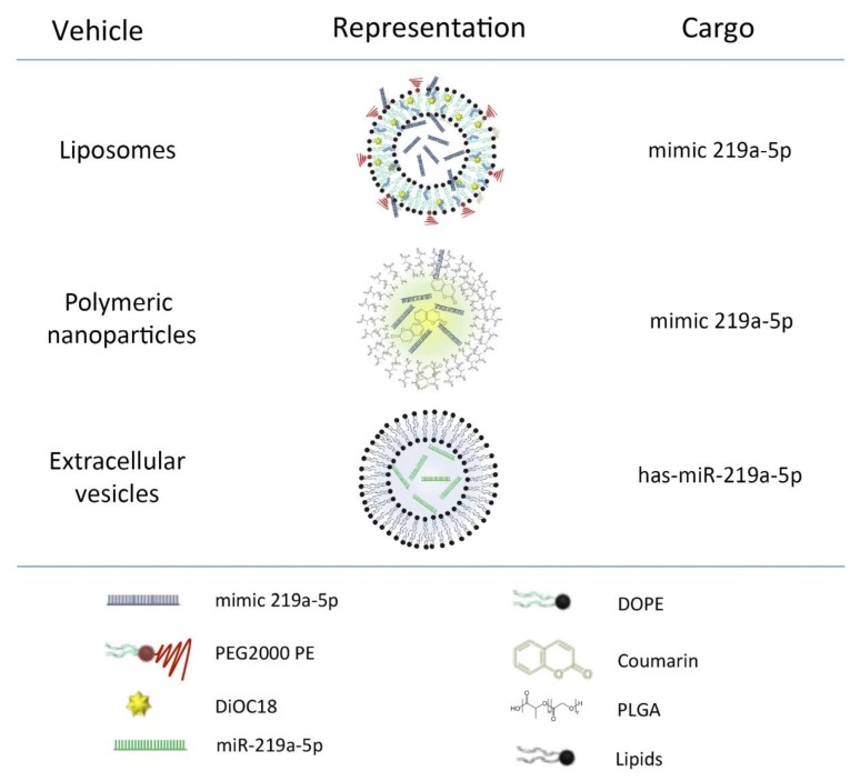
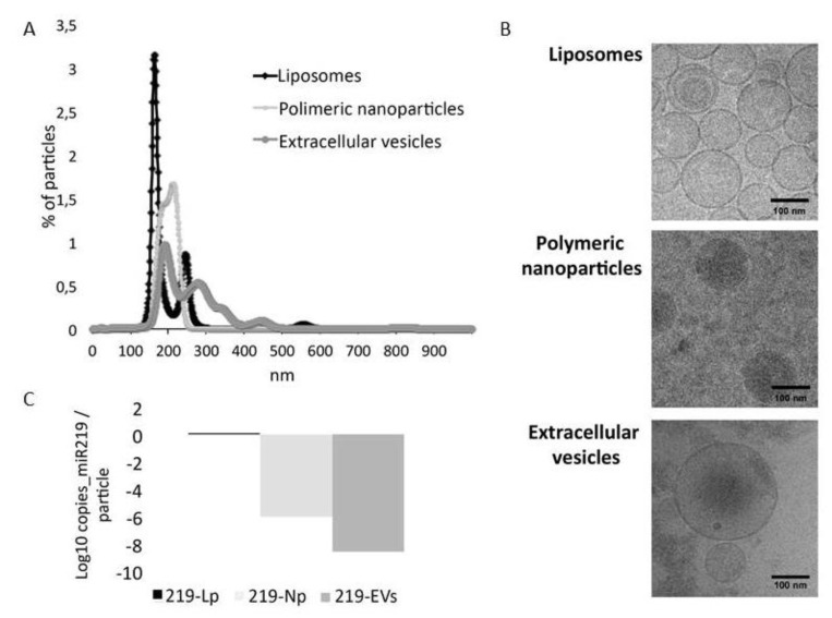
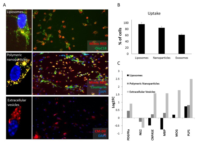
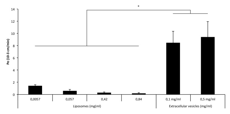
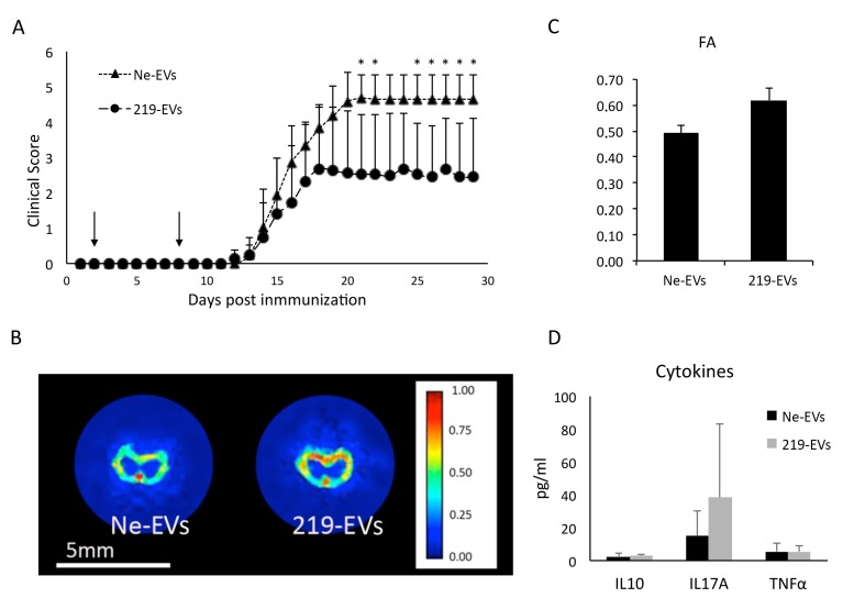
Remyelination is a key aspect in multiple sclerosis pathology and a special effort is being made to promote it. However, there is still no available treatment to regenerate myelin and several strategies are being scrutinized. Myelination is naturally performed by oligodendrocytes and microRNAs have been postulated as a promising tool to induce oligodendrocyte precursor cell differentiation and therefore remyelination. Herein, DSPC liposomes and PLGA nanoparticles were studied for miR-219a-5p encapsulation, release and remyelination promotion. In parallel, they were compared with biologically engineered extracellular vesicles overexpressing miR-219a-5p. Interestingly, extracellular vesicles showed the highest oligodendrocyte precursor cell differentiation levels and were more effective than liposomes and polymeric nanoparticles crossing the blood–brain barrier. Finally, extracellular vesicles were able to improve EAE animal model clinical evolution. Our results indicate that the use of extracellular vesicles as miR-219a-5p delivery system can be a feasible and promising strategy to induce remyelination in multiple sclerosis patients.
Overview
The claims and statistics of this paper haven’t been checked yet.
Run the check — free, no account needed. It reads the open-access text and checks the sources it cites.
Integrity signals
Registry
Cites
Statistics
Figures
Verification
Claims the paper makes
The claims and statistics of this paper haven’t been checked yet.
Run the check — free, no account needed. It reads the open-access text and checks the sources it cites.
Claims other papers make about this one
Each is checked against this paper’s own text: does it say what the citing paper says it does?
In the citing paper
“There are various delivery systems for miRNAs, such as poly (lactic-co-glycolic acid) (PLGA) nanoparticles, extracellular vesicles (EVs), and liposomes. These delivery systems are noninvasive and efficient and can protect miRNA from degradation and promote its uptake by cells of the CNS, most likely oligodendrocytes (Osorio-Querejeta et al. 2020).”
From MiR-124 and MiR-155 as Therapeutic Targets in Microglia-Mediated Inflammation in Multiple Sclerosis (2025) · cites this paper as Osorio-Querejeta et al. 2020
Self-check
Percentages, sums and reported differences re-computed from the paper’s own numbers.
The self-check needs the paper’s full text.
Citations
Cites
Regenerating CNS myelin — from mechanisms to experimental medicines
Franklin, R. J. M., & ffrench-Constant, C. (2017) · Nature Reviews Neuroscience · doi:10.1038/nrn.2017.136 · Cited by 647
miR-219 Cooperates with miR-338 in Myelination and Promotes Myelin Repair in the CNS
Wang, H., Moyano, A. L., Ma, Z., Deng, Y., Lin, Y., Zhao, C., Zhang, L., Jiang, M., He, X., Ma, Z., et al. (2017) · Developmental Cell · doi:10.1016/j.devcel.2017.03.001 · Cited by 174
Regulation of miRNA 219 and miRNA Clusters 338 and 17-92 in Oligodendrocytes
de Faria Jr., O., Cui, Q.-L., Bin, J. M., Bull, S.-J., Kennedy, T. E., Bar-Or, A., Antel, J. P., Colman, D. R., & Dhaunchak, A. S. (2012) · Frontiers in Genetics · doi:10.3389/fgene.2012.00046 · Cited by 54
Nazari, B., Soleimani, M., Ebrahimi-Barough, S., Enderami, S. E., Kazemi, M., Negahdari, B., Sadroddiny, E., & Ai, J. (2018) · Journal of Chemical Neuroanatomy · doi:10.1016/j.jchemneu.2018.03.001 · Cited by 24
Research works printed but not linked: 27
- Agulla, 2014, In vivo theranostics at the peri-infarct region in cerebral ischemia, Theranostics, 4, 90, 10.7150/thno.7088 · DOI
- Alberro, A., Sáenz-Cuesta, M., Muñoz-Culla, M., Mateo-Abad, M., Gonzalez, E., Carrasco-Garcia, E., Araúzo-Bravo, M.J., Matheu, A., Vergara, I., and Otaegui, D. (2016). Inflammaging and frailty status do not result in an increased extracellular vesicle concentration in circulation. Int. J. Mol. Sci., 17. · DOI
- Buyens, 2009, Elucidating the encapsulation of short interfering RNA in PEGylated cationic liposomes, Langmuir, 25, 4886, 10.1021/la803973p · DOI
- Dugas, 2010, Dicer1 and miR-219 Are Required for Normal Oligodendrocyte Differentiation and Myelination, Neuron, 65, 597, 10.1016/j.neuron.2010.01.027 · DOI · Paper page
- Dulamea, 2017, The contribution of oligodendrocytes and oligodendrocyte progenitor cells to central nervous system repair in multiple sclerosis: Perspectives for remyelination therapeutic strategies, Neural Regen. Res., 12, 1939, 10.4103/1673-5374.221146 · DOI
- Fan, 2017, Transplanted miR-219-overexpressing oligodendrocyte precursor cells promoted remyelination and improved functional recovery in a chronic demyelinated model, Sci. Rep., 7, 41407, 10.1038/srep41407 · DOI
- Fletcher, 2010, T cells in multiple sclerosis and experimental autoimmune encephalomyelitis, Clin. Exp. Immunol., 162, 1, 10.1111/j.1365-2249.2010.04143.x · DOI
- Franklin, 2002, Why does remyelination fail in multiple sclerosis?, Nat. Rev. Neurosci., 3, 705, 10.1038/nrn917 · DOI
- Goldman, 2014, So many progenitors, so little myelin, Nat. Neurosci., 17, 483, 10.1038/nn.3685 · DOI
- Julián, P., Milon, P., Agirrezabala, X., Lasso, G., Gil, D., Rodnina, M.V., and Valle, M. (2011). The Cryo-EM Structure of a Complete 30S Translation Initiation Complex from Escherichia coli. PLoS Biol., 9. · DOI
- Kurte, 2015, Intravenous Administration of Bone Marrow-Derived Mesenchymal Stem Cells Induces a Switch from Classical to Atypical Symptoms in Experimental Autoimmune Encephalomyelitis, Stem Cells Int., 2015, 140170, 10.1155/2015/140170 · DOI
- Liu, 2017, miR-219 attenuates demyelination in cuprizone-induced demyelinated mice by regulating monocarboxylate transporter 1, Eur. J. Neurosci., 45, 249, 10.1111/ejn.13485 · DOI
- Livak, 2001, Analysis of relative gene expression data using real-time quantitative PCR and the 2(-Delta Delta C(T)) Method, Methods, 25, 402, 10.1006/meth.2001.1262 · DOI · Paper page
- Lunavat, 2016, RNAi delivery by exosome-mimetic nanovesicles—Implications for targeting c-Myc in cancer, Biomaterials, 102, 231, 10.1016/j.biomaterials.2016.06.024 · DOI
- Medina-Rodríguez, E.M., Arenzana, F.J., Bribián, A., and de Castro, F. (2013). Protocol to isolate a large amount of functional oligodendrocyte precursor cells from the cerebral cortex of adult mice and humans. PLoS ONE, 8. · DOI
- Melo, 2014, Cancer Exosomes Perform Cell-Independent MicroRNA Biogenesis and Promote Tumorigenesis, Cancer Cell, 26, 707, 10.1016/j.ccell.2014.09.005 · DOI
- Fuentes, 2015, White matter injury restoration after stem cell administration in subcortical ischemic stroke, Sci. Rep., 6, 44433
- Pusic, 2014, Youth and environmental enrichment generate serum exosomes containing miR-219 that promote CNS myelination, Glia, 62, 284, 10.1002/glia.22606 · DOI
- Raposo, 2013, Extracellular vesicles: Exosomes, microvesicles, and friends, J. Cell Biol., 200, 373, 10.1083/jcb.201211138 · DOI · Paper page
- Sork, 2018, Heterogeneity and interplay of the extracellular vesicle small RNA transcriptome and proteome, Sci. Rep., 8, 10813, 10.1038/s41598-018-28485-9 · DOI · Paper page
- Boussac, 2001, Proteomic Analysis of Dendritic Cell-Derived Exosomes: A Secreted Subcellular Compartment Distinct from Apoptotic Vesicles, J. Immunol., 166, 7309, 10.4049/jimmunol.166.12.7309 · DOI
- Tian, 2014, A doxorubicin delivery platform using engineered natural membrane vesicle exosomes for targeted tumor therapy, Biomaterials, 35, 2383, 10.1016/j.biomaterials.2013.11.083 · DOI
- Valadi, 2007, Exosome-mediated transfer of mRNAs and microRNAs is a novel mechanism of genetic exchange between cells, Nat. Cell Biol., 9, 654, 10.1038/ncb1596 · DOI · Paper page
- Ventola, 2017, Progress in Nanomedicine: Approved and Investigational Nanodrugs, Pharm. Ther., 42, 742
- Willms, 2018, Extracellular Vesicle Heterogeneity: Subpopulations, Isolation Techniques, and Diverse Functions in Cancer Progression, Front. Immunol., 9, 738, 10.3389/fimmu.2018.00738 · DOI
- Yu, X., Odenthal, M., and Fries, J.W.U. (2016). Exosomes as miRNA carriers: Formation-function-future. Int. J. Mol. Sci., 17. · DOI
- Zhuang, 2011, Treatment of brain inflammatory diseases by delivering exosome encapsulated anti-inflammatory drugs from the nasal region to the brain, Mol. Ther., 19, 1769, 10.1038/mt.2011.164 · DOI · Paper page
Cited by
Sorted by citations
Extracellular Vesicles as an Emerging Frontier in Spinal Cord Injury Pathobiology and Therapy
Dutta, D., Khan, N., Wu, J., & Jay, S. M. (2021) · Trends in Neurosciences · doi:10.1016/j.tins.2021.01.003 · Cited by 112
Chivero, E. T., Liao, K., Niu, F., Tripathi, A., Tian, C., Buch, S., & Hu, G. (2020) · Frontiers in Cell and Developmental Biology · doi:10.3389/fcell.2020.00573 · Cited by 69
Exosomes and Biomaterials: In Search of a New Therapeutic Strategy for Multiple Sclerosis
Ojeda-Hernández, D. D., Hernández-Sapiéns, M. A., Reza-Zaldívar, E. E., Canales-Aguirre, A., Matías-Guiu, J. A., Matías-Guiu, J., Mateos-Díaz, J. C., Gómez-Pinedo, U., & Sancho-Bielsa, F. (2022) · Life · doi:10.3390/life12091417 · Cited by 25
MiR-124 and MiR-155 as Therapeutic Targets in Microglia-Mediated Inflammation in Multiple Sclerosis
Asl, E. R., Hosseini, S. E., Tahmasebi, F., Bolandi, N., & Barati, S. (2025) · Cellular and Molecular Neurobiology · doi:10.1007/s10571-025-01578-6 · Cited by 19
Metadata
- Authors
- Osorio-Querejeta, I., Carregal-Romero, S., Ayerdi-Izquierdo, A., Mäger, I., Nash, L. A., Wood, M., Egimendia, A., Betanzos, M., Alberro, A., Iparraguirre, L., Moles, L., Llarena, I., Möller, M., Goñi-de-Cerio, F., Bijelic, G., Ramos-Cabrer, P., Muñoz-Culla, M., & Otaegui, D.
- Venue
- Pharmaceutics
- Publisher
- MDPI AG
- Published
- 2020-02-21
- Volume
- 12
- Issue
- 2
- Pages
- 186
- ISSN
- 1999-4923
- PMC
- PMC7076664
- Cited by
- 85
- References
- 4
- Language
- en
- Funders
- Instituto de Salud Carlos III (PI17/00189 and DTS15/00069)
- Gipuzkoa Regional Council (DFG 15/006)
- European Regional Development Fund (PI17/00189 and DTS15/00069)
- Eusko Jaurlaritza (RIS3/DTS/2018222025)
- Spanish Estate Reserach Agency (SAF2017-87670-R)
About these checks
Registry, retraction, Cites and statistics results are computed, not judged, with one exception: a printed reference that no index lists is read once by a model before it counts as a citation error. Claims are extracted by a model and kept only when their quote appears verbatim in the text; each cited source is then checked, with a model’s help, for whether it says what the claim relies on. Those verdicts can be wrong — read the claim’s quote and the cited source before relying on one.
Statistics and arithmetic are recomputed, not judged.
Confidence bands are shown only for fields calibrated on our annotated test set. None is calibrated yet, so no band is shown.
What counts as a claim ›How we measure confidence ›How we check this ›
Are you an author, or do you see a problem with this page? Request a review
