

The DOI matches its Crossref record; 2 of the 2 references it cites were found in a registry or an index.
Chitosan Nanoparticles at the Biological Interface: Implications for Drug Delivery
Abstract
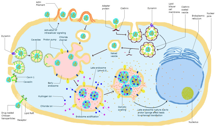
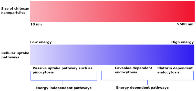
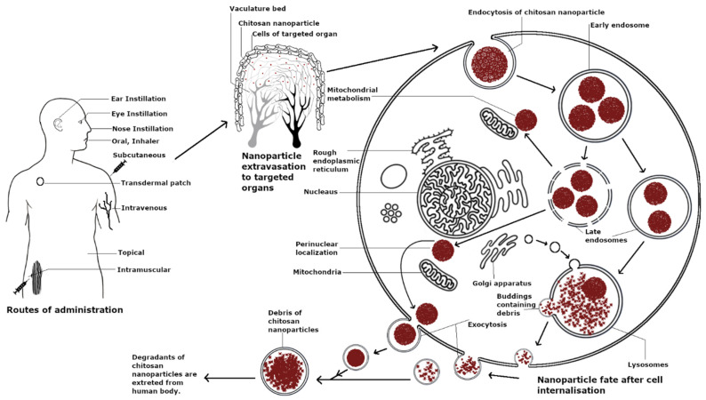
The unique properties of chitosan make it a useful choice for various nanoparticulate drug delivery applications. Although chitosan is biocompatible and enables cellular uptake, its interactions at cellular and systemic levels need to be studied in more depth. This review focuses on the various physical and chemical properties of chitosan that affect its performance in biological systems. We aim to analyze recent research studying interactions of chitosan nanoparticles (NPs) upon their cellular uptake and their journey through the various compartments of the cell. The positive charge of chitosan enables it to efficiently attach to cells, increasing the probability of cellular uptake. Chitosan NPs are taken up by cells via different pathways and escape endosomal degradation due to the proton sponge effect. Furthermore, we have reviewed the interaction of chitosan NPs upon in vivo administration. Chitosan NPs are immediately surrounded by a serum protein corona in systemic circulation upon intravenous administration, and their biodistribution is mainly to the liver and spleen indicating RES uptake. However, the evasion of RES system as well as the targeting ability and bioavailability of chitosan NPs can be improved by utilizing specific routes of administration and covalent modifications of surface properties. Ongoing clinical trials of chitosan formulations for therapeutic applications are paving the way for the introduction of chitosan into the pharmaceutical market and for their toxicological evaluation. Chitosan provides specific biophysical properties for effective and tunable cellular uptake and systemic delivery for a wide range of applications.
Overview
The claims and statistics of this paper haven’t been checked yet.
Run the check — free, no account needed. It reads the open-access text and checks the sources it cites.
Integrity signals
Registry
Cites
Statistics
Figures
Verification
Claims the paper makes
The claims and statistics of this paper haven’t been checked yet.
Run the check — free, no account needed. It reads the open-access text and checks the sources it cites.
Claims other papers make about this one
Each is checked against this paper’s own text: does it say what the citing paper says it does?
In the citing paper
“Moreover, chitosan is known for transiently opening the tight junction between epithelial cells, which allows the permeation of the hydrophilic loaded drug.128”
From Current Advances in Chitosan Nanoparticles Based Oral Drug Delivery for Colorectal Cancer Treatment (2022) · cites this paper as [128]
Self-check
Percentages, sums and reported differences re-computed from the paper’s own numbers.
The self-check needs the paper’s full text.
Citations
Cites
Bertrand, N., Grenier, P., Mahmoudi, M., Lima, E. M., Appel, E. A., Dormont, F., Lim, J.-M., Karnik, R., Langer, R., & Farokhzad, O. C. (2017) · Nature Communications · doi:10.1038/s41467-017-00600-w · Cited by 708
Lien, C.-F., Molnár, É., Toman, P., Tsibouklis, J., Pilkington, G. J., Górecki, D. C., & Barbu, E. (2012) · Biomacromolecules · doi:10.1021/bm201790s · Cited by 42
Research works printed but not linked: 175
- Calvo, 1997, Novel Hydrophilic Chitosan-Polyethylene Oxide Nanoparticles as Protein Carriers, J. Appl. Polym. Sci., 63, 125, 10.1002/(SICI)1097-4628(19970103)63:1<125::AID-APP13>3.0.CO;2-4 · DOI
- Calvo, 1997, Evaluation of Cationic Polymer-Coated Nanocapsules as Ocular Drug Carriers, Int. J. Pharm., 153, 41, 10.1016/S0378-5173(97)00083-5 · DOI
- Berthold, 1996, Preparation and Characterization of Chitosan Microspheres as Drug Carrier for Prednisolone Sodium Phosphate as Model for Anti-Inflammatory Drugs, J. Control Release, 39, 17, 10.1016/0168-3659(95)00129-8 · DOI
- Ojeda-Hernández, D.D., Canales-Aguirre, A.A., Matias-Guiu, J., Gomez-Pinedo, U., and Mateos-Díaz, J.C. (2020). Potential of Chitosan and Its Derivatives for Biomedical Applications in the Central Nervous System. Front. Bioeng. Biotechnol., 8. · DOI
- Wang, W., Meng, Q., Li, Q., Liu, J., Zhou, M., Jin, Z., and Zhao, K. (2020). Chitosan Derivatives and Their Application in Biomedicine. Int. J. Mol. Sci., 21. · DOI
- Jhaveri, J., Raichura, Z., Khan, T., Momin, M., and Omri, A. (2021). Chitosan Nanoparticles-Insight into Properties, Functionalization and Applications in Drug Delivery and Theranostics. Molecules, 26. · DOI
- Moraru, C., Mincea, M., Menghiu, G., and Ostafe, V. (2020). Understanding the Factors Influencing Chitosan-Based Nanoparticles-Protein Corona Interaction and Drug Delivery Applications. Molecules, 25. · DOI
- Sharma, P.D. (2017). Cellular Screening Methods for the Study of Nanoparticle- Induced Lysosomal Damage. Lysosomes—Associated Diseases and Methods to Study Their Function, InTech. · DOI
- Jiang, 2017, Intracellular Disposition of Chitosan Nanoparticles in Macrophages: Intracellular Uptake, Exocytosis, and Intercellular Transport, Int. J. Nanomed., 12, 6383, 10.2147/IJN.S142060 · DOI
- Pilipenko, I., Korzhikov-Vlakh, V., Sharoyko, V., Zhang, N., Schäfer-Korting, M., Rühl, E., Zoschke, C., and Tennikova, T. (2019). PH-Sensitive Chitosan–Heparin Nanoparticles for Effective Delivery of Genetic Drugs into Epithelial Cells. Pharmaceutics, 11. · DOI
- Nam, 2016, Target Gene Delivery from Targeting Ligand Conjugated Chitosan–PEI Copolymer for Cancer Therapy, Carbohydr. Polym., 135, 153, 10.1016/j.carbpol.2015.08.053 · DOI
- Chen, 2017, Stepwise PH-Responsive Nanoparticles for Enhanced Cellular Uptake and on-Demand Intracellular Release of Doxorubicin, Int. J. Nanomed., 12, 4241, 10.2147/IJN.S129748 · DOI
- Chen, 2018, N-Trimethyl Chitosan Nanoparticles and CSKSSDYQC Peptide: N-Trimethyl Chitosan Conjugates Enhance the Oral Bioavailability of Gemcitabine to Treat Breast Cancer, J. Control Release, 277, 142, 10.1016/j.jconrel.2018.03.013 · DOI
- Garg, 2019, Current Advances in Chitosan Nanoparticles Based Drug Delivery and Targeting, Adv. Pharm. Bull., 9, 195, 10.15171/apb.2019.023 · DOI · Paper page
- Yang, 2015, Getting Across the Cell Membrane: An Overview for Small Molecules, Peptides, and Proteins, Site-Specif. Protein Labeling, 1266, 29, 10.1007/978-1-4939-2272-7_3 · DOI · Paper page
- Magalhaes, 2010, Pivotal Advance: Phospholipids Determine Net Membrane Surface Charge Resulting in Differential Localization of Active Rac1 and Rac2, J. Leukoc. Biol., 87, 545, 10.1189/jlb.0609390 · DOI
- Schipper, 1996, Chitosans as Absorption Enhancers for Poorly Absorbable Drugs. 1: Influence of Molecular Weight and Degree of Acetylation on Drug Transport Across Human Intestinal Epithelial (Caco-2) Cells, Pharm. Res., 13, 1686, 10.1023/A:1016444808000 · DOI
- Verhoef, 1997, Chitosans for Enhanced Delivery of Therapeutic Peptides across Intestinal Epithelia: In Vitro Evaluation in Caco-2 Cell Monolayers, Int. J. Pharm., 159, 243, 10.1016/S0378-5173(97)00287-1 · DOI
- Yue, 2011, Surface Charge Affects Cellular Uptake and Intracellular Trafficking of Chitosan-Based Nanoparticles, Biomacromolecules, 12, 2440, 10.1021/bm101482r · DOI · Paper page
- Boyles, 2015, Chitosan Functionalisation of Gold Nanoparticles Encourages Particle Uptake and Induces Cytotoxicity and Pro-Inflammatory Conditions in Phagocytic Cells, as Well as Enhancing Particle Interactions with Serum Components, J. Nanobiotechnol., 13, 84, 10.1186/s12951-015-0146-9 · DOI
- Hu, 2012, Influence of Charge on FITC-BSA-Loaded Chondroitin Sulfate-Chitosan Nanoparticles upon Cell Uptake in Human Caco-2 Cell Monolayers, Int. J. Nanomed., 7, 4861
- Kalliola, 2018, Carboxymethyl Chitosan and Its Hydrophobically Modified Derivative as PH-Switchable Emulsifiers, Langmuir, 34, 2800, 10.1021/acs.langmuir.7b03959 · DOI
- Nogueira, 2013, In Vitro Antitumor Activity of Methotrexate via PH-Sensitive Chitosan Nanoparticles, Biomaterials, 34, 2758, 10.1016/j.biomaterials.2013.01.005 · DOI
- Amoozgar, 2012, Low Molecular-Weight Chitosan as a PH-Sensitive Stealth Coating for Tumor-Specific Drug Delivery, Mol. Pharm., 9, 1262, 10.1021/mp2005615 · DOI
- Khalili, 2015, A Review of Cell Adhesion Studies for Biomedical and Biological Applications, Int. J. Mol. Sci., 16, 18149, 10.3390/ijms160818149 · DOI
- Chen, 2012, Control of Cell Attachment on PH-Responsive Chitosan Surface by Precise Adjustment of Medium PH, Biomaterials, 33, 1336, 10.1016/j.biomaterials.2011.10.048 · DOI
- Taketa, 2019, Tailored Chitosan/Hyaluronan Coatings for Tumor Cell Adhesion: Effects of Topography, Charge Density and Surface Composition, Appl. Surf. Sci., 486, 508, 10.1016/j.apsusc.2019.04.227 · DOI
- Noriega, 2011, Consequences of Neutralization on the Proliferation and Cytoskeletal Organization of Chondrocytes on Chitosan-Based Matrices, Int. J. Carbohydr. Chem., 2011, 1, 10.1155/2011/809743 · DOI
- Zhang, 2018, A Proteomics Study to Explore the Role of Adsorbed Serum Proteins for PC12 Cell Adhesion and Growth on Chitosan and Collagen/Chitosan Surfaces, Regen. Biomater., 5, 261, 10.1093/rb/rby017 · DOI
- Ibrahim, F., Osman, N.A.A., Usman, J., and Kadri, N.A. (2006, January 11–14). Cell Adhesion and Degradation Behaviors of Acetylated Chitosan Films. Proceedings of the 3rd Kuala Lumpur International Conference on Biomedical Engineering 2006, Kuala Lumpur, Malaysia. · DOI
- Foster, L.J.R., Ho, S., Hook, J., Basuki, M., and Marçal, H. (2015). Chitosan as a Biomaterial: Influence of Degree of Deacetylation on Its Physiochemical, Material and Biological Properties. PLoS ONE, 10. · DOI
- 2015, Chitosan and Its Potential Use as a Scaffold for Tissue Engineering in Regenerative Medicine, BioMed Res. Int., 2015, 821279
- Boecker, A., Daeschler, S.C., Kneser, U., and Harhaus, L. (2019). Relevance and Recent Developments of Chitosan in Peripheral Nerve Surgery. Front. Cell. Neurosci., 13. · DOI
- Dixit, 2017, Immunological Challenges Associated with Artificial Skin Grafts: Available Solutions and Stem Cells in Future Design of Synthetic Skin, J. Biol. Eng., 11, 49, 10.1186/s13036-017-0089-9 · DOI
- Salati, 2011, Design and Fabrication of Artificial Skin: Chitosan and Gelatin Immobilization on Silicone by Poly Acrylic Acid Graft Using a Plasma Surface Modification Method, J. Macromol. Sci. Part B, 50, 1972, 10.1080/00222348.2010.549438 · DOI
- Vijayan, A., Sabareeswaran, A., and Kumar, G.S.V. (2019). PEG Grafted Chitosan Scaffold for Dual Growth Factor Delivery for Enhanced Wound Healing. Sci. Rep., 9. · DOI
- Liu, 2018, A Functional Chitosan-Based Hydrogel as a Wound Dressing and Drug Delivery System in the Treatment of Wound Healing, RSC Adv., 8, 7533, 10.1039/C7RA13510F · DOI
- Cho, M.O., Li, Z., Shim, H.-E., Cho, I.-S., Nurunnabi, M., Park, H., Lee, K.Y., Moon, S.-H., Kim, K.-S., and Kang, S.-W. (2016). Bioinspired Tuning of Glycol Chitosan for 3D Cell Culture. NPG Asia Mater., 8. · DOI
- Kafi, 2020, Engineered Chitosan for Improved 3D Tissue Growth through Paxillin-FAK-ERK Activation, Regen. Biomater., 7, 141, 10.1093/rb/rbz034 · DOI
- Yeh, 2011, Mechanism and Consequence of Chitosan-Mediated Reversible Epithelial Tight Junction Opening, Biomaterials, 32, 6164, 10.1016/j.biomaterials.2011.03.056 · DOI
- Sonaje, 2011, Effects of Chitosan-Nanoparticle-Mediated Tight Junction Opening on the Oral Absorption of Endotoxins, Biomaterials, 32, 8712, 10.1016/j.biomaterials.2011.07.086 · DOI
- Lee, 2018, Tight Junction in the Intestinal Epithelium: Its Association with Diseases and Regulation by Phytochemicals, J. Immunol. Res., 2018, 1, 10.1155/2018/2645465 · DOI
- Bhat, A.A., Uppada, S., Achkar, I.W., Hashem, S., Yadav, S.K., Shanmugakonar, M., Al-Naemi, H.A., Haris, M., and Uddin, S. (2019). Tight Junction Proteins and Signaling Pathways in Cancer and Inflammation: A Functional Crosstalk. Front. Physiol., 9. · DOI
- Zihni, 2016, Tight Junctions: From Simple Barriers to Multifunctional Molecular Gates, Nat. Rev. Mol. Cell Biol., 17, 564, 10.1038/nrm.2016.80 · DOI
- Wang, 2017, Mechanism of Surface Charge Triggered Intestinal Epithelial Tight Junction Opening upon Chitosan Nanoparticles for Insulin Oral Delivery, Carbohydr. Polym., 157, 596, 10.1016/j.carbpol.2016.10.021 · DOI
- Srinivasan, 2015, TEER Measurement Techniques for In Vitro Barrier Model Systems, J. Lab. Autom., 20, 107, 10.1177/2211068214561025 · DOI
- Smith, 2004, Effect of Chitosan on Epithelial Cell Tight Junctions, Pharm. Res., 21, 43, 10.1023/B:PHAM.0000012150.60180.e3 · DOI
- Vllasaliu, 2010, Tight Junction Modulation by Chitosan Nanoparticles: Comparison with Chitosan Solution, Int. J. Pharm., 400, 183, 10.1016/j.ijpharm.2010.08.020 · DOI
- Foroozandeh, 2018, Insight into Cellular Uptake and Intracellular Trafficking of Nanoparticles, Nanoscale Res. Lett., 13, 339, 10.1186/s11671-018-2728-6 · DOI
- Li, 2017, Transporting Carriers for Intracellular Targeting Delivery via Non-Endocytic Uptake Pathways, Drug Deliv., 24, 45, 10.1080/10717544.2017.1391889 · DOI
- Juliano, 2015, Cellular Uptake and Intracellular Trafficking of Oligonucleotides, Adv. Drug Deliv. Rev., 87, 35, 10.1016/j.addr.2015.04.005 · DOI
- Malatesta, M., Grecchi, S., Chiesa, E., Cisterna, B., Costanzo, M., and Zancanaro, C. (2015). Internalized Chitosan Nanoparticles Persist for Long Time in Cultured Cells. Eur. J. Histochem., 59. · DOI
- Huang, 2002, Uptake of FITC-Chitosan Nanoparticles by A549 Cells, Pharm. Res., 19, 1488, 10.1023/A:1020404615898 · DOI
- Park, 2007, In Vitro Cellular Uptake and Cytotoxicity of Paclitaxel-Loaded Glycol Chitosan Self-Assembled Nanoparticles, Macromol. Res., 15, 513, 10.1007/BF03218824 · DOI
- Tahara, 2009, Improved Cellular Uptake of Chitosan-Modified PLGA Nanospheres by A549 Cells, Int. J. Pharm., 382, 198, 10.1016/j.ijpharm.2009.07.023 · DOI
- Koppolu, 2013, The Effect of Antigen Encapsulation in Chitosan Particles on Uptake, Activation and Presentation by Antigen Presenting Cells, Biomaterials, 34, 2359, 10.1016/j.biomaterials.2012.11.066 · DOI
- Tammam, 2015, Chitosan Nanoparticles for Nuclear Targeting: The Effect of Nanoparticle Size and Nuclear Localization Sequence Density, Mol. Pharm., 12, 4277, 10.1021/acs.molpharmaceut.5b00478 · DOI
- 2012, The Role of Surface Charge in Cellular Uptake and Cytotoxicity of Medical Nanoparticles, Int. J. Nanomed., 7, 5577
- He, 2010, Effects of Particle Size and Surface Charge on Cellular Uptake and Biodistribution of Polymeric Nanoparticles, Biomaterials, 31, 3657, 10.1016/j.biomaterials.2010.01.065 · DOI · Paper page
- Liu, 2010, Hydrophobic Modifications of Cationic Polymers for Gene Delivery, Prog. Polym. Sci., 35, 1144, 10.1016/j.progpolymsci.2010.04.007 · DOI
- Nam, 2009, Cellular Uptake Mechanism and Intracellular Fate of Hydrophobically Modified Glycol Chitosan Nanoparticles, J. Control. Release, 135, 259, 10.1016/j.jconrel.2009.01.018 · DOI
- Chiu, 2010, The Characteristics, Cellular Uptake and Intracellular Trafficking of Nanoparticles Made of Hydrophobically-Modified Chitosan, J. Control. Release, 146, 152, 10.1016/j.jconrel.2010.05.023 · DOI
- Layek, 2014, Hexanoic Acid and Polyethylene Glycol Double Grafted Amphiphilic Chitosan for Enhanced Gene Delivery: Influence of Hydrophobic and Hydrophilic Substitution Degree, Mol. Pharm., 11, 982, 10.1021/mp400633r · DOI
- Wang, 2011, Effects of Hydrophobic and Hydrophilic Modifications on Gene Delivery of Amphiphilic Chitosan Based Nanocarriers, Biomaterials, 32, 4630, 10.1016/j.biomaterials.2011.03.003 · DOI
- Zhan, J., Ting, X.L., and Zhu, J. (2017). The Research Progress of Targeted Drug Delivery Systems. IOP Conf. Ser. Mater. Sci. Eng., 207. · DOI
- Wang, 2010, Norcantharidin-Associated Galactosylated Chitosan Nanoparticles for Hepatocyte-Targeted Delivery, Nanomed. Nanotechnol. Biol. Med., 6, 371, 10.1016/j.nano.2009.07.006 · DOI
- Liu, 2014, TAT-LHRH Conjugated Low Molecular Weight Chitosan as a Gene Carrier Specific for Hepatocellular Carcinoma Cells, Int. J. Nanomed., 9, 2879, 10.2147/IJN.S61392 · DOI
- Han, 2015, Dual-Targeting and PH/Redox-Responsive Multi-Layered Nanocomplexes for Smart Co-Delivery of Doxorubicin and SiRNA, Biomaterials, 60, 42, 10.1016/j.biomaterials.2015.05.001 · DOI
- Shi, 2017, Enhanced Antitumor Immunity by Targeting Dendritic Cells with Tumor Cell Lysate-Loaded Chitosan Nanoparticles Vaccine, Biomaterials, 113, 191, 10.1016/j.biomaterials.2016.10.047 · DOI
- Nascimento, 2017, Overcoming Cisplatin Resistance in Non-Small Cell Lung Cancer with Mad2 Silencing SiRNA Delivered Systemically Using EGFR-Targeted Chitosan Nanoparticles, Acta Biomater., 47, 71, 10.1016/j.actbio.2016.09.045 · DOI
- Yang, 2018, Estrone-Modified PH-Sensitive Glycol Chitosan Nanoparticles for Drug Delivery in Breast Cancer, Acta Biomater., 73, 400, 10.1016/j.actbio.2018.04.020 · DOI
- Jiang, 2017, A Smart PH-Responsive Nano-Carrier as a Drug Delivery System for the Targeted Delivery of Ursolic Acid: Suppresses Cancer Growth and Metastasis by Modulating P53/MMP-9/PTEN/CD44 Mediated Multiple Signaling Pathways, Nanoscale, 9, 9428, 10.1039/C7NR01677H · DOI
- Hefnawy, 2020, Dual-Ligand Functionalized Core-Shell Chitosan-Based Nanocarrier for Hepatocellular Carcinoma-Targeted Drug Delivery, Int. J. Nanomed., 15, 821, 10.2147/IJN.S240359 · DOI
- Zhu, 2011, Folate-Modified Chitosan Micelles with Enhanced Tumor Targeting Evaluated by near Infrared Imaging System, Carbohydr. Polym., 86, 1118, 10.1016/j.carbpol.2011.05.061 · DOI
- Ramya, A.N., Joseph, M.M., Maniganda, S., Karunakaran, V., Sreelekha, T.T., and Maiti, K.K. (2017). Emergence of Gold-Mesoporous Silica Hybrid Nanotheranostics: Dox-Encoded, Folate Targeted Chemotherapy with Modulation of SERS Fingerprinting for Apoptosis Toward Tumor Eradication. Small, 13. · DOI
- Verma, 2016, Vitamin B12 Functionalized Layer by Layer Calcium Phosphate Nanoparticles: A Mucoadhesive and PH Responsive Carrier for Improved Oral Delivery of Insulin, Acta Biomater., 31, 288, 10.1016/j.actbio.2015.12.017 · DOI
- Rekha, 2009, Synthesis and Evaluation of Lauryl Succinyl Chitosan Particles towards Oral Insulin Delivery and Absorption, J. Control. Release, 135, 144, 10.1016/j.jconrel.2009.01.011 · DOI
- Zhao, 2017, Simultaneous Inhibition of Growth and Metastasis of Hepatocellular Carcinoma by Co-Delivery of Ursolic Acid and Sorafenib Using Lactobionic Acid Modified and PH-Sensitive Chitosan-Conjugated Mesoporous Silica Nanocomplex, Biomaterials, 143, 1, 10.1016/j.biomaterials.2017.07.030 · DOI
- Zhu, R., Zhang, C., Liu, Y., Yuan, Z., Chen, W., Yang, S., Li, J., Zhu, W., Zhou, X., and You, B. (2015). CD147 Monoclonal Antibody Mediated by Chitosan Nanoparticles Loaded with α-Hederin Enhances Antineoplastic Activity and Cellular Uptake in Liver Cancer Cells. Sci. Rep., 5. · DOI
- Shen, 2012, A Novel Carboxymethyl Chitosan-Based Folate/Fe3O4/CdTe Nanoparticle for Targeted Drug Delivery and Cell Imaging, Carbohydr. Polym., 88, 239, 10.1016/j.carbpol.2011.11.087 · DOI
- Mathew, 2010, Folate Conjugated Carboxymethyl Chitosan–Manganese Doped Zinc Sulphide Nanoparticles for Targeted Drug Delivery and Imaging of Cancer Cells, Carbohydr. Polym., 80, 442, 10.1016/j.carbpol.2009.11.047 · DOI
- Guan, 2012, N-Trimethyl Chitosan Nanoparticle-Encapsulated Lactosyl-Norcantharidin for Liver Cancer Therapy with High Targeting Efficacy, Nanomed. Nanotechnol. Biol. Med., 8, 1172, 10.1016/j.nano.2012.01.009 · DOI
- Zhu, 2014, Low-Density Lipoprotein-Coupled N-Succinyl Chitosan Nanoparticles Co-Delivering SiRNA and Doxorubicin for Hepatocyte-Targeted Therapy, Biomaterials, 35, 5965, 10.1016/j.biomaterials.2014.03.088 · DOI
- Chou, 2011, Strategies for the Intracellular Delivery of Nanoparticles, Chem. Soc. Rev., 40, 233, 10.1039/C0CS00003E · DOI
- Cao, Y., Tan, Y.F., Wong, Y.S., Liew, M.W.J., and Venkatraman, S. (2019). Recent Advances in Chitosan-Based Carriers for Gene Delivery. Mar. Drugs, 17. · DOI
- Lai, 2018, Design of Polymeric Gene Carriers for Effective Intracellular Delivery, Trends Biotechnol., 36, 713, 10.1016/j.tibtech.2018.02.006 · DOI
- Zhang, 2018, Convenient Preparation of Charge-Adaptive Chitosan Nanomedicines for Extended Blood Circulation and Accelerated Endosomal Escape, Nano Res., 11, 4278, 10.1007/s12274-018-2014-z · DOI
- Moreira, 2009, Improving Chitosan-Mediated Gene Transfer by the Introduction of Intracellular Buffering Moieties into the Chitosan Backbone, Acta Biomater., 5, 2995, 10.1016/j.actbio.2009.04.021 · DOI
- Chang, 2010, Efficient Gene Transfection by Histidine-Modified Chitosan through Enhancement of Endosomal Escape, Bioconj. Chem., 21, 1087, 10.1021/bc1000609 · DOI
- Huang, G., Chen, Q., Wu, W., Wang, J., Chu, P.K., Bai, H., and Tang, G. (2020). Reconstructed Chitosan with Alkylamine for Enhanced Gene Delivery by Promoting Endosomal Escape. Carbohydr. Polym., 227. · DOI
- Rathore, 2019, Nanomaterial Designing Strategies Related to Cell Lysosome and Their Biomedical Applications: A Review, Biomaterials, 211, 25, 10.1016/j.biomaterials.2019.05.002 · DOI
- Manshian, 2018, The Impact of Nanoparticle-Driven Lysosomal Alkalinization on Cellular Functionality, J. Nanobiotechnol., 16, 85, 10.1186/s12951-018-0413-7 · DOI
- Fong, 2017, Lysosomal Rupture Induced by Structurally Distinct Chitosans Either Promotes a Type 1 IFN Response or Activates the Inflammasome in Macrophages, Biomaterials, 129, 127, 10.1016/j.biomaterials.2017.03.022 · DOI
- Thibault, 2011, Excess Polycation Mediates Efficient Chitosan-Based Gene Transfer by Promoting Lysosomal Release of the Polyplexes, Biomaterials, 32, 4639, 10.1016/j.biomaterials.2011.03.010 · DOI
- Seijo, 2011, Intracellular Trafficking of Hyaluronic Acid-Chitosan Oligomer-Based Nanoparticles in Cultured Human Ocular Surface Cells, Mol. Vis., 17, 279
- Yameen, 2014, Insight into Nanoparticle Cellular Uptake and Intracellular Targeting, J. Control Release, 190, 485, 10.1016/j.jconrel.2014.06.038 · DOI
- Tammam, 2016, How Successful Is Nuclear Targeting by Nanocarriers?, J. Control Release, 229, 140, 10.1016/j.jconrel.2016.03.022 · DOI
- Opanasopit, 2009, Nuclear Localization Signal Peptides Enhance Transfection Efficiency of Chitosan/DNA Complexes, Int. J. Pharm., 382, 291, 10.1016/j.ijpharm.2009.08.029 · DOI
- Barbieri, 2013, Lecithin/Chitosan Controlled Release Nanopreparations of Tamoxifen Citrate: Loading, Enzyme-Trigger Release and Cell Uptake, J. Control Release, 167, 276, 10.1016/j.jconrel.2013.02.009 · DOI
- Lee, 2009, Tumor Specificity and Therapeutic Efficacy of Photosensitizer-Encapsulated Glycol Chitosan-Based Nanoparticles in Tumor-Bearing Mice, Biomaterials, 30, 2929, 10.1016/j.biomaterials.2009.01.058 · DOI
- Zorova, 2018, Functional Significance of the Mitochondrial Membrane Potential, Biochem. Mosc. Suppl. Ser. Membr. Cell Biol., 12, 20
- Qi, 2005, In Vitro Effects of Chitosan Nanoparticles on Proliferation of Human Gastric Carcinoma Cell Line MGC803 Cells, World J. Gastroenterol., 11, 5136
- Jiang, 2019, Chitosan Nanoparticles Induced the Antitumor Effect in Hepatocellular Carcinoma Cells by Regulating ROS-Mediated Mitochondrial Damage and Endoplasmic Reticulum Stress, Artif. Cells Nanomed. Biotechnol., 47, 747, 10.1080/21691401.2019.1577876 · DOI
- Chen, 2015, Hierarchical Targeted Hepatocyte Mitochondrial Multifunctional Chitosan Nanoparticles for Anticancer Drug Delivery, Biomaterials, 52, 240, 10.1016/j.biomaterials.2015.02.001 · DOI
- Hou, 2017, Triphenyl Phosphine-Functionalized Chitosan Nanoparticles Enhanced Antitumor Efficiency Through Targeted Delivery of Doxorubicin to Mitochondria, Nanoscale Res. Lett., 12, 158, 10.1186/s11671-017-1931-1 · DOI
- Wang, 2017, Hyaluronic Acid-Coated Chitosan Nanoparticles Induce ROS-Mediated Tumor Cell Apoptosis and Enhance Antitumor Efficiency by Targeted Drug Delivery via CD44, J. Nanobiotechnol., 15, 7, 10.1186/s12951-016-0245-2 · DOI
- Mallick, 2019, Self-Assembled Nanoparticles Composed of Glycol Chitosan-Dequalinium for Mitochondria-Targeted Drug Delivery, Int. J. Biol. Macromol., 132, 451, 10.1016/j.ijbiomac.2019.03.215 · DOI
- He, 2018, Carboxymethylated Chitosan Protects Schwann Cells against Hydrogen Peroxide-Induced Apoptosis by Inhibiting Oxidative Stress and Mitochondria Dependent Pathway, Eur. J. Pharmacol., 825, 48, 10.1016/j.ejphar.2018.02.024 · DOI
- Shukla, 2015, In Vitro Toxicity Assessment of Chitosan Oligosaccharide Coated Iron Oxide Nanoparticles, Toxicol. Rep., 2, 27, 10.1016/j.toxrep.2014.11.002 · DOI
- Park, 2014, Endocytosis and Exocytosis of Nanoparticles in Mammalian Cells, Int. J. Nanomed., 9, 51, 10.2147/IJN.S26592 · DOI
- Sakhtianchi, 2013, Exocytosis of Nanoparticles from Cells: Role in Cellular Retention and Toxicity, Adv. Colloid Interface Sci., 201–202, 18, 10.1016/j.cis.2013.10.013 · DOI
- Park, 2006, N-Acetyl Histidine-Conjugated Glycol Chitosan Self-Assembled Nanoparticles for Intracytoplasmic Delivery of Drugs: Endocytosis, Exocytosis and Drug Release, J. Control Release, 115, 37, 10.1016/j.jconrel.2006.07.011 · DOI
- Tahara, 2012, The Suppression of IgE-Mediated Histamine Release from Mast Cells Following Exocytic Exclusion of Biodegradable Polymeric Nanoparticles, Biomaterials, 33, 343, 10.1016/j.biomaterials.2011.09.043 · DOI
- Bakhru, 2012, Enhanced Cellular Uptake and Long-Term Retention of Chitosan-Modified Iron-Oxide Nanoparticles for MRI-Based Cell Tracking, Int. J. Nanomed., 7, 4613, 10.2147/IJN.S28294 · DOI
- Jin, 2012, PH-Sensitive Chitosan-Derived Nanoparticles as Doxorubicin Carriers for Effective Anti-Tumor Activity: Preparation and in Vitro Evaluation, Colloids Surf. B Biointerfaces, 94, 184, 10.1016/j.colsurfb.2012.01.032 · DOI
- Trif, 2015, Cytotoxicity and Intracellular Fate of PLGA and Chitosan-Coated PLGA Nanoparticles in Madin-Darby Bovine Kidney (MDBK) and Human Colorectal Adenocarcinoma (Colo 205) Cells, J. Biomed. Mater. Res. A, 103, 3599, 10.1002/jbm.a.35498 · DOI
- Durán, V., Yasar, H., Becker, J., Thiyagarajan, D., Loretz, B., Kalinke, U., and Lehr, C.-M. (2019). Preferential Uptake of Chitosan-Coated PLGA Nanoparticles by Primary Human Antigen Presenting Cells. Nanomed. Nanotechnol. Biol. Med., 21. · DOI
- Grenha, 2007, Chitosan Nanoparticles Are Compatible with Respiratory Epithelial Cells in Vitro, Eur. J. Pharm. Sci., 31, 73, 10.1016/j.ejps.2007.02.008 · DOI
- Rajam, 2011, Chitosan Nanoparticles as a Dual Growth Factor Delivery System for Tissue Engineering Applications, Int. J. Pharm., 410, 145, 10.1016/j.ijpharm.2011.02.065 · DOI
- Zhang, 2017, Amino Acid-Modified Chitosan Nanoparticles for Cu2+ Chelation to Suppress CuO Nanoparticle Cytotoxicity, J. Mater. Chem. B, 5, 3521, 10.1039/C7TB00344G · DOI
- Nafee, 2009, Relevance of the Colloidal Stability of Chitosan/PLGA Nanoparticles on Their Cytotoxicity Profile, Int. J. Pharm., 381, 130, 10.1016/j.ijpharm.2009.04.049 · DOI
- Sarangapani, 2018, Chitosan Nanoparticles’ Functionality as Redox Active Drugs through Cytotoxicity, Radical Scavenging and Cellular Behaviour, Integr. Biol., 10, 313, 10.1039/C8IB00038G · DOI
- Lin, 2019, Chitosan Nanoparticles Strengthen Vγ9Vδ2 T-Cell Cytotoxicity through Upregulation of Killing Molecules and Cytoskeleton Polarization, Int. J. Nanomed., 14, 9325, 10.2147/IJN.S212898 · DOI
- Richardson, 1999, Potential of Low Molecular Mass Chitosan as a DNA Delivery System: Biocompatibility, Body Distribution and Ability to Complex and Protect DNA, Int. J. Pharm., 178, 231, 10.1016/S0378-5173(98)00378-0 · DOI
- Suzuki, 1998, Biodistribution and Kinetics of Holmium-166-Chitosan Complex (DW-166HC) in Rats and Mice, J. Nucl. Med., 39, 2161
- Onishi, 1999, Biodegradation and Distribution of Water-Soluble Chitosan in Mice, Biomaterials, 20, 175, 10.1016/S0142-9612(98)00159-8 · DOI
- Banerjee, 2002, Preparation, Characterization and Biodistribution of Ultrafine Chitosan Nanoparticles, Int. J. Pharm., 243, 93, 10.1016/S0378-5173(02)00267-3 · DOI
- Kean, 2010, Biodegradation, Biodistribution and Toxicity of Chitosan, Adv. Drug Deliv. Rev., 62, 3, 10.1016/j.addr.2009.09.004 · DOI · Paper page
- Kadam, 2012, Nano-Advantage in Enhanced Drug Delivery with Biodegradable Nanoparticles: Contribution of Reduced Clearance, Drug Metab. Dispos., 40, 1380, 10.1124/dmd.112.044925 · DOI
- Termsarasab, 2014, Polyethylene Glycol-Modified Arachidyl Chitosan-Based Nanoparticles for Prolonged Blood Circulation of Doxorubicin, Int. J. Pharm., 464, 127, 10.1016/j.ijpharm.2014.01.015 · DOI
- Wu, 2017, Facile Fabrication of Poly(Acrylic Acid) Coated Chitosan Nanoparticles with Improved Stability in Biological Environments, Eur. J. Pharm. Biopharm., 112, 148, 10.1016/j.ejpb.2016.11.020 · DOI
- Zhang, 2009, O-Carboxymethyl-Chitosan/Organosilica Hybrid Nanoparticles as Non-Viral Vectors for Gene Delivery, Mater. Sci. Eng. C, 29, 2045, 10.1016/j.msec.2009.04.003 · DOI
- Katas, 2006, Development and Characterisation of Chitosan Nanoparticles for SiRNA Delivery, J. Control Release, 115, 216, 10.1016/j.jconrel.2006.07.021 · DOI
- Shi, S., Zhang, Z., Luo, Z., Yu, J., Liang, R., Li, X., and Chen, H. (2015). Chitosan Grafted Methoxy Poly(Ethylene Glycol)-Poly(ε-Caprolactone) Nanosuspension for Ocular Delivery of Hydrophobic Diclofenac. Sci. Rep., 5. · DOI
- Sun, 2014, Biocompatible Glycol Chitosan-Coated Gold Nanoparticles for Tumor-Targeting CT Imaging, Pharm. Res., 31, 1418, 10.1007/s11095-013-1142-0 · DOI
- Duan, 2018, Chitosan-Stabilized Self-Assembled Fluorescent Gold Nanoclusters for Cell Imaging and Biodistribution In Vivo, ACS Biomater. Sci. Eng., 4, 1055, 10.1021/acsbiomaterials.7b00975 · DOI
- Mahmoudi, 2016, Emerging Understanding of the Protein Corona at the Nano-Bio Interfaces, Nano Today, 11, 817, 10.1016/j.nantod.2016.10.005 · DOI · Paper page
- Forest, 2017, Preferential Binding of Positive Nanoparticles on Cell Membranes Is Due to Electrostatic Interactions: A Too Simplistic Explanation That Does Not Take into Account the Nanoparticle Protein Corona, Mater. Sci. Eng. C, 70, 889, 10.1016/j.msec.2016.09.016 · DOI
- Almalik, 2013, Hyaluronic Acid-Coated Chitosan Nanoparticles: Molecular Weight-Dependent Effects on Morphology and Hyaluronic Acid Presentation, J. Control Release, 172, 1142, 10.1016/j.jconrel.2013.09.032 · DOI
- Almalik, A., Benabdelkamel, H., Masood, A., Alanazi, I.O., Alradwan, I., Majrashi, M.A., Alfadda, A.A., Alghamdi, W.M., Alrabiah, H., and Tirelli, N. (2017). Hyaluronic Acid Coated Chitosan Nanoparticles Reduced the Immunogenicity of the Formed Protein Corona. Sci. Rep., 7. · DOI
- Basu, 2019, Biopolymer Nanoparticle Surface Chemistry Dictates the Nature and Extent of Protein Hard Corona, J. Mol. Liq., 282, 169, 10.1016/j.molliq.2019.03.016 · DOI
- Abouelmagd, 2015, Low Molecular Weight Chitosan-Coated Polymeric Nanoparticles for Sustained and PH-Sensitive Delivery of Paclitaxel, J. Drug Target., 23, 725, 10.3109/1061186X.2015.1054829 · DOI
- Tekie, 2020, Controlling Evolution of Protein Corona: A Prosperous Approach to Improve Chitosan-Based Nanoparticle Biodistribution and Half-Life, Sci. Rep., 10, 9664, 10.1038/s41598-020-66572-y · DOI
- Chen, 2009, The Characteristics, Biodistribution and Bioavailability of a Chitosan-Based Nanoparticulate System for the Oral Delivery of Heparin, Biomaterials, 30, 6629, 10.1016/j.biomaterials.2009.08.030 · DOI
- Raj, 2018, Biodistribution and Targeting Potential Assessment of Mucoadhesive Chitosan Nanoparticles Designed for Ulcerative Colitis via Scintigraphy, RSC Adv., 8, 20809, 10.1039/C8RA01898G · DOI
- Navarro, 2014, Biodistribution of PLGA and PLGA/Chitosan Nanoparticles after Repeat-Dose Oral Delivery in F344 Rats for 7 Days, Ther. Deliv., 5, 1191, 10.4155/tde.14.79 · DOI
- Gartziandia, 2015, Chitosan Coated Nanostructured Lipid Carriers for Brain Delivery of Proteins by Intranasal Administration, Colloids Surf. B Biointerfaces, 134, 304, 10.1016/j.colsurfb.2015.06.054 · DOI
- Yousfan, 2020, Preparation and Characterisation of PHT-Loaded Chitosan Lecithin Nanoparticles for Intranasal Drug Delivery to the Brain, RSC Adv., 10, 28992, 10.1039/D0RA04890A · DOI
- Franca, J.R., Foureaux, G., Fuscaldi, L.L., Ribeiro, T.G., Rodrigues, L.B., Bravo, R., Castilho, R.O., Yoshida, M.I., Cardoso, V.N., and Fernandes, S.O. (2014). Bimatoprost-Loaded Ocular Inserts as Sustained Release Drug Delivery Systems for Glaucoma Treatment: In Vitro and In Vivo Evaluation. PLoS ONE, 9. · DOI
- Franca, 2020, Use of Chitosan as Pharmaceutical Excipient in Ocular Drug Delivery Systems: Sterilization and Pharmacokinetics, J. Biomed. Mater. Res. B Appl. Biomater., 108, 2227, 10.1002/jbm.b.34560 · DOI
- Kamiyama, 1999, Biodisposition Characteristics of N-Succinyl-Chitosan and Glycol-Chitosan in Normal and Tumor-Bearing Mice, Biol. Pharm. Bull., 22, 179, 10.1248/bpb.22.179 · DOI
- Li, 2020, Biodistribution and Biocompatibility of Glycyrrhetinic Acid and Galactose-Modified Chitosan Nanoparticles as a Novel Targeting Vehicle for Hepatocellular Carcinoma, Nanomedicine, 15, 145, 10.2217/nnm-2018-0455 · DOI
- Niu, 2019, A Chitosan-Based Cascade-Responsive Drug Delivery System for Triple-Negative Breast Cancer Therapy, J. Nanobiotechnol., 17, 95, 10.1186/s12951-019-0529-4 · DOI
- Cordeiro, 2021, Carboxymethyl-β-Glucan/Chitosan Nanoparticles: New Thermostable and Efficient Carriers for Antigen Delivery, Drug Deliv. Transl. Res., 11, 1689, 10.1007/s13346-021-00968-9 · DOI
- Zhang, 2008, Nasal Absorption Enhancement of Insulin Using PEG-Grafted Chitosan Nanoparticles, Eur. J. Pharm. Biopharm., 68, 526, 10.1016/j.ejpb.2007.08.009 · DOI
- Zu, 2016, Preparation and In Vitro/In Vivo Evaluation of Resveratrol-Loaded Carboxymethyl Chitosan Nanoparticles, Drug Deliv., 23, 971, 10.3109/10717544.2014.924167 · DOI
- Wang, 2014, In Vitro and In Vivo Evaluation of Chitosan Graft Glyceryl Monooleate as Peroral Delivery Carrier of Enoxaparin, Int. J. Pharm., 471, 391, 10.1016/j.ijpharm.2014.05.050 · DOI
- Asasutjarit, 2015, Development and Evaluation of Diclofenac Sodium Loaded-N-Trimethyl Chitosan Nanoparticles for Ophthalmic Use, AAPS PharmSciTech, 16, 1013, 10.1208/s12249-015-0290-4 · DOI
- Narayanan, 2013, In Vitro and In Vivo Evaluation of Osteoporosis Therapeutic Peptide PTH 1–34 Loaded PEGylated Chitosan Nanoparticles, Mol. Pharm., 10, 4159, 10.1021/mp400184v · DOI
- Feng, 2013, Chitosan/o-Carboxymethyl Chitosan Nanoparticles for Efficient and Safe Oral Anticancer Drug Delivery: In Vitro and In Vivo Evaluation, Int. J. Pharm., 457, 158, 10.1016/j.ijpharm.2013.07.079 · DOI
- Liu, 2011, In Vivo Evaluation of Novel Chitosan Graft Polymeric Micelles for Delivery of Paclitaxel, Drug Deliv., 18, 181, 10.3109/10717544.2010.520355 · DOI
- Braz, 2017, Chitosan/Sulfated Locust Bean Gum Nanoparticles: In Vitro and In Vivo Evaluation towards an Application in Oral Immunization, Int. J. Biol. Macromol., 96, 786, 10.1016/j.ijbiomac.2016.12.076 · DOI
- Yu, 2009, Polymeric Nanoparticles of Cholesterol-Modified Glycol Chitosan for Doxorubicin Delivery: Preparation and in-Vitro and in-Vivo Characterization, J. Pharm. Pharmacol., 61, 713, 10.1211/jpp.61.06.0003 · DOI
- Zhang, 2008, Pharmacokinetics, Biodistribution, Efficacy and Safety of N-Octyl-O-Sulfate Chitosan Micelles Loaded with Paclitaxel, Biomaterials, 29, 1233, 10.1016/j.biomaterials.2007.11.029 · DOI
- Bachir, 2018, Effects of PEG Surface Density and Chain Length on the Pharmacokinetics and Biodistribution of Methotrexate-Loaded Chitosan Nanoparticles, Int. J. Nanomed., 13, 5657, 10.2147/IJN.S167443 · DOI
- Dong, 2010, Pharmacokinetics and Biodegradation Mechanisms of a Versatile Carboxymethyl Derivative of Chitosan in Rats: In Vivo and In Vitro Evaluation, Biomacromolecules, 11, 1527, 10.1021/bm100158p · DOI
- Sonin, D., Pochkaeva, E., Zhuravskii, S., Postnov, V., Korolev, D., Vasina, L., Kostina, D., Mukhametdinova, D., Zelinskaya, I., and Skorik, Y. (2020). Biological Safety and Biodistribution of Chitosan Nanoparticles. Nanomaterials, 10. · DOI
- Nadesh, 2013, Hematotoxicological Analysis of Surface-Modified and -Unmodified Chitosan Nanoparticles, J. Biomed. Mater. Res. A, 101, 2957, 10.1002/jbm.a.34591 · DOI
- Sonaje, 2009, In Vivo Evaluation of Safety and Efficacy of Self-Assembled Nanoparticles for Oral Insulin Delivery, Biomaterials, 30, 2329, 10.1016/j.biomaterials.2008.12.066 · DOI
- Shan, 2017, Safety and Toxicology of the Intravenous Administration of Ang2-SiRNA Plasmid Chitosan Magnetic Nanoparticles, Mol. Med. Rep., 15, 736, 10.3892/mmr.2016.6090 · DOI
- Islam, N., Dmour, I., and Taha, M.O. (2019). Degradability of Chitosan Micro/Nanoparticles for Pulmonary Drug Delivery. Heliyon, 5. · DOI
- Lichtfouse, 2019, Applications of Chitosan in Food, Pharmaceuticals, Medicine, Cosmetics, Agriculture, Textiles, Pulp and Paper, Biotechnology, and Environmental Chemistry, Environ. Chem. Lett., 17, 1667, 10.1007/s10311-019-00904-x · DOI
- Marques, C., Som, C., Schmutz, M., Borges, O., and Borchard, G. (2020). How the Lack of Chitosan Characterization Precludes Implementation of the Safe-by-Design Concept. Front. Bioeng. Biotechnol., 8. · DOI
- Pasetti, 2010, Adjuvanted Intranasal Norwalk Virus-Like Particle Vaccine Elicits Antibodies and Antibody-Secreting Cells That Express Homing Receptors for Mucosal and Peripheral Lymphoid Tissues, J. Infect. Dis., 202, 1649, 10.1086/657087 · DOI
- Mann, A.J., Noulin, N., Catchpole, A., Stittelaar, K.J., de Waal, L., Kroeze, E.J.B.V., Hinchcliffe, M., Smith, A., Montomoli, E., and Piccirella, S. (2014). Intranasal H5N1 Vaccines, Adjuvanted with Chitosan Derivatives, Protect Ferrets against Highly Pathogenic Influenza Intranasal and Intratracheal Challenge. PLoS ONE, 9. · DOI
Cited by
Sorted by citations
Preparation and application of pH-responsive drug delivery systems
Ding, H., Tan, P., Fu, S., Tian, X., Zhang, H., Ma, X., Gu, Z., & Luo, K. (2022) · Journal of Controlled Release · doi:10.1016/j.jconrel.2022.05.056 · Cited by 645
Current Advances in Chitosan Nanoparticles Based Oral Drug Delivery for Colorectal Cancer Treatment
Choukaife, H., Seyam, S., Alallam, B., Doolaanea, A. A., & Alfatama, M. (2022) · International Journal of Nanomedicine · doi:10.2147/ijn.s375229 · Cited by 127
Metadata
- Authors
- Aibani, N., Rai, R., Patel, P., Cuddihy, G., & Wasan, E. K.
- Venue
- Pharmaceutics
- Publisher
- MDPI AG
- Published
- 2021-10-14
- Volume
- 13
- Issue
- 10
- Pages
- 1686
- ISSN
- 1999-4923
- PMC
- PMC8540112
- Cited by
- 419
- References
- 2
- Language
- en
About these checks
Registry, retraction, Cites and statistics results are computed, not judged, with one exception: a printed reference that no index lists is read once by a model before it counts as a citation error. Claims are extracted by a model and kept only when their quote appears verbatim in the text; each cited source is then checked, with a model’s help, for whether it says what the claim relies on. Those verdicts can be wrong — read the claim’s quote and the cited source before relying on one.
Statistics and arithmetic are recomputed, not judged.
Confidence bands are shown only for fields calibrated on our annotated test set. None is calibrated yet, so no band is shown.
What counts as a claim ›How we measure confidence ›How we check this ›
Are you an author, or do you see a problem with this page? Request a review
