Cited by 88

The DOI matches its Crossref record; 2 of the 2 references it cites were found in a registry or an index.

Exosomal microRNA‑146a derived from mesenchymal stem cells increases the sensitivity of ovarian cancer cells to docetaxel and taxane via a LAMC2‑mediated PI3K/Akt axis

Qiu, Liya, Wang, Jiakun, Chen, Mei, Chen, Fengyun, Tu, Wenluo·2020·International Journal of Molecular Medicine

Overview

The claims and statistics of this paper haven’t been checked yet.

Run the check — free, no account needed. It reads the open-access text and checks the sources it cites.

A few free checks a day · papers already verified open instantly · results are public

Integrity signals

Registry

Crossref · 10.3892/ijmm.2020.4634

Cites

2 of 2 references verified0 citation errors · 0 not checkable

Statistics

Figures

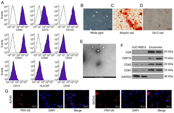
Figure 1 Identification of hUCMSCs and their derived exosomes. (A) Expression of MSCs surface markers, including CD29, CD44, CD73, CD90, CD105, CD14, CD34, CD45 and HLA-DR, analyzed by flow cytometry. (B) hUCMSC morphology at passage 3 observed under a light microscope. Scale bar, 100 µm. (C) Representative images of osteogenic differentiation of hUCMSCs using Alizarin Red staining. Scale bar, 100 µm. (D) Representative images of adipogenic differentiation of hUCMSCs using Oil red O staining. Scale bar, 100 µm. (E) Morphological analysis of hUCMSC by transmission electronic microscopy. Scale bar, 200 nm. (F) Detection of exosomal marker expression in hUCMSC-released exosomes by western blotting. (G) Internalization of PKH26-labeled exosomes by SKOV3 and A2780 cells was observed under a fluorescence microscope. Scale bar, 50 µm. hUCMSCs, human umbilical cord mesenchymal stem cells.
Figure 1 Identification of hUCMSCs and their derived exosomes. (A) Expression of MSCs surface markers, including CD29, CD44, CD73, CD90, CD105, CD14, CD34, CD45 and HLA-DR, analyzed by flow cytometry. (B) hUCMSC morphology at passage 3 observed under a light microscope. Scale bar, 100 µm. (C) Representative images of osteogenic differentiation of hUCMSCs using Alizarin Red staining. Scale bar, 100 µm. (D) Representative images of adipogenic differentiation of hUCMSCs using Oil red O staining. Scale bar, 100 µm. (E) Morphological analysis of hUCMSC by transmission electronic microscopy. Scale bar, 200 nm. (F) Detection of exosomal marker expression in hUCMSC-released exosomes by western blotting. (G) Internalization of PKH26-labeled exosomes by SKOV3 and A2780 cells was observed under a fluorescence microscope. Scale bar, 50 µm. hUCMSCs, human umbilical cord mesenchymal stem cells.CC BY-NC-ND · PubMed Central
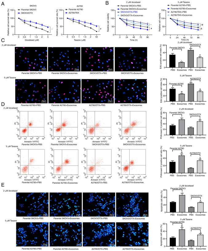
Figure 2 hUCMSC-derived exosomes inhibit OC cell drug resistance. SKOV3/DTX and A2780/TAX cells were induced by the exposure to gradient concentrations of drugs. (A) Cell survival rate examined by CCK-8 cell survival assays. Subsequently, the parental or drug-resistant OC cells were treated with 20 µg/ml MSCs-derived exosomes or PBS as a negative control. Exosome-treated parental or drug-resistant OC cells were exposed to 2 µM DTX or 5 µM TAX for 2 h, respectively. *P<0.05 vs. parental SKOV3 cells, #P<0.05 vs. parental A2780 cells. (B) Cell viability evaluated by CCK-8 cell viability assays. (C) EdU staining for cell proliferation. Scale bar, 25 µm. (D) Flow cytometry analysis for cells labelled with PI/Annexin V. (E) Hoechst 33258 staining for cell apoptosis. Scale bar, 25 µm. Data are presented as the mean ± SD. One-way (panels C, D and E) or two-way (panel A) ANOVA followed by Tukey's multiple comparisons test was used to determine statistical significance. Each assessment was performed in triplicate with three repetitions to ensure minimum deviation. In panels B, C, D and E: *P<0.05 vs. parental SKOV3 cells treated with PBS; #P<0.05 vs. SKOV3/DTX cells treated with PBS; @P<0.05 vs. parental A2780 cells treated with PBS, &P<0.05 vs. A2780/TAX cells treated with PBS. CCK-8, Cell Counting Kit-8; DTX, docetaxel; hUCMSCs, human umbilical cord mesenchymal stem cells; OC, ovarian cancer; PI, propidium iodide; TAX, taxane.
Figure 2 hUCMSC-derived exosomes inhibit OC cell drug resistance. SKOV3/DTX and A2780/TAX cells were induced by the exposure to gradient concentrations of drugs. (A) Cell survival rate examined by CCK-8 cell survival assays. Subsequently, the parental or drug-resistant OC cells were treated with 20 µg/ml MSCs-derived exosomes or PBS as a negative control. Exosome-treated parental or drug-resistant OC cells were exposed to 2 µM DTX or 5 µM TAX for 2 h, respectively. *P<0.05 vs. parental SKOV3 cells, #P<0.05 vs. parental A2780 cells. (B) Cell viability evaluated by CCK-8 cell viability assays. (C) EdU staining for cell proliferation. Scale bar, 25 µm. (D) Flow cytometry analysis for cells labelled with PI/Annexin V. (E) Hoechst 33258 staining for cell apoptosis. Scale bar, 25 µm. Data are presented as the mean ± SD. One-way (panels C, D and E) or two-way (panel A) ANOVA followed by Tukey's multiple comparisons test was used to determine statistical significance. Each assessment was performed in triplicate with three repetitions to ensure minimum deviation. In panels B, C, D and E: *P<0.05 vs. parental SKOV3 cells treated with PBS; #P<0.05 vs. SKOV3/DTX cells treated with PBS; @P<0.05 vs. parental A2780 cells treated with PBS, &P<0.05 vs. A2780/TAX cells treated with PBS. CCK-8, Cell Counting Kit-8; DTX, docetaxel; hUCMSCs, human umbilical cord mesenchymal stem cells; OC, ovarian cancer; PI, propidium iodide; TAX, taxane.CC BY-NC-ND · PubMed Central
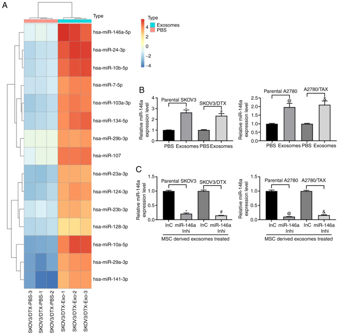
Figure 3 miR-146a is enriched in hUCMSC-derived exosomes. (A) Heatmap of 15 ectopic expressed miRNAs in hUCMSC-derived exosomes. (B) miR-146a expression in exosome-treated ovarian cancer cells. *P<0.05 vs. parental SKOV3 cells treated with PBS; #P<0.05 vs. SKOV3/DTX cells treated with PBS; @P<0.05 vs. parental A2780 cells treated with PBS; &P<0.05 vs. A2780/TAX cells treated with PBS. (C) miR-146a expression pattern in SKOV3 and A2780 cells treated with miR-146a inhibitor or inhibitor control and co-cultured with exosomes extracted from hUCMSCs. *P<0.05 vs. exosomes co-cultured with parental SKOV3 cells transfected with InC; #P<0.05 vs. exosomes co-cultured with SKOV3/DTX cells transfected with InC; @P<0.05 vs. exosomes co-cultured with parental A2780 cells transfected with InC; &P<0.05 vs. exosomes co-cultured with A2780/TAX cells transfected with InC. Data are presented as the mean ± SD. One-way ANOVA and Tukey's multiple comparisons test were used to determine statistical significance. Each assessment was performed in triplicate with three repetitions to ensure minimum deviation. hUCMSCs, human umbilical cord mesenchymal stem cells; inhi, inhibitor; InC, inhibitor control; miR, microRNA.
Figure 3 miR-146a is enriched in hUCMSC-derived exosomes. (A) Heatmap of 15 ectopic expressed miRNAs in hUCMSC-derived exosomes. (B) miR-146a expression in exosome-treated ovarian cancer cells. *P<0.05 vs. parental SKOV3 cells treated with PBS; #P<0.05 vs. SKOV3/DTX cells treated with PBS; @P<0.05 vs. parental A2780 cells treated with PBS; &P<0.05 vs. A2780/TAX cells treated with PBS. (C) miR-146a expression pattern in SKOV3 and A2780 cells treated with miR-146a inhibitor or inhibitor control and co-cultured with exosomes extracted from hUCMSCs. *P<0.05 vs. exosomes co-cultured with parental SKOV3 cells transfected with InC; #P<0.05 vs. exosomes co-cultured with SKOV3/DTX cells transfected with InC; @P<0.05 vs. exosomes co-cultured with parental A2780 cells transfected with InC; &P<0.05 vs. exosomes co-cultured with A2780/TAX cells transfected with InC. Data are presented as the mean ± SD. One-way ANOVA and Tukey's multiple comparisons test were used to determine statistical significance. Each assessment was performed in triplicate with three repetitions to ensure minimum deviation. hUCMSCs, human umbilical cord mesenchymal stem cells; inhi, inhibitor; InC, inhibitor control; miR, microRNA.CC BY-NC-ND · PubMed Central
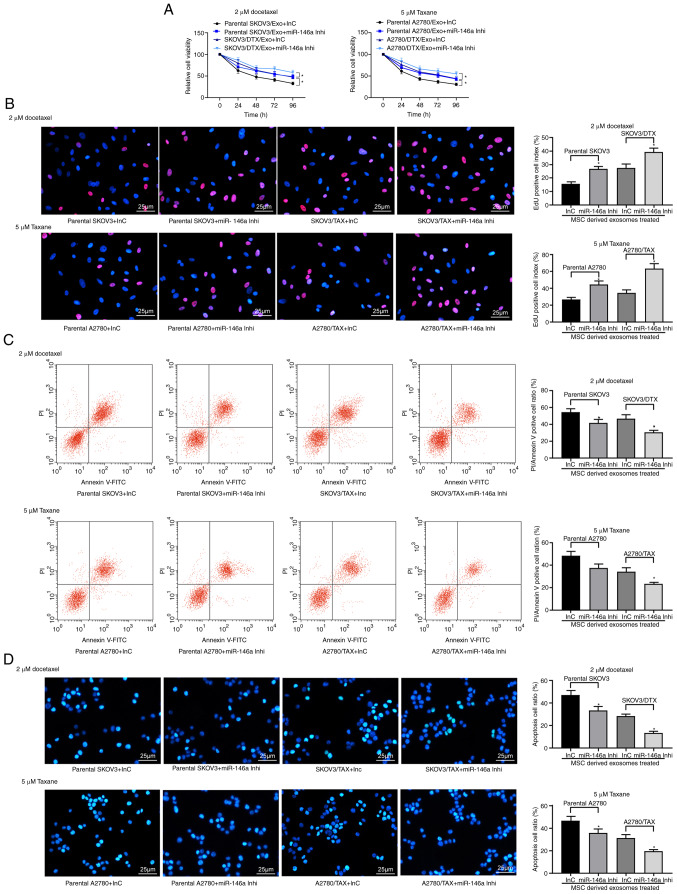
Figure 4 Exosomes from hUCMSCs promote OC cell chemosensitivity partly by transferring miR-146a. hUCMSC-derived exosome-treated parental or drug resistant OC cells were exposed to 2 µM DTX or 5 µM TAX for 2 h, respectively. (A) Cell viability assessed by Cell Counting Kit-8 cell viability assays. (B) EdU staining for cell proliferation. Scale bar, 25 µm. (C) Flow cytometry analysis for cells labelled with PI/Annexin V. (D) Hoechst 33258 staining for cell apoptosis. Scale bar, 25 µm. Data are presented as the mean ± SD. One-way (panels C, D and E) or two-way (panel A) ANOVA followed by Tukey's post hoc test was used to determine statistical significance. Each assessment was performed in triplicate with three repetitions to ensure minimum deviation. *P<0.05 vs. parental cells co-cultured with exosomes derived from InC-transfected MSCs. DTX, docetaxel; hUCMSCs, human umbilical cord mesenchymal stem cells; inhi, inhibitor; InC, inhibitor control; miR, microRNA; OC, ovarian cancer; PI, propidium iodide; TAX, taxane.
Figure 4 Exosomes from hUCMSCs promote OC cell chemosensitivity partly by transferring miR-146a. hUCMSC-derived exosome-treated parental or drug resistant OC cells were exposed to 2 µM DTX or 5 µM TAX for 2 h, respectively. (A) Cell viability assessed by Cell Counting Kit-8 cell viability assays. (B) EdU staining for cell proliferation. Scale bar, 25 µm. (C) Flow cytometry analysis for cells labelled with PI/Annexin V. (D) Hoechst 33258 staining for cell apoptosis. Scale bar, 25 µm. Data are presented as the mean ± SD. One-way (panels C, D and E) or two-way (panel A) ANOVA followed by Tukey's post hoc test was used to determine statistical significance. Each assessment was performed in triplicate with three repetitions to ensure minimum deviation. *P<0.05 vs. parental cells co-cultured with exosomes derived from InC-transfected MSCs. DTX, docetaxel; hUCMSCs, human umbilical cord mesenchymal stem cells; inhi, inhibitor; InC, inhibitor control; miR, microRNA; OC, ovarian cancer; PI, propidium iodide; TAX, taxane.CC BY-NC-ND · PubMed Central
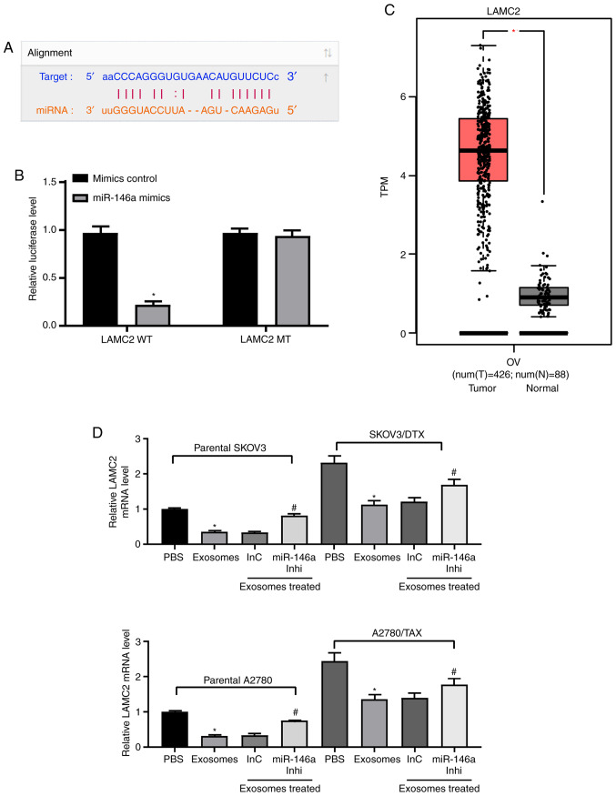
Figure 5 miR-146a blocks the PI3K signaling pathway by inhibiting LAMC2. (A) Possible miR-146 binding sites in the LAMC2 3' untranslated region. (B) Impact of miR-146a on the luciferase activity of LAMC2-WT and LAMC2-MT evaluated by dual luciferase assays. *P<0.05 vs. LAMC2 MT. (C) LAMC2 expression in The Cancer Genome Atlas-ovarian cancer dataset (n=426 for tumor samples and n=88 for normal tissues) predicted by gene expression profiling interactive analysis. *P<0.05. (D) LAMC2 expression in SKOV3 and A2780 cells determined by RT-qPCR. *P<0.05 vs. cells treated with PBS; #P<0.05 vs. cells co-cultured with exosomes extracted from InC-treated MSCs. (E) Protein expression levels of PI3K measured by ELISA. *P<0.05 vs. cells treated with PBS; #P<0.05 vs. cells co-cultured with exosomes extracted from InC-treated MSCs. (F) LAMC2 expression in SKOV3/DTX and A2780/TAX cells determined by RT-qPCR. Data are presented as the mean ± SD. *P<0.05 vs. cells transfected with EV. One-way (panels D, E and F) or two-way (panel B) ANOVA and Tukey's multiple comparisons test were used to determine statistical significance. Each assessment was performed in triplicate with three repetitions to ensure minimum deviation. DTX, docetaxel; InC, inhibitor control; inhi, inhibitor; LAMC2, laminin γ2; miR, microRNA; MSCs, mesenchymal stem cells; MT, mutant; RT-qPCR, reverse transcription-quantitative PCR; TAX, taxane; WT, wild type.
Figure 5 miR-146a blocks the PI3K signaling pathway by inhibiting LAMC2. (A) Possible miR-146 binding sites in the LAMC2 3' untranslated region. (B) Impact of miR-146a on the luciferase activity of LAMC2-WT and LAMC2-MT evaluated by dual luciferase assays. *P<0.05 vs. LAMC2 MT. (C) LAMC2 expression in The Cancer Genome Atlas-ovarian cancer dataset (n=426 for tumor samples and n=88 for normal tissues) predicted by gene expression profiling interactive analysis. *P<0.05. (D) LAMC2 expression in SKOV3 and A2780 cells determined by RT-qPCR. *P<0.05 vs. cells treated with PBS; #P<0.05 vs. cells co-cultured with exosomes extracted from InC-treated MSCs. (E) Protein expression levels of PI3K measured by ELISA. *P<0.05 vs. cells treated with PBS; #P<0.05 vs. cells co-cultured with exosomes extracted from InC-treated MSCs. (F) LAMC2 expression in SKOV3/DTX and A2780/TAX cells determined by RT-qPCR. Data are presented as the mean ± SD. *P<0.05 vs. cells transfected with EV. One-way (panels D, E and F) or two-way (panel B) ANOVA and Tukey's multiple comparisons test were used to determine statistical significance. Each assessment was performed in triplicate with three repetitions to ensure minimum deviation. DTX, docetaxel; InC, inhibitor control; inhi, inhibitor; LAMC2, laminin γ2; miR, microRNA; MSCs, mesenchymal stem cells; MT, mutant; RT-qPCR, reverse transcription-quantitative PCR; TAX, taxane; WT, wild type.CC BY-NC-ND · PubMed Central
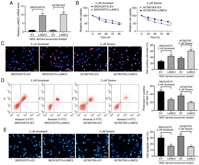
Figure 6 LAMC2 is involved in miR-146a-mediated OC cell sensitivity. hUCMSCs-derived exosome-treated parental or drug resistant OC cells were exposed to 2 µM DTX or 5 µM TAX for 2 h, respectively. (A) LAMC2 mRNA expression measured by reverse transcription-quantitative PCR. (B) Cell viability examined by Cell Counting Kit-8 cell viability assays. (C) EdU staining of OC cells. Scale bar, 25 µm. (D) Flow cytometry analysis for cells labelled with propidium iodide/Annexin V. Scale bar, 25 µm. (E) Hoechst 33258 staining for cell apoptosis. Data are presented as the mean ± SD. One-way (panels A, C, D and E) or two-way (panel B) ANOVA followed by Tukey's multiple comparisons test was used. *P<0.05 vs. cells transfected with EV. Each assessment was performed in triplicate with three repetitions to ensure minimum deviation. DTX, docetaxel; EV, empty vector; hUCMSCs, human umbilical cord mesenchymal stem cells; LAMC2, laminin γ2; miR, microRNA; OC, ovarian cancer; TAX, taxane.
Figure 6 LAMC2 is involved in miR-146a-mediated OC cell sensitivity. hUCMSCs-derived exosome-treated parental or drug resistant OC cells were exposed to 2 µM DTX or 5 µM TAX for 2 h, respectively. (A) LAMC2 mRNA expression measured by reverse transcription-quantitative PCR. (B) Cell viability examined by Cell Counting Kit-8 cell viability assays. (C) EdU staining of OC cells. Scale bar, 25 µm. (D) Flow cytometry analysis for cells labelled with propidium iodide/Annexin V. Scale bar, 25 µm. (E) Hoechst 33258 staining for cell apoptosis. Data are presented as the mean ± SD. One-way (panels A, C, D and E) or two-way (panel B) ANOVA followed by Tukey's multiple comparisons test was used. *P<0.05 vs. cells transfected with EV. Each assessment was performed in triplicate with three repetitions to ensure minimum deviation. DTX, docetaxel; EV, empty vector; hUCMSCs, human umbilical cord mesenchymal stem cells; LAMC2, laminin γ2; miR, microRNA; OC, ovarian cancer; TAX, taxane.CC BY-NC-ND · PubMed Central
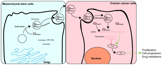
Figure 7 Regulatory mechanism of hUCMSC-derived exosomal miR-146a involved in OC cell proliferation and drug resistance. hUCMSC-derived exosomal miR-146a impeded the proliferation and drug resistance of OC cells by targeting LAMC2 to regulate the PI3K/Akt signaling pathway. hUCMSCs, human umbilical cord mesenchymal stem cells; miR, microRNA; OC, ovarian cancer.
Figure 7 Regulatory mechanism of hUCMSC-derived exosomal miR-146a involved in OC cell proliferation and drug resistance. hUCMSC-derived exosomal miR-146a impeded the proliferation and drug resistance of OC cells by targeting LAMC2 to regulate the PI3K/Akt signaling pathway. hUCMSCs, human umbilical cord mesenchymal stem cells; miR, microRNA; OC, ovarian cancer.CC BY-NC-ND · PubMed Central

Verification

Claims the paper makes

The claims and statistics of this paper haven’t been checked yet.

Run the check — free, no account needed. It reads the open-access text and checks the sources it cites.

A few free checks a day · papers already verified open instantly · results are public

Claims other papers make about this one

Each is checked against this paper’s own text: does it say what the citing paper says it does?

Self-check

Percentages, sums and reported differences re-computed from the paper’s own numbers.

The self-check needs the paper’s full text.

Citations

Cites

Show all 2 references

Research works printed but not linked: 34

  1. Tawde, 2016, Evaluation of microparticulate ovarian cancer vaccine via transdermal route of delivery, J Control Release, 235, 147, 10.1016/j.jconrel.2016.05.058 · DOI
  2. Zahedi, 2011, Combination drug delivery strategy for the treatment of multi-drug resistant ovarian cancer, Mol Pharm, 8, 260, 10.1021/mp100323z · DOI
  3. Lohse, 2019, Ovarian cancer treatment stratification using ex vivo drug sensitivity testing, Anticancer Res, 39, 4023, 10.21873/anticanres.13558 · DOI
  4. Secord, 2019, Ovarian cancer: Time to move beyond one size fits all, Lancet Oncol, 20, 754, 10.1016/S1470-2045(19)30285-2 · DOI
  5. Borley, 2015, Epigenetic mechanisms and therapeutic targets of chemotherapy resistance in epithelial ovarian cancer, Ann Med, 47, 359, 10.3109/07853890.2015.1043140 · DOI
  6. Ridge, 2017, Mesenchymal stem cells: Key players in cancer progression, Mol Cancer, 16, 31, 10.1186/s12943-017-0597-8 · DOI
  7. Li, 2019, Mesenchymal stem cells: From regeneration to cancer, Pharmacol Ther, 200, 42, 10.1016/j.pharmthera.2019.04.005 · DOI
  8. Ding, 2012, Human umbilical cord mesenchymal stem cells support nontu-morigenic expansion of human embryonic stem cells, Cell Transplant, 21, 1515, 10.3727/096368912X647199 · DOI
  9. Houthuijzen, 2012, The role of mesenchymal stem cells in anti-cancer drug resistance and tumour progression, Br J Cancer, 106, 1901, 10.1038/bjc.2012.201 · DOI
  10. Sundararajan, 2018, Correction to: The versatile role of exosomes in cancer progression: Diagnostic and therapeutic implications, Cell Oncol (Dordr), 41, 463, 10.1007/s13402-018-0396-2 · DOI
  11. Halkein, 2013, MicroRNA-146a is a therapeutic target and biomarker for peri-partum cardiomyopathy, J Clin Invest, 123, 2143, 10.1172/JCI64365 · DOI
  12. Roos, 2016, miR-146a-mediated suppression of the inflammatory response in human adipocytes, Sci Rep, 6, 38339, 10.1038/srep38339 · DOI
  13. Yang, 2019, Anti-inflammatory effects of mesenchymal stem cell-derived exosomal microRNA-146a-5p and microRNA-548e-5p on human trophoblast cells, Mol Hum Reprod, 25, 755, 10.1093/molehr/gaz054 · DOI
  14. Cheng, 2019, RAB27B-activated secretion of stem-like tumor exosomes delivers the biomarker microRNA-146a-5p which promotes tumorigenesis and associates with an immunosuppressive tumor microenvironment in colorectal cancer, Int J Cancer, 145, 2209, 10.1002/ijc.32338 · DOI
  15. Zhang, 2019, LAMC2 regulated by microRNA-125a-5p accelerates the progression of ovarian cancer via activating p38 MAPK signalling, Life Sci, 232, 116648, 10.1016/j.lfs.2019.116648 · DOI
  16. Hu, 2020, miR-29c-3p inhibits autophagy and cisplatin resistance in ovarian cancer by regulating FOXP1/ATG14 pathway, Cell Cycle, 19, 193, 10.1080/15384101.2019.1704537 · DOI
  17. Livak, 2001, Analysis of relative gene expression data using real-time quantitative PCR and the 2(-Delta Delta C(T)) method, Methods, 25, 402, 10.1006/meth.2001.1262 · DOI · Paper page
  18. Dominici, 2006, Minimal criteria for defining multipotent mesen-chymal stromal cells. The international society for cellular therapy position statement, Cytotherapy, 8, 315, 10.1080/14653240600855905 · DOI · Paper page
  19. Zhang, 2015, Exosome and exosomal microRNA: Trafficking, sorting, and function, Genomics Proteomics Bioinformatics, 13, 17, 10.1016/j.gpb.2015.02.001 · DOI · Paper page
  20. Asare-Werehene, 2020, The exosome-mediated autocrine and paracrine actions of plasma gelsolin in ovarian cancer chemoresistance, Oncogene, 39, 1600, 10.1038/s41388-019-1087-9 · DOI
  21. Guo, 2019, Cancer-associated fibroblast-derived exosomal microRNA-98-5p promotes cisplatin resistance in ovarian cancer by targeting CDKN1A, Cancer Cell Int, 19, 347, 10.1186/s12935-019-1051-3 · DOI
  22. Hu, 2020, Exosomal miR-146a-5p from treponema pallidum-stimulated macrophages reduces endothelial cells permeability and mono-cyte transendothelial migration by targeting JAM-C, Exp Cell Res, 388, 111823, 10.1016/j.yexcr.2020.111823 · DOI
  23. McCann, 2019, A miRNA signature in endothelial cell-derived extracellular vesicles in tumor-bearing mice, Sci Rep, 9, 16743, 10.1038/s41598-019-52466-1 · DOI
  24. Cui, 2016, miR-146a inhibits proliferation and enhances chemosensitivity in epithelial ovarian cancer via reduction of SOD2, Oncol Res, 23, 275, 10.3727/096504016X14562725373798 · DOI
  25. Ding, 2019, Exosomes derived from human umbilical cord mesen-chymal stromal cells deliver exogenous miR-145-5p to inhibit pancreatic ductal adenocarcinoma progression, Cancer Lett, 442, 351, 10.1016/j.canlet.2018.10.039 · DOI · Paper page
  26. Yuan, 2019, Exosomes derived from MicroRNA-148b-3p-overexpressing human umbilical cord mesenchymal stem cells restrain breast cancer progression, Front Oncol, 9, 1076, 10.3389/fonc.2019.01076 · DOI
  27. Kamada, 2015, Urinary laminin-g2 is a novel biomarker of non-muscle invasive urothelial carcinoma, Cancer Sci, 106, 1730, 10.1111/cas.12832 · DOI
  28. Korbakis, 2015, Serum LAMC2 enhances the prognostic value of a multi-parametric panel in non-small cell lung cancer, Br J Cancer, 113, 484, 10.1038/bjc.2015.171 · DOI
  29. Kosanam, 2013, Laminin, gamma 2 (LAMC2): A promising new putative pancreatic cancer biomarker identified by proteomic analysis of pancreatic adenocarcinoma tissues, Mol Cell Proteomics, 12, 2820, 10.1074/mcp.M112.023507 · DOI
  30. Govaere, 2016, Laminin-332 sustains chemo-resistance and quiescence as part of the human hepatic cancer stem cell niche, J Hepatol, 64, 609, 10.1016/j.jhep.2015.11.011 · DOI
  31. Liang, 2018, LncRNA CASC9 promotes esophageal squamous cell carcinoma metastasis through upregulating LAMC2 expression by interacting with the CREB-binding protein, Cell Death Differ, 25, 1980, 10.1038/s41418-018-0084-9 · DOI
  32. Binju, 2019, Therapeutic inducers of apoptosis in ovarian cancer, 1786
  33. Zhang, 2019, The exosome-mediated PI3k/Akt/mTOR signaling pathway in cervical cancer, Int J Clin Exp Pathol, 12, 2474
  34. Li, LncRNA MALAT1 mediates proliferation of LPS treated-articular chondrocytes by targeting the miR-146a-PI3K/Akt/mTOR axis, Life Sci

Cited by

Sorted by citations

See all 88

Metadata

Authors
Qiu, L., Wang, J., Chen, M., Chen, F., & Tu, W.
Venue
International Journal of Molecular Medicine
Published
2020-06-05
Volume
46
Issue
2
Pages
609-620
ISSN
  • 1107-3756
  • 1791-244X
Cited by
88
References
2

About these checks

Registry, retraction, Cites and statistics results are computed, not judged, with one exception: a printed reference that no index lists is read once by a model before it counts as a citation error. Claims are extracted by a model and kept only when their quote appears verbatim in the text; each cited source is then checked, with a model’s help, for whether it says what the claim relies on. Those verdicts can be wrong — read the claim’s quote and the cited source before relying on one.

Statistics and arithmetic are recomputed, not judged.

Confidence bands are shown only for fields calibrated on our annotated test set. None is calibrated yet, so no band is shown.

What counts as a claim ›How we measure confidence ›How we check this ›

Are you an author, or do you see a problem with this page? Request a review

How to cite

Cite CoChat for the checks on this page, and the paper for its own findings.

This page
CoChat. (2026). Citation check: Exosomal microRNA‑146a derived from mesenchymal stem cells increases the sensitivity of ovarian cancer cells to docetaxel and taxane via a LAMC2‑mediated PI3K/Akt axis. https://app.cochat.ai/papers/10.3892/ijmm.2020.4634
The paper
Qiu, L., Wang, J., Chen, M., Chen, F., & Tu, W. (2020). Exosomal microRNA‑146a derived from mesenchymal stem cells increases the sensitivity of ovarian cancer cells to docetaxel and taxane via a LAMC2‑mediated PI3K/Akt axis. International Journal of Molecular Medicine. https://doi.org/10.3892/ijmm.2020.4634