

The DOI matches its Crossref record; 7 of the 7 references it cites were found in a registry or an index.
Neural stem cell-derived exosome as a nano-sized carrier for BDNF delivery to a rat model of ischemic stroke
Abstract
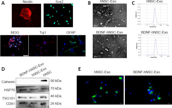
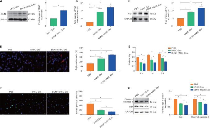
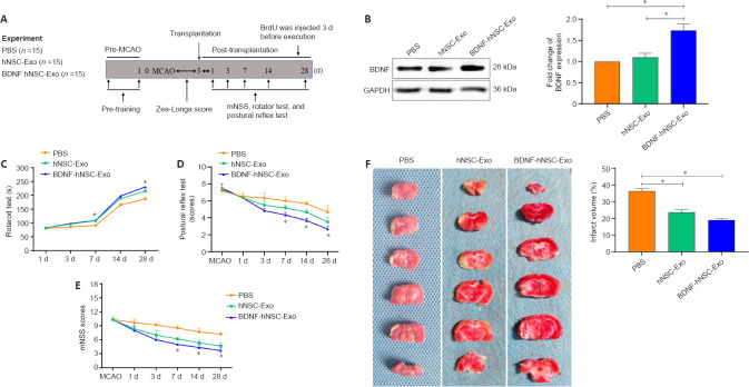
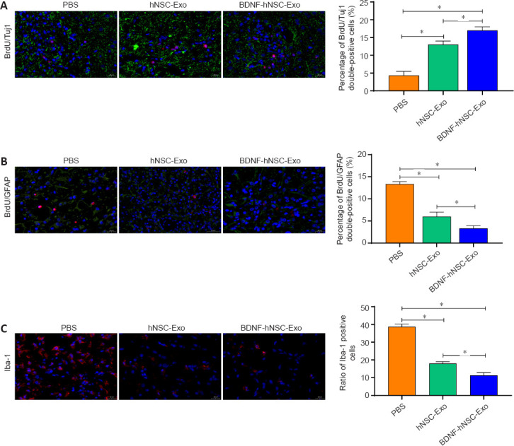
JOURNAL/nrgr/04.03/01300535-202302000-00042/figure1/v/2026-05-26T054653Z/r/image-tiff Our previous study demonstrated the potential therapeutic role of human neural stem cell-derived exosomes (hNSC-Exo) in ischemic stroke. Here, we loaded brain-derived neurotrophic factor (BDNF) into exosomes derived from NSCs to construct engineered exosomes (BDNF-hNSC-Exo) and compared their effects with those of hNSC-Exo on ischemic stroke both in vitro and in vivo . In a model of H 2 O 2 -induced oxidative stress in NSCs, BDNF-hNSC-Exo markedly enhanced cell survival. In a rat middle cerebral artery occlusion model, BDNF-hNSC-Exo not only inhibited the activation of microglia, but also promoted the differentiation of endogenous NSCs into neurons. These results suggest that BDNF can improve the function of NSC-derived exosomes in the treatment of ischemic stroke. Our research may support the clinical use of other neurotrophic factors for central nervous system diseases.
Overview
The claims and statistics of this paper haven’t been checked yet.
Run the check — free, no account needed. It reads the open-access text and checks the sources it cites.
Integrity signals
Registry
Cites
Statistics
Figures
Verification
Claims the paper makes
The claims and statistics of this paper haven’t been checked yet.
Run the check — free, no account needed. It reads the open-access text and checks the sources it cites.
Claims other papers make about this one
Each is checked against this paper’s own text: does it say what the citing paper says it does?
In the citing paper
“microglia and NSC serete sEVs which facilitate their long-distance communication [50, 51].”
From M2 microglia-derived small extracellular vesicles modulate NSC fate after ischemic stroke via miR-25-3p/miR-93-5p-TGFBR/PTEN/FOXO3 axis (2025) · cites this paper as [51]
Self-check
Percentages, sums and reported differences re-computed from the paper’s own numbers.
The self-check needs the paper’s full text.
Citations
Cites
The biology , function , and biomedical applications of exosomes
Kalluri, R., & LeBleu, V. S. (2020) · Science · doi:10.1126/science.aau6977 · Cited by 10,770
The ARRIVE guidelines 2.0: Updated guidelines for reporting animal research
Percie du Sert, N., Hurst, V., Ahluwalia, A., Alam, S., Avey, M. T., Baker, M., Browne, W. J., Clark, A., Cuthill, I. C., Dirnagl, U., et al. (2020) · PLOS Biology · doi:10.1371/journal.pbio.3000410 · Cited by 5,939
The Glial Nature of Embryonic and Adult Neural Stem Cells
Kriegstein, A., & Alvarez-Buylla, A. (2009) · Annual Review of Neuroscience · doi:10.1146/annurev.neuro.051508.135600 · Cited by 2,475
Xu, X., Liang, Y., Li, X., Ouyang, K., Wang, M., Cao, T., Li, W., Liu, J., Xiong, J., Li, B., et al. (2021) · Biomaterials · doi:10.1016/j.biomaterials.2020.120539 · Cited by 377
Extracellular vesicles: A bright star of nanomedicine
Wu, P., Zhang, B., Ocansey, D. K. W., Xu, W., & Qian, H. (2021) · Biomaterials · doi:10.1016/j.biomaterials.2020.120467 · Cited by 368
Nanotechnology: A Promising Approach for Delivery of Neuroprotective Drugs
Naqvi, S., Panghal, A., & Flora, S. J. S. (2020) · Frontiers in Neuroscience · doi:10.3389/fnins.2020.00494 · Cited by 305 · cited 1×
“However, exogenous administration of neurotrophic factors for stroke treatment is limited by their poor half-life, lack of blood-brain barrier permeability, and rapid degradation (Yuan et al., 2017; Naqvi et al., 2020).”
BackgroundNeural stem cells traffic functional mitochondria via extracellular vesicles
Peruzzotti-Jametti, L., Bernstock, J. D., Willis, C. M., Manferrari, G., Rogall, R., Fernandez-Vizarra, E., Williamson, J. C., Braga, A., van den Bosch, A., Leonardi, T., et al. (2021) · PLOS Biology · doi:10.1371/journal.pbio.3001166 · Cited by 263
Research works printed but not linked: 35
- Ahmad, 2022, Novel therapeutic interventions for combating Parkinson's disease and prospects of Nose-to-Brain drug delivery, Biochem Pharmacol, 195, 114849, 10.1016/j.bcp.2021.114849 · DOI
- Arumugam, 2006, Gamma secretase-mediated Notch signaling worsens brain damage and functional outcome in ischemic stroke, Nat Med, 12, 621, 10.1038/nm1403 · DOI
- Belayev, 2017, Neuroprotectin D1 upregulates Iduna expression and provides protection in cellular uncompensated oxidative stress and in experimental ischemic stroke, Cell Death Differ, 24, 1091, 10.1038/cdd.2017.55 · DOI
- Chen, 2020, Exosome-shuttled circSHOC2 from IPASs regulates neuronal autophagy and ameliorates ischemic brain injury via the miR-7670-3p/SIRT1 axis, Mol Ther Nucleic Acids, 22, 657, 10.1016/j.omtn.2020.09.027 · DOI
- Fang, 2020, Small extracellular vesicles derived from human mesenchymal stromal cells prevent group 2 innate lymphoid cell-dominant allergic airway inflammation through delivery of miR-146a-5p, J Extracell Vesicles, 9, 1723260, 10.1080/20013078.2020.1723260 · DOI
- Feng, 2021, Emerging exosomes and exosomal miRNAs in spinal cord injury, Front Cell Dev Biol, 9, 703989, 10.3389/fcell.2021.703989 · DOI
- Fu, 2019, CAR exosomes derived from effector CAR-T cells have potent antitumour effects and low toxicity, Nat Commun, 10, 4355, 10.1038/s41467-019-12321-3 · DOI
- Goldstein, 2007, Acute ischemic stroke treatment in 2007, Circulation, 116, 1504, 10.1161/CIRCULATIONAHA.106.670885 · DOI
- Hamblin, 2022, Modulation of gene expression on a transcriptome-wide level following human neural stem cell transplantation in aged mouse stroke brains, Exp Neurol, 347, 113913, 10.1016/j.expneurol.2021.113913 · DOI
- Hao, 2021, Memantine, NMDA receptor antagonist, attenuates ox-LDL-induced inflammation and oxidative stress via activation of BDNF/TrkB signaling pathway in HUVECs, Inflammation, 44, 659, 10.1007/s10753-020-01365-z · DOI
- Huang, 2021, A conductive dual-network hydrogel composed of oxidized dextran and hyaluronic-hydrazide as BDNF delivery systems for potential spinal cord injury repair, Int J Biol Macromol, 167, 434, 10.1016/j.ijbiomac.2020.11.206 · DOI
- Hughes, 2020, Association of blood pressure lowering with incident dementia or cognitive impairment:a systematic review and meta-analysis, Jama, 323, 1934, 10.1001/jama.2020.4249 · DOI · Paper page
- Kim, 2020, Exosomes:cell-derived nanoplatforms for the delivery of cancer therapeutics, Int J Mol Sci, 22, 14, 10.3390/ijms22010014 · DOI
- Lai, 2020, Restoration of L-OPA1 alleviates acute ischemic stroke injury in rats via inhibiting neuronal apoptosis and preserving mitochondrial function, Redox Biol, 34, 101503, 10.1016/j.redox.2020.101503 · DOI
- Leira, 2020, Preserving stroke care during the COVID-19 pandemic:Potential issues and solutions, Neurology, 95, 124, 10.1212/WNL.0000000000009713 · DOI
- Li, 2010, Coexistence of quiescent and active adult stem cells in mammals, Science, 327, 542, 10.1126/science.1180794 · DOI · Paper page
- Li, 2021, Recovery of post-stroke cognitive and motor deficiencies by Shuxuening injection via regulating hippocampal BDNF-mediated neurotrophin/Trk signaling, Biomed Pharmacother, 141, 111828, 10.1016/j.biopha.2021.111828 · DOI
- Lv, 2021, Progress in clinical trials of stem cell therapy for cerebral palsy, Neural Regen Res, 16, 1377, 10.4103/1673-5374.300979 · DOI
- Ma, 2021, Temporal trend and attributable risk factors of stroke burden in China, 1990-2019: an analysis for the Global Burden of Disease Study 2019, Lancet Public Health, 6, e897, 10.1016/S2468-2667(21)00228-0 · DOI
- Marko, 2020, Trends of r-tPA (recombinant tissue-type plasminogen activator) treatment and treatment-influencing factors in acute ischemic stroke, Stroke, 51, 1240, 10.1161/STROKEAHA.119.027921 · DOI
- Muheremu, 2021, Sustained delivery of neurotrophic factors to treat spinal cord injury, Transl Neurosci, 12, 494, 10.1515/tnsci-2020-0200 · DOI
- Song, 2013, Apoptosis signal-regulating kinase 1 (ASK1) is linked to neural stem cell differentiation after ischemic brain injury, Exp Mol Med, 45, e69, 10.1038/emm.2013.134 · DOI
- Tian, 2021, Targeted delivery of neural progenitor cell-derived extracellular vesicles for anti-inflammation after cerebral ischemia, Theranostics, 11, 6507, 10.7150/thno.56367 · DOI
- Vogel, 2018, Neural stem cell derived extracellular vesicles:Attributes and prospects for treating neurodegenerative disorders, EBioMedicine, 38, 273, 10.1016/j.ebiom.2018.11.026 · DOI
- Xiong, 2021, Brain-derived neurotrophic factor and its related enzymes and receptors play important roles after hypoxic-ischemic brain damage, Neural Regen Res, 16, 1453, 10.4103/1673-5374.303033 · DOI
- Xiong, 2016, Functions and mechanisms of microglia/macrophages in neuroinflammation and neurogenesis after stroke, Prog Neurobiol, 142, 23, 10.1016/j.pneurobio.2016.05.001 · DOI
- Yuan, 2017, Macrophage exosomes as natural nanocarriers for protein delivery to inflamed brain, Biomaterials, 142, 1, 10.1016/j.biomaterials.2017.07.011 · DOI · Paper page
- Yuan, 2021, Neural stem cell-derived exosomes regulate neural stem cell differentiation through miR-9-Hes1 axis, Front Cell Dev Biol, 9, 601600, 10.3389/fcell.2021.601600 · DOI
- Zhang, 2018, Neural stem cells alleviate inflammation via neutralization of IFN-γnegative effect in ischemic stroke model, J Biomed Nanotechnol, 14, 1178, 10.1166/jbn.2018.2568 · DOI
- Zhang, 2020, Exosomes derived from human neural stem cells stimulated by interferon gamma improve therapeutic ability in ischemic stroke model, J Adv Res, 24, 435, 10.1016/j.jare.2020.05.017 · DOI
- Zhang, 2016, Mesenchymal stromal cell therapy in ischemic stroke, J Neurorestoratol, 4, 79, 10.2147/JN.S119874 · DOI
- Zhang, 2019, Sevoflurane prevents miR-181a-induced cerebral ischemia/reperfusion injury, Chem Biol Interact, 308, 332, 10.1016/j.cbi.2019.06.008 · DOI
- Zhang, 2016, Neural stem cells and ischemic brain, J Stroke, 18, 267, 10.5853/jos.2016.00206 · DOI
- Zhang, 2016, Exosomes in stroke pathogenesis and therapy, J Clin Invest, 126, 1190, 10.1172/JCI81133 · DOI
- Zhong, 2020, Neural stem cell-derived exosomes facilitate spinal cord functional recovery after injury by promoting angiogenesis, Exp Biol Med (Maywood), 245, 54, 10.1177/1535370219895491 · DOI
Cited by
Sorted by citations
Clinical applications of stem cell-derived exosomes
Tan, F., Li, X., Wang, Z., Li, J., Shahzad, K., & Zheng, J. (2024) · Signal Transduction and Targeted Therapy · doi:10.1038/s41392-023-01704-0 · Cited by 708
Zhong, L., Wang, J., Wang, P., Liu, X., Liu, P., Cheng, X., Cao, L., Wu, H., Chen, J., & Zhou, L. (2023) · Stem Cell Research & Therapy · doi:10.1186/s13287-023-03409-1 · Cited by 110
Zhang, Q., Yi, Y., Chen, T., Ai, Y., Chen, Z., Liu, G., Tang, Z., Chen, J., Xu, T., Chen, X., et al. (2025) · Journal of Nanobiotechnology · doi:10.1186/s12951-025-03390-2 · Cited by 27
Metadata
- Authors
- Zhu, Z.-H., Jia, F., Ahmed, W., Zhang, G.-L., Wang, H., Lin, C.-Q., Chen, W.-H., & Chen, L.-K.
- Affiliations
- Department of Neurosurgery, School of Medicine, Southeast University, Nanjing, Jiangsu Province, China
- Department of Neurosurgery, Neuroscience Center, Cancer Center, Integrated Hospital of Traditional Chinese Medicine, Southern Medical University, Guangzhou, Guangdong Province, China
- Department of Neurosurgery, The Second Affiliated Hospital of Guangzhou Medical University, Guangzhou, Guangdong Province, China
- Department of Neurosurgery, Shanghai Ninth People’s Hospital, Shanghai Jiao Tong University School of Medicine, Shanghai, China
- ORCID
- Chen, Lu-Kui
- Venue
- Neural Regeneration Research
- Published
- 2022-06-02
- Volume
- 18
- Issue
- 2
- Pages
- 404-409
- ISSN
- 1673-5374
- 1876-7958
- PMC
- PMC9396474
- Cited by
- 132
- References
- 7
- Language
- en
About these checks
Registry, retraction, Cites and statistics results are computed, not judged, with one exception: a printed reference that no index lists is read once by a model before it counts as a citation error. Claims are extracted by a model and kept only when their quote appears verbatim in the text; each cited source is then checked, with a model’s help, for whether it says what the claim relies on. Those verdicts can be wrong — read the claim’s quote and the cited source before relying on one.
Statistics and arithmetic are recomputed, not judged.
Confidence bands are shown only for fields calibrated on our annotated test set. None is calibrated yet, so no band is shown.
What counts as a claim ›How we measure confidence ›How we check this ›
Are you an author, or do you see a problem with this page? Request a review
