

The DOI matches its Crossref record; 167 of the 171 references it cites were found in a registry or an index.
The PI3K/AKT pathway in obesity and type 2 diabetes
Open accessAbstract
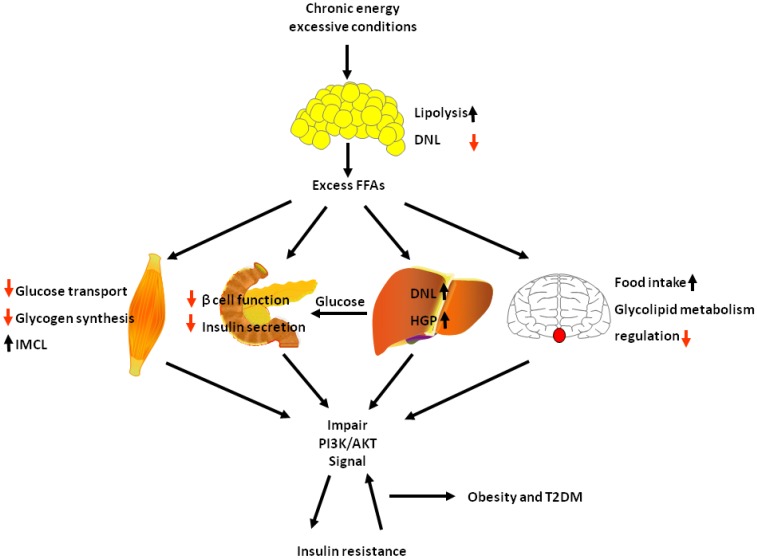
Obesity and type 2 diabetes mellitus are complicated metabolic diseases that affect multiple organs and are characterized by hyperglycaemia. Currently, stable and effective treatments for obesity and type 2 diabetes mellitus are not available. Therefore, the mechanisms leading to obesity and diabetes and more effective ways to treat obesity and diabetes should be identified. Based on accumulated evidences, the PI3K/AKT signalling pathway is required for normal metabolism due to its characteristics, and its imbalance leads to the development of obesity and type 2 diabetes mellitus. This review focuses on the role of PI3K/AKT signalling in the skeletal muscle, adipose tissue, liver, brain and pancreas, and discusses how this signalling pathway affects the development of the aforementioned diseases. We also summarize evidences for recently identified therapeutic targets of the PI3K/AKT pathway as treatments for obesity and type 2 diabetes mellitus. PI3K/AKT pathway damaged in various tissues of the body leads to obesity and type 2 diabetes as the result of insulin resistance, and in turn, insulin resistance exacerbates the PI3K/AKT pathway, forming a vicious circle.
Overview
The claims and statistics of this paper haven’t been checked yet.
Run the check — free, no account needed. It reads the open-access text and checks the sources it cites.
Integrity signals
Registry
Cites
Statistics
Figures
Verification
Claims the paper makes
The claims and statistics of this paper haven’t been checked yet.
Run the check — free, no account needed. It reads the open-access text and checks the sources it cites.
Claims other papers make about this one
Each is checked against this paper’s own text: does it say what the citing paper says it does?
The cited source says what the claim attributes to it.
In the citing paper
“AKT activation leads to increased glucose transporter 4 (GLUT4) expression and translocation, ultimately leading to increased glucose uptake.”
From Non-Diabetic Hypoglycemia: Evaluation and Management in Adults (2025) · cites this paper as [48]
The cited source says what the claim attributes to it.
In the citing paper
“PI3K is an enzyme that activates protein kinase B (AKT), which regulates glucose metabolism, among other intracellular functions [48].”
From Non-Diabetic Hypoglycemia: Evaluation and Management in Adults (2025) · cites this paper as [48]
Self-check
Percentages, sums and reported differences re-computed from the paper’s own numbers.
The self-check needs the paper’s full text.
Citations
Cites
mTOR Signaling in Growth Control and Disease
Laplante, M., & Sabatini, D. M. (2012) · Cell · doi:10.1016/j.cell.2012.03.017 · Cited by 8,509 · cited 1×
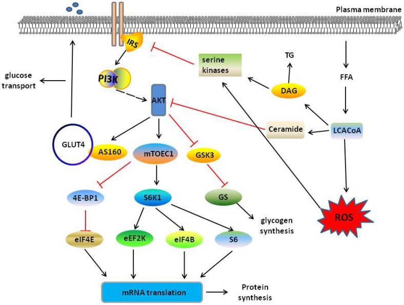
“AKT-mediated mTORC1 activation regulates a number of downstream effectors proteins through phosphorylation, including the well-characterized effectors p70 ribosomal S6 protein kinase-1 (S6K1) and eukaryotic translation initiation factor-4E (eIF4E)binding protein-1 (4E-BP1), to increase protein synthesis [43, 44].”
BackgroundPhosphorylation and regulation of Akt/PKB by the rictor-mTOR complex
Sarbassov, D. D., Guertin, D. A., Ali, S. M., & Sabatini, D. M. (2005) · Science · doi:10.1126/science.1106148 · Cited by 6,216 · cited 1×
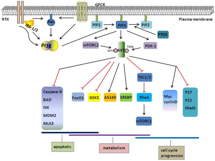
“First, phosphorylation of the threonine 308 (AKT1) in the kinase domain by phosphoinositide-dependent protein kinase 1 (PDK1) initiates the activation process [19], subsequent phosphorylation at serine 473 (AKT1) in the carboxy-terminal regulatory domain through mTOR complex 2 (mTORC2)[17, 20], which is activated by a…”
BackgroundAKT/PKB signaling: navigating downstream
Manning, B. D., & Cantley, L. C. (2007) · Cell · doi:10.1016/j.cell.2007.06.009 · Cited by 6,115 · cited 1×
“AKT regulates two processes by glycolysis Glucose 6-phosphate and glycogen synthase kinase 3 (GSK3) to produce cellular energy via glycolysis and promotes glycogen production by inhibiting [30].”
BackgroundMechanisms linking obesity to insulin resistance and type 2 diabetes
Kahn, S. E., Hull, R. L., & Utzschneider, K. M. (2006) · Nature · doi:10.1038/nature05482 · Cited by 5,448 · cited 1×
“In obese subjects, excess levels of circulating FFAs, which are caused by a blunted response of adipose tissues, impairs the function of β cells [150].”
BackgroundInhibition of glycogen synthase kinase-3 by insulin mediated by protein kinase B
Cross, D. A. E., Alessi, D. R., Cohen, P., Andjelkovich, M., & Hemmings, B. A. (1995) · Nature · doi:10.1038/378785a0 · Cited by 5,244 · cited 8×
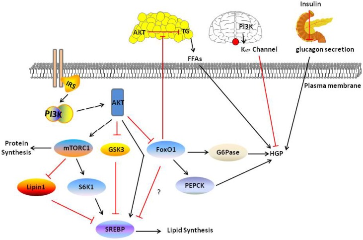
“In the feeding state, SREBP-1c is regulated by four pathways: activated AKT stimulates liver X receptor (LXR), which is require for SREBP-1c transcription [64]; activated AKT stimulates mTORC1 to activate SREBP-1c transcription, and mTORC1 also inhibits Lipin-1, which decreases the half-life of nuclear SREBP-1c [65, 6…”
BackgroundInfluentialAKT/PKB Signaling: Navigating the Network
Manning, B. D., & Toker, A. (2017) · Cell · doi:10.1016/j.cell.2017.04.001 · Cited by 3,772 · cited 2×
“Similar phosphorylation events are observed at corresponding residues in AKT2 (T309 and S474) and AKT3 (T305 and S472) [22].”
BackgroundRole of leptin in the neuroendocrine response to fasting
Ahima, R. S., Prabakaran, D., Mantzoros, C., Qu, D., Lowell, B., Maratos-Flier, E., & Flier, J. S. (1996) · Nature · doi:10.1038/382250a0 · Cited by 3,117 · cited 1×
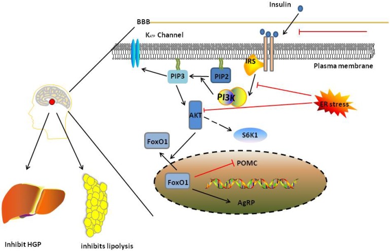
“In the presence of persistent excess energy availability, leptin signalling activates other pathways, such as STAT3 or SH2-containing tyrosine phosphatase, to predominantly regulate food intake in POMC neurons [90, 113, 119].”
BackgroundAlessi, D. R., James, S. R., Downes, C. P., Holmes, A. B., Gaffney, P. R. J., Reese, C. B., & Cohen, P. (1997) · Current Biology · doi:10.1016/s0960-9822(06)00122-9 · Cited by 2,874 · cited 1×
“First, phosphorylation of the threonine 308 (AKT1) in the kinase domain by phosphoinositide-dependent protein kinase 1 (PDK1) initiates the activation process [19], subsequent phosphorylation at serine 473 (AKT1) in the carboxy-terminal regulatory domain through mTOR complex 2 (mTORC2)[17, 20], which is activated by a…”
BackgroundEndoplasmic reticulum stress and the inflammatory basis of metabolic disease
Hotamisligil, G. S. (2010) · Cell · doi:10.1016/j.cell.2010.02.034 · Cited by 2,802 · cited 1×
“Chronic metabolic inflammation and endoplasmic reticulum (ER) stress also impair hepatic insulin-AKT signalling and subsequently causes insulin resistance [106, 107].”
BackgroundHealth and economic burden of the projected obesity trends in the USA and the UK
Wang, Y. C., McPherson, K., Marsh, T., Gortmaker, S. L., & Brown, M. (2011) · The Lancet · doi:10.1016/s0140-6736(11)60814-3 · Cited by 2,775 · cited 1×
“The prevalence of obesity has grown at an alarming rate although specific public health policies and treatment efforts have been developed to resist the obesity epidemic [1], potentially leading to increased numbers of patients affected by complications of obesity, such as the most devastating type 2 diabetes mellitus…”
Background
Research works printed but not linked: 5
- [98]Yecies Jessica L Zhang Hui H Menon S Liu S Yecies D Lipovsky Alex I Akt Stimulates Hepatic SREBP1c and Lipogenesis through Parallel mTORC1-Dependent and Independent Pathways Cell metabolism 2011 14 1 21 32 21723501 PMC3652544
- [120]Al-Qassab H Smith MA Irvine EE Guillermet-Guibert J Claret M Choudhury AI Dominant role of the p110beta isoform of PI3K over p110alpha in energy homeostasis regulation by POMC and AgRP neurons Cell metabolism 2009 10 5 343 54 19883613 PMC2806524
- [144]Bernal-Mizrachi E Wen W Stahlhut S Welling CM Permutt MA Islet beta cell expression of constitutively active Akt1/PKB alpha induces striking hypertrophy, hyperplasia, and hyperinsulinemia J Clin Invest 2001 108 11 1631 8 11733558 PMC200992
- [147]Wang HW Mizuta M Saitoh Y Noma K Ueno H Nakazato M Glucagon-like peptide-1 and candesartan additively improve glucolipotoxicity in pancreatic beta-cells Metabolism 2011 60 8 1081 9 21255808
- [161]Villani V Milanesi A Sedrakyan S Da Sacco S Angelow S Conconi MT Amniotic fluid stem cells prevent beta-cell injury Cytotherapy 2014 16 1 41 55 24210784 PMC4374982
Cited by
Sorted by citations
Items 1,001–1,008 of 1,008
Li, Y., Gao, S., Yang, Y., & Yin, G. (2023) · International Journal of Diabetes in Developing Countries · doi:10.1007/s13410-023-01240-1
“Thus the dysregulation of PI3K/AKT signalling pathway is considered to be one of the mechanisms underlying the pathogenesis of type-2 diabetes mellitus [6].”
InfluentialHasan, H. J., Jouda, J., & Saihood, Y. D. (2023) · Al-Kufa University Journal for Biology · doi:10.36320/ajb/v15.i2.13329
“This is explained by the recently discovered fact that adipose tissues are neither inert nor restricted to serving as an energy storage organ but instead are a major endocrine organ and play a substantial role in the etiology of T2DM and insulin resistance (IR) [7].”
BackgroundYu, D., Wang, Y., Ma, F., Guan, Y., Ye, S., Shen, G.-M., & You, L. (2023) · All Life · doi:10.1080/26895293.2023.2220537 · Cited by 1
Liang, Y., Chen, Q., Zhou, E., Bi, J.-P., Wang, J., Li, Y., & Li, L. (2023) · Journal of Functional Foods · doi:10.1016/j.jff.2023.105568
Gou, H., Zhou, H., Zhao, M., Zhang, X.-J., & Zhao, Q. (2023) · Translational Pediatrics · doi:10.21037/tp-2026-1-0108
“Under physiological conditions, insulin-induced activation of PI3K/Akt maintains metabolic homeostasis; however, chronic energy excess and elevated free fatty acid levels disrupt this pathway, leading to insulin resistance and promoting obesity and type 2 diabetes (14,15).”
Thyroid gland cancer and insulin resistance: a modern view of the problem
Pidchenko, N. S. (2022) · Український радіологічний та онкологічний журнал · doi:10.46879/ukroj.3.2022.79-92
“PI3K/Akt/mTOR сприяє реалізації основних метаболічних ефектів інсуліну, а саме: стимулює синтез білка, глікогену, ліпідів, пригнічує глюконеогенез і ліполіз [68, 69].”
BackgroundThottappillil, A., Sahoo, S., Chakraborty, A., Kouser, S., Ravi, R., Garawadmath, S., Banvi, P. G., Kukkupuni, S. K., Suma Mohan, S., & Vishnuprasad, C. N. (2022) · bioRxiv (Cold Spring Harbor Laboratory) · doi:10.1101/2022.07.15.500175 · Cited by 1
“Proinflammatory markers like NFKB1 and TNFα, characteristic of the insulin resistance were present in the cluster along with lipid regulatory and obesity-linked PPARα and PPARγ [2628] The other critical markers were insulin signalling molecules like AKT1 and PI3K, demonstrating that these core pathways involved in dia…”
BackgroundOlder Women with lower lean mass values have hypermethylated sites in the PI3K-Akt pathwayPreprint
Correia, I. M., Rodrigues, G. D. S., Noronha, N., de Almeida, M. L., Sobrinho, A. C. D. S., Nonino, C., & Bueno, C. (2022) · bioRxiv · doi:10.1101/2022.10.14.512202
“However, excess energy, as in obesity, can impair the signaling of this pathway, causing insulin resistance (61).”
Metadata
- Authors
- Huang, X., Liu, G., Guo, J., & Su, Z.
- Affiliations
- Guangdong Pharmaceutical University (CN)
- Shenzhen Center for Disease Control and Prevention (CN)
- Corresponding author
- Guo, Jiao
- Su, Zhengquan
- Venue
- International Journal of Biological Sciences
- Publisher
- Ivyspring International Publisher
- Published
- 2018
- Volume
- 14
- Issue
- 11
- Pages
- 1483-1496
- ISSN
- 1449-2288 (ISSN-L)
- PMC
- PMC6158718
- OpenAlex
- W2886504687
- Cited by
- 1,673
- References
- 171
- Citations by year
- 2026: 197 · 2025: 296 · 2024: 282 · 2023: 258 · 2022: 246 · 2021: 211 · 2020: 136 · 2019: 44 · 2018: 3
- FWCI
- 9.71
- Type
- journal article
- Language
- en
- Open access
- gold
- doi.org
- Licence
- cc-by-nc
- Locations
- International Journal of Biological Sciences (publishedVersion, cc-by-nc)
- Europe PMC (PubMed Central) (publishedVersion, other-oa)
- PubMed Central (publishedVersion, cc-by-nc)
- PubMed (publishedVersion)
- Funders
- National Natural Science Foundation of China (2017-02, 81173107)
- Topics
- Life Sciences › Biochemistry, Genetics and Molecular Biology › Molecular Biology › Metabolism, Diabetes, and Cancer
- Life Sciences › Biochemistry, Genetics and Molecular Biology › Molecular Biology › PI3K/AKT/mTOR signaling in cancer
- Health Sciences › Medicine › Endocrinology, Diabetes and Metabolism › Adipokines, Inflammation, and Metabolic Diseases
- Keywords
- PI3K/Akt signaling, obesity, type 2 diabetes, insulin resistance, hyperglycemia, skeletal muscle, adipose tissue, liver, pancreas, therapeutic target, metabolic disorders
- MeSH terms
- Animals, Diabetes Mellitus, Type 2, Humans, Insulin Resistance, Obesity, Signal Transduction, Phosphatidylinositol 3-Kinases, Proto-Oncogene Proteins c-akt
About these checks
Registry, retraction, Cites and statistics results are computed, not judged, with one exception: a printed reference that no index lists is read once by a model before it counts as a citation error. Claims are extracted by a model and kept only when their quote appears verbatim in the text; each cited source is then checked, with a model’s help, for whether it says what the claim relies on. Those verdicts can be wrong — read the claim’s quote and the cited source before relying on one.
Statistics and arithmetic are recomputed, not judged.
Confidence bands are shown only for fields calibrated on our annotated test set. None is calibrated yet, so no band is shown.
What counts as a claim ›How we measure confidence ›How we check this ›
Are you an author, or do you see a problem with this page? Request a review
