Cited by 136Alzheimer's disease research and treatmentsGenomic variations and chromosomal abnormalitiesDementia and Cognitive Impairment Research

The DOI matches its Crossref record; 86 of the 87 references it cites were found in a registry or an index.

Genomic mosaicism with increased amyloid precursor protein (APP) gene copy number in single neurons from sporadic Alzheimer's disease brains

Open access

Abstract

Previous reports have shown that individual neurons of the brain can display somatic genomic mosaicism of unknown function. In this study, we report altered genomic mosaicism in single, sporadic Alzheimer's disease (AD) neurons characterized by increases in DNA content and amyloid precursor protein (APP) gene copy number. AD cortical nuclei displayed large variability with average DNA content increases of ∼8% over non-diseased controls that were unrelated to trisomy 21. Two independent single-cell copy number analyses identified amplifications at the APP locus. The use of single-cell qPCR identified up to 12 copies of APP in sampled neurons. Peptide nucleic acid (PNA) probes targeting APP, combined with super-resolution microscopy detected primarily single fluorescent signals of variable intensity that paralleled single-cell qPCR analyses. These data identify somatic genomic changes in single neurons, affecting known and unknown loci, which are increased in sporadic AD, and further indicate functionality for genomic mosaicism in the CNS.

Overview

The claims and statistics of this paper haven’t been checked yet.

Run the check — free, no account needed. It reads the open-access text and checks the sources it cites.

A few free checks a day · papers already verified open instantly · results are public

Integrity signals

Registry

Crossref · 10.7554/elife.05116

Cites

86 of 86 references verified0 citation errors · 0 not checkable

Statistics

Figures

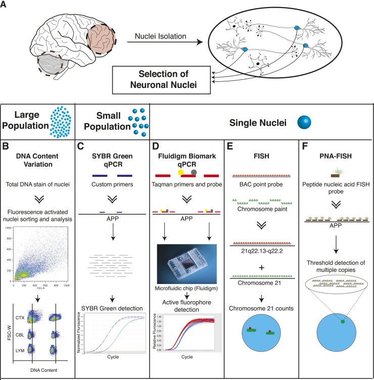
Figure 1. Methodologies used in assessing genomically mosaic AD. (A) Neuronal nuclei were isolated from the prefrontal cortex and cerebellum of postmortem human brain (see ‘Materials and methods’ for samples used) as described (Westra et al., 2008). (B) Nuclear DNA was stained with propidium iodide (PI) and DNA content was quantified using flow cytometric analysis. (C) APP copy number variations were analyzed in small populations of nuclei (∼75 genomes) using custom primers for exon 14 of APP. (D) Single-cell qPCR assessed APP copy number variations in individual neuronal nuclei via TaqMan probes and a modified Biomark integrated fluidic chip system (Fluidigm Corporation, South San Francisco). (E) FISH paints against the whole q arm of chromosome 21 and a point probe against a region on the q arm of 21 (21q22.13-q22.2) were used to double-label and call aneusomies in AD samples. (F) Peptide nucleic acid (PNA) FISH was combined with super resolution microscopy for threshold detection of APP copy number above ∼2 occurring at a single locus. DOI: http://dx.doi.org/10.7554/eLife.05116.003
Figure 1. Methodologies used in assessing genomically mosaic AD. (A) Neuronal nuclei were isolated from the prefrontal cortex and cerebellum of postmortem human brain (see ‘Materials and methods’ for samples used) as described (Westra et al., 2008). (B) Nuclear DNA was stained with propidium iodide (PI) and DNA content was quantified using flow cytometric analysis. (C) APP copy number variations were analyzed in small populations of nuclei (∼75 genomes) using custom primers for exon 14 of APP. (D) Single-cell qPCR assessed APP copy number variations in individual neuronal nuclei via TaqMan probes and a modified Biomark integrated fluidic chip system (Fluidigm Corporation, South San Francisco). (E) FISH paints against the whole q arm of chromosome 21 and a point probe against a region on the q arm of 21 (21q22.13-q22.2) were used to double-label and call aneusomies in AD samples. (F) Peptide nucleic acid (PNA) FISH was combined with super resolution microscopy for threshold detection of APP copy number above ∼2 occurring at a single locus. DOI: http://dx.doi.org/10.7554/eLife.05116.003CC BY · PubMed Central
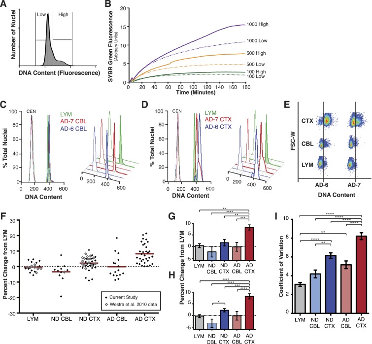
Figure 2. AD cortical nuclei show increased DNA content variation (DCV) by flow cytometry. (A) Histogram displaying gating parameters used in sorting ‘high’ and ‘low’ DNA content populations for validation of DNA content. (B) Validation of DNA content analyses using semi-quantitative MDA whole-genome amplification (WGA) on ‘high’ and ‘low’ DNA content populations of 1000, 500, and 100 nuclei. (C and D) Representative DNA content histograms for lymphocytes (LYM), AD cerebellum (CBL), and AD prefrontal cortex (CTX). Each colored histogram represents a separate sample in each set; CTX and CBL samples are from paired brains. Chicken erythrocyte nuclei (CEN) were used as internal calibration controls. (E) Representative orthogonal view of DNA content vs forward scatter width (FSC-W). For each brain sample, the area to the right of the vertical line indicates a DNA content increase of the population of nuclei. AD-6 CTX is a representative right-hand peak shift and AD-7 is a representative right-hand shoulder (see Figure 3A for more examples). (F) DNA content changes for all human LYM, ND, and AD brain samples examined (AD CTX N = 32, AD CBL N = 16, LYM N = 15 [20 meta analysis], ND CTX = 21 [36 meta analysis], ND CBL = 11 [12 meta analysis]). Red bars denote average for each group relative to lymphocytes. Averages are as follows (including metadata from Westra et al. (2010)): AD CTX 8.219%; AD CBL −0.1104%; LYM −0.2915%; ND CTX 2.239%; ND CBL −3.358%. (G) DNA content changes of the current study (AVOVA p < 0.0001). (H) DNA content changes of the current study combined with metadata from Westra et al. (2010) (ANOVA p < 0.0001). (I) Comparison of mean coefficient of variation (CV statistic from FlowJo of the population, included metadata from Westra et al., 2010) demonstrates that there is an average increase in the variation of AD samples (ANOVA p < 0.0001). *p = 0.05, **p = 0.01, ***p = 0.001, ****p < 0.0001, See Figure 2—source data 1 for exact p values. See Figure 2—figure supplement 1 for age, PMI and Braak score correlations. See Figure 2—figure supplement 2 for control of nuclear size analysis. DOI: http://dx.doi.org/10.7554/eLife.05116.004
Figure 2. AD cortical nuclei show increased DNA content variation (DCV) by flow cytometry. (A) Histogram displaying gating parameters used in sorting ‘high’ and ‘low’ DNA content populations for validation of DNA content. (B) Validation of DNA content analyses using semi-quantitative MDA whole-genome amplification (WGA) on ‘high’ and ‘low’ DNA content populations of 1000, 500, and 100 nuclei. (C and D) Representative DNA content histograms for lymphocytes (LYM), AD cerebellum (CBL), and AD prefrontal cortex (CTX). Each colored histogram represents a separate sample in each set; CTX and CBL samples are from paired brains. Chicken erythrocyte nuclei (CEN) were used as internal calibration controls. (E) Representative orthogonal view of DNA content vs forward scatter width (FSC-W). For each brain sample, the area to the right of the vertical line indicates a DNA content increase of the population of nuclei. AD-6 CTX is a representative right-hand peak shift and AD-7 is a representative right-hand shoulder (see Figure 3A for more examples). (F) DNA content changes for all human LYM, ND, and AD brain samples examined (AD CTX N = 32, AD CBL N = 16, LYM N = 15 [20 meta analysis], ND CTX = 21 [36 meta analysis], ND CBL = 11 [12 meta analysis]). Red bars denote average for each group relative to lymphocytes. Averages are as follows (including metadata from Westra et al. (2010)): AD CTX 8.219%; AD CBL −0.1104%; LYM −0.2915%; ND CTX 2.239%; ND CBL −3.358%. (G) DNA content changes of the current study (AVOVA p < 0.0001). (H) DNA content changes of the current study combined with metadata from Westra et al. (2010) (ANOVA p < 0.0001). (I) Comparison of mean coefficient of variation (CV statistic from FlowJo of the population, included metadata from Westra et al., 2010) demonstrates that there is an average increase in the variation of AD samples (ANOVA p < 0.0001). *p = 0.05, **p = 0.01, ***p = 0.001, ****p < 0.0001, See Figure 2—source data 1 for exact p values. See Figure 2—figure supplement 1 for age, PMI and Braak score correlations. See Figure 2—figure supplement 2 for control of nuclear size analysis. DOI: http://dx.doi.org/10.7554/eLife.05116.004CC BY · PubMed Central
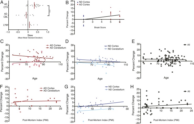
Figure 2—figure supplement 1. DNA content shows no correlation with age or post-mortem index (PMI). (A) Comparison of mean skew values for each sample group, skew determined as: (Mean − Mode/Standard Deviation of the diploid DNA content peak). (B) No correlation was observed between DNA content and Braak score. (C–E) No correlation was observed between DNA content and age across all brains analyzed. (F–H) No correlation was observed between DNA content and post-mortem index across all brains analyzed. DOI: http://dx.doi.org/10.7554/eLife.05116.006
Figure 2—figure supplement 1. DNA content shows no correlation with age or post-mortem index (PMI). (A) Comparison of mean skew values for each sample group, skew determined as: (Mean − Mode/Standard Deviation of the diploid DNA content peak). (B) No correlation was observed between DNA content and Braak score. (C–E) No correlation was observed between DNA content and age across all brains analyzed. (F–H) No correlation was observed between DNA content and post-mortem index across all brains analyzed. DOI: http://dx.doi.org/10.7554/eLife.05116.006CC BY · PubMed Central
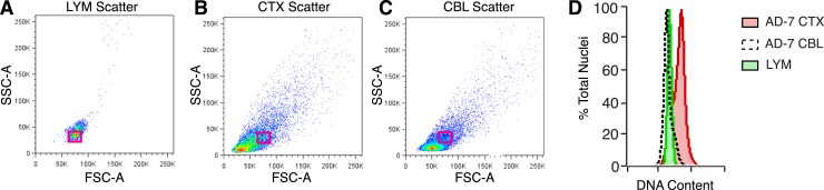
Figure 2—figure supplement 2. Analysis of nuclear size and DNA content. (A–C) Representative flow cytometry scatter plots of nuclei. (A) Lymphocytes (LYM), (B) CTX nuclei, (C) CBL nuclei. (D) Overlay of red boxes shown in (A–C), demonstrating that cortical nuclei similar in size to LYM and CBL consistently display a DNA content shift. DOI: http://dx.doi.org/10.7554/eLife.05116.007
Figure 2—figure supplement 2. Analysis of nuclear size and DNA content. (A–C) Representative flow cytometry scatter plots of nuclei. (A) Lymphocytes (LYM), (B) CTX nuclei, (C) CBL nuclei. (D) Overlay of red boxes shown in (A–C), demonstrating that cortical nuclei similar in size to LYM and CBL consistently display a DNA content shift. DOI: http://dx.doi.org/10.7554/eLife.05116.007CC BY · PubMed Central
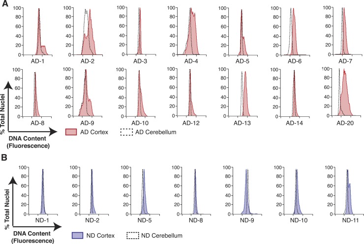
Figure 3. Pairwise DNA content analyses in AD cortical nuclei vs AD cerebellum. (A) Pairwise analysis of overlaid DNA content histograms (CTX = solid red, CBL = black dashed lines) in the same AD individual (each graph represents a unique AD individual). (B) Pairwise analysis of overlaid DNA content histograms (CTX = solid blue, CBL = black dashed lines) in the same ND individual. DOI: http://dx.doi.org/10.7554/eLife.05116.008
Figure 3. Pairwise DNA content analyses in AD cortical nuclei vs AD cerebellum. (A) Pairwise analysis of overlaid DNA content histograms (CTX = solid red, CBL = black dashed lines) in the same AD individual (each graph represents a unique AD individual). (B) Pairwise analysis of overlaid DNA content histograms (CTX = solid blue, CBL = black dashed lines) in the same ND individual. DOI: http://dx.doi.org/10.7554/eLife.05116.008CC BY · PubMed Central
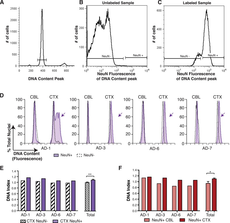
Figure 4. DNA content increases observed in AD cortical nuclei are attributable to neurons. (A–C) The gating procedure used for NeuN-positive flow cytometry analysis. (A) DNA content peak for identified nuclei. (B) A sample of unlabeled neuronal nuclei that display no NeuN-positive signal. (C) Selection for NeuN-positive nuclei for downstream analysis. (D) DNA content histograms of four AD samples displaying NeuN-positive nuclei (solid purple) vs NeuN-negative nuclei (black dashed line). NeuN-positive populations display distinct cortical histograms with prominent right-shifted peaks (arrows). (E) Comparison of DNA index (DI) increases from NeuN-positive nuclei (solid purple) vs NeuN-negative nuclei (black dashes) from AD CTX samples. NeuN-positive nuclei (DI = 1.10) showed an average gain of 9% over NeuN-negative nuclei (DI = 1.01), **p = 0.0011. (F) Comparison of DNA content in NeuN-positive nuclei from AD CTX (DI = 1.10) (red) vs AD CBL (DI = 0.94) (pink) from the same individual; CTX nuclei displayed a 15.6% gain over CBL nuclei, *p = 0.0335. Statistics are paired two-tailed t-test. Bars indicate ± SEM. DOI: http://dx.doi.org/10.7554/eLife.05116.009
Figure 4. DNA content increases observed in AD cortical nuclei are attributable to neurons. (A–C) The gating procedure used for NeuN-positive flow cytometry analysis. (A) DNA content peak for identified nuclei. (B) A sample of unlabeled neuronal nuclei that display no NeuN-positive signal. (C) Selection for NeuN-positive nuclei for downstream analysis. (D) DNA content histograms of four AD samples displaying NeuN-positive nuclei (solid purple) vs NeuN-negative nuclei (black dashed line). NeuN-positive populations display distinct cortical histograms with prominent right-shifted peaks (arrows). (E) Comparison of DNA index (DI) increases from NeuN-positive nuclei (solid purple) vs NeuN-negative nuclei (black dashes) from AD CTX samples. NeuN-positive nuclei (DI = 1.10) showed an average gain of 9% over NeuN-negative nuclei (DI = 1.01), **p = 0.0011. (F) Comparison of DNA content in NeuN-positive nuclei from AD CTX (DI = 1.10) (red) vs AD CBL (DI = 0.94) (pink) from the same individual; CTX nuclei displayed a 15.6% gain over CBL nuclei, *p = 0.0335. Statistics are paired two-tailed t-test. Bars indicate ± SEM. DOI: http://dx.doi.org/10.7554/eLife.05116.009CC BY · PubMed Central
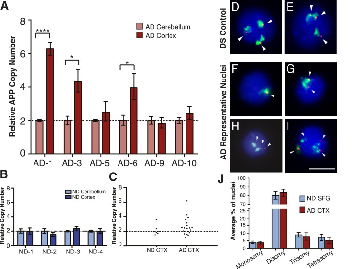
Figure 5. Mosaic amplification of the APP locus in small cohorts of AD cortical neurons unrelated to trisomy 21. (A) Comparison of relative copy number of APP in CBL and CTX fractions from six AD brains. APP locus-specific amplification was determined relative to reference gene SEMA4A; paired CBL nuclei were used as a calibrator sample for each brain, normalized to 2.00 for a diploid cell. Differences in ΔΔCt ± SEM of APP in the cortex vs cerebellum were assessed in each individual using an unpaired, two-tailed t-test (****p = 0.0001, *p = 0.0165, *p = 0.0489) (B) Comparison of relative copy number of APP in CBL and CTX fractions from 4 non-diseased brains. (C) Average relative copy number in non-diseased vs AD brains. Control genes and DS individuals were also examined (Figure 5—figure supplement 1). (D–J) FISH strategy of chromosome 21 counting through simultaneous labeling using chr 21 q arm ‘whole’ chromosome paint (WCP, green) and chr 21 regional FISH probe for 21q22.13-q22.2 (red) (see Figure 5—source data 1 for raw counts). (D and E) The ability to detect aneuploidy was validated using interphase nuclei from a human trisomy 21 brain, where three regional spots (red, encompassing the APP gene) were seen, despite WCP spatial variation (see also Rehen et al. (2005)). (F–I) Chromosome 21 aneusomy was examined in prefrontal cortical nuclei. Examples of chr 21 (F) monosomy, (G) disomy, (H) trisomy, and (I) tetrasomy (please note tetrasomy is not an example of aneuploidy). (J) Quantification of individual FISH signals showed no significant differences in monosomy, disomy, trisomy, or tetrasomy. 5 control brains and 9 AD brains were used. At least 450 nuclei were quantified per brain sample. Scale bar = 10 um. 4974 total nuclei examined. DOI: http://dx.doi.org/10.7554/eLife.05116.010
Figure 5. Mosaic amplification of the APP locus in small cohorts of AD cortical neurons unrelated to trisomy 21. (A) Comparison of relative copy number of APP in CBL and CTX fractions from six AD brains. APP locus-specific amplification was determined relative to reference gene SEMA4A; paired CBL nuclei were used as a calibrator sample for each brain, normalized to 2.00 for a diploid cell. Differences in ΔΔCt ± SEM of APP in the cortex vs cerebellum were assessed in each individual using an unpaired, two-tailed t-test (****p = 0.0001, *p = 0.0165, *p = 0.0489) (B) Comparison of relative copy number of APP in CBL and CTX fractions from 4 non-diseased brains. (C) Average relative copy number in non-diseased vs AD brains. Control genes and DS individuals were also examined (Figure 5—figure supplement 1). (D–J) FISH strategy of chromosome 21 counting through simultaneous labeling using chr 21 q arm ‘whole’ chromosome paint (WCP, green) and chr 21 regional FISH probe for 21q22.13-q22.2 (red) (see Figure 5—source data 1 for raw counts). (D and E) The ability to detect aneuploidy was validated using interphase nuclei from a human trisomy 21 brain, where three regional spots (red, encompassing the APP gene) were seen, despite WCP spatial variation (see also Rehen et al. (2005)). (F–I) Chromosome 21 aneusomy was examined in prefrontal cortical nuclei. Examples of chr 21 (F) monosomy, (G) disomy, (H) trisomy, and (I) tetrasomy (please note tetrasomy is not an example of aneuploidy). (J) Quantification of individual FISH signals showed no significant differences in monosomy, disomy, trisomy, or tetrasomy. 5 control brains and 9 AD brains were used. At least 450 nuclei were quantified per brain sample. Scale bar = 10 um. 4974 total nuclei examined. DOI: http://dx.doi.org/10.7554/eLife.05116.010CC BY · PubMed Central
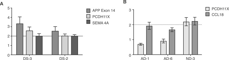
Figure 5—figure supplement 1. Controls for small population qPCR. (A) Reference genes validated in small population qPCR via examination of APP exon 14 in Down Syndrome (DS) nuclei as a positive control. (B) Representative males (AD-1 and AD-6) displayed reduced copy number of PCDH11X, a gene located on the X chromosome, while a representative female demonstrates two copies of PCDH11X and CCL18, a second single copy control gene. DOI: http://dx.doi.org/10.7554/eLife.05116.012
Figure 5—figure supplement 1. Controls for small population qPCR. (A) Reference genes validated in small population qPCR via examination of APP exon 14 in Down Syndrome (DS) nuclei as a positive control. (B) Representative males (AD-1 and AD-6) displayed reduced copy number of PCDH11X, a gene located on the X chromosome, while a representative female demonstrates two copies of PCDH11X and CCL18, a second single copy control gene. DOI: http://dx.doi.org/10.7554/eLife.05116.012CC BY · PubMed Central
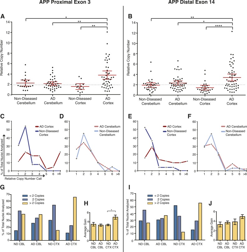
Figure 6. Mosaic APP locus amplification in single neurons from AD brains. (A) Single nuclei relative copy numbers for exon 3 of APP from non-diseased (ND) CBL, ND CTX, AD CBL, and AD CTX; each black diamond represents one neuron. For each group, the mean is displayed in red and bars represent 95% confidence intervals. AD CTX showed a mean APP copy number of 3.80; this is significantly higher than AD CBL (2.23), ND CTX (1.60), and ND CBL (2.28). *p = 0.0147, **p = 0.0015, **p < 0.0012, ANOVA p < 0.0001 (see Figure 6—source data 1 for raw numbers and statistics). (B) Single nuclei relative copy numbers for exon 14 of APP, similar to (A). The two exons showed a high concordance (Figure 6—figure supplement 1) where the AD CTX showed a mean APP copy number of 3.40 while the AD CBL (2.34), ND CTX (1.44), and ND CBL (1.92) remained closer to 2 copies. *p = 0.0163, **p = 0.0016, ****p < 0.0001, ANOVA p < 0.0001. (C–F) Distribution of copy number calls for exon 3 (C and D) and exon 14 (E and F) binned by relative copy number. The AD CTX for both exons displayed unique distributions, with more nuclei falling into the high copy number bins. (G and I) Distribution of nuclei with copy numbers less than, equal to, and greater than two copies. (H and J) Average copy number increases in nuclei binned with greater than two copies (gold columns in G) (AD CTX: Exon 3 = 5.01, Exon 14 = 4.96, *p = 0.0361). All statistics represent an ANOVA with a Tukey's multiple comparison test. Bars indicate ± SEM. DOI: http://dx.doi.org/10.7554/eLife.05116.013
Figure 6. Mosaic APP locus amplification in single neurons from AD brains. (A) Single nuclei relative copy numbers for exon 3 of APP from non-diseased (ND) CBL, ND CTX, AD CBL, and AD CTX; each black diamond represents one neuron. For each group, the mean is displayed in red and bars represent 95% confidence intervals. AD CTX showed a mean APP copy number of 3.80; this is significantly higher than AD CBL (2.23), ND CTX (1.60), and ND CBL (2.28). *p = 0.0147, **p = 0.0015, **p < 0.0012, ANOVA p < 0.0001 (see Figure 6—source data 1 for raw numbers and statistics). (B) Single nuclei relative copy numbers for exon 14 of APP, similar to (A). The two exons showed a high concordance (Figure 6—figure supplement 1) where the AD CTX showed a mean APP copy number of 3.40 while the AD CBL (2.34), ND CTX (1.44), and ND CBL (1.92) remained closer to 2 copies. *p = 0.0163, **p = 0.0016, ****p < 0.0001, ANOVA p < 0.0001. (C–F) Distribution of copy number calls for exon 3 (C and D) and exon 14 (E and F) binned by relative copy number. The AD CTX for both exons displayed unique distributions, with more nuclei falling into the high copy number bins. (G and I) Distribution of nuclei with copy numbers less than, equal to, and greater than two copies. (H and J) Average copy number increases in nuclei binned with greater than two copies (gold columns in G) (AD CTX: Exon 3 = 5.01, Exon 14 = 4.96, *p = 0.0361). All statistics represent an ANOVA with a Tukey's multiple comparison test. Bars indicate ± SEM. DOI: http://dx.doi.org/10.7554/eLife.05116.013CC BY · PubMed Central
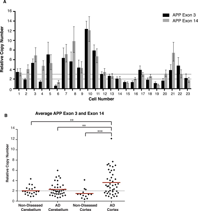
Figure 6—figure supplement 1. Concordance of APP exon 3 and 14 from single cell qPCR. (A) Relative copy numbers (RCN) for APP exon 3 and APP exon 14 displaying concordance between exons. 100 of 115 nuclei examined for both exons display copy numbers within one copy number call. 10 of the 15 remaining nuclei, while more than one copy number apart, were both called as gains. Bars represent RCN Min and RCN Max. (B) Scatter plot of average relative copy numbers. The data remain consistent with those displayed for individual exons (Figure 6A,B). Statistics represent an ANOVA with a Tukey's multiple comparison test. **p < 0.01, ***p < 0.001. DOI: http://dx.doi.org/10.7554/eLife.05116.015
Figure 6—figure supplement 1. Concordance of APP exon 3 and 14 from single cell qPCR. (A) Relative copy numbers (RCN) for APP exon 3 and APP exon 14 displaying concordance between exons. 100 of 115 nuclei examined for both exons display copy numbers within one copy number call. 10 of the 15 remaining nuclei, while more than one copy number apart, were both called as gains. Bars represent RCN Min and RCN Max. (B) Scatter plot of average relative copy numbers. The data remain consistent with those displayed for individual exons (Figure 6A,B). Statistics represent an ANOVA with a Tukey's multiple comparison test. **p < 0.01, ***p < 0.001. DOI: http://dx.doi.org/10.7554/eLife.05116.015CC BY · PubMed Central
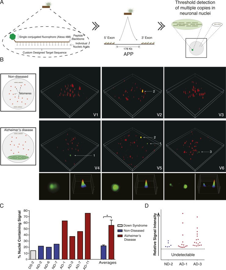
Figure 7. Visualization of APP copy number increases in neuronal nuclei from AD brain samples. (A) Peptide nucleic acid probes (PNA) were developed against nine separate sites on APP (4 sites within exon 3 and 5 sites within exon 14). Each PNA probe consists of a peptide backbone conjugated to a single fluorophore, with separately conjugated nucleotides, substantially increasing specificity (Lansdorp et al., 1996). Single copies of APP are not detectable because of fluorophore detection limits. Detection of increased copy number by PNA probes can be visualized as copies of APP increase (Figure 7—figure supplement 1B,C). Positive internal controls using PNA probes directed against telomere sequences were simultaneously hybridized. (B) Visualization of copy number increases in neuronal nuclei. Green puncta (arrow 1, insets) indicate visualized APP increases. Telomere labeling (red puncta) was present in all nuclei, demonstrating probe accessibility and template fidelity. Lipofuscin (arrow 2, orange puncta) was detected in nuclei, visualized by extensive fluorescence signal in all channels, but was eliminated from quantifications. Limited nuclei displayed two green puncta (arrow 3). V1-6 Refers to the supplemental videos where 3-D projections can be visualized. (C) Graphic representation of non-diseased (blue) and DS (grey) brains displayed limited numbers of threshold-detected increases in APP (Figure 7—source data 1). AD (red) brains displayed significant and consistent threshold-detected increases in APP. (D) Individual threshold-detected APP increases were quantified and plotted on a relative intensity scale (blue diamonds: non-diseased, red diamonds: AD). Dotted line represents the threshold below which APP copy number was undetectable, only limited puncta were identified in non-diseased nuclei. Bars indicate ± SEM, *p < 0.05. DOI: http://dx.doi.org/10.7554/eLife.05116.016
Figure 7. Visualization of APP copy number increases in neuronal nuclei from AD brain samples. (A) Peptide nucleic acid probes (PNA) were developed against nine separate sites on APP (4 sites within exon 3 and 5 sites within exon 14). Each PNA probe consists of a peptide backbone conjugated to a single fluorophore, with separately conjugated nucleotides, substantially increasing specificity (Lansdorp et al., 1996). Single copies of APP are not detectable because of fluorophore detection limits. Detection of increased copy number by PNA probes can be visualized as copies of APP increase (Figure 7—figure supplement 1B,C). Positive internal controls using PNA probes directed against telomere sequences were simultaneously hybridized. (B) Visualization of copy number increases in neuronal nuclei. Green puncta (arrow 1, insets) indicate visualized APP increases. Telomere labeling (red puncta) was present in all nuclei, demonstrating probe accessibility and template fidelity. Lipofuscin (arrow 2, orange puncta) was detected in nuclei, visualized by extensive fluorescence signal in all channels, but was eliminated from quantifications. Limited nuclei displayed two green puncta (arrow 3). V1-6 Refers to the supplemental videos where 3-D projections can be visualized. (C) Graphic representation of non-diseased (blue) and DS (grey) brains displayed limited numbers of threshold-detected increases in APP (Figure 7—source data 1). AD (red) brains displayed significant and consistent threshold-detected increases in APP. (D) Individual threshold-detected APP increases were quantified and plotted on a relative intensity scale (blue diamonds: non-diseased, red diamonds: AD). Dotted line represents the threshold below which APP copy number was undetectable, only limited puncta were identified in non-diseased nuclei. Bars indicate ± SEM, *p < 0.05. DOI: http://dx.doi.org/10.7554/eLife.05116.016CC BY · PubMed Central
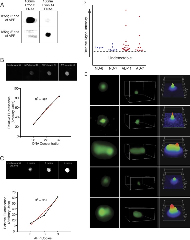
Figure 7—figure supplement 1. PNA FISH controls. (A) PNA probe specificity was verified via dot blots of APP sequence followed by PNA probe hybridization and immunoblotting against the Alexa-488 fluorophor. Probes designed against exon 3 exhibited specific binding to the 5′ region of APP, and probes designed against exon 14 exhibited specific binding to the 3′ region of APP, while probes did not display significant binding to non-specific sequences. (B) Plasmids containing all 9 APP PNA binding sites were blotted at 1× (1.8 µg), 2× (3.6 µg), and 3× (5.4 µg) DNA concentration, and PNA probes were hybridized and an empty plasmid at 1.8 μg was used for a negative control in lane 1. Fluorescent output demonstrated a linear increase with increasing DNA concentration. (C) 10 μg of plasmids containing 0, 3, 6, and 9 copies of the APP PNA binding sites was blotted onto membrane and probes were hybridized. Fluorescent output showed an expected linear increase with the number of PNA probe binding sites. (D) Quantification of the variable APP signal increases observed across four brains. (E) Representative APP signals visualized and verified using super-resolution 3D projections displayed a range of variable intensities. DOI: http://dx.doi.org/10.7554/eLife.05116.018
Figure 7—figure supplement 1. PNA FISH controls. (A) PNA probe specificity was verified via dot blots of APP sequence followed by PNA probe hybridization and immunoblotting against the Alexa-488 fluorophor. Probes designed against exon 3 exhibited specific binding to the 5′ region of APP, and probes designed against exon 14 exhibited specific binding to the 3′ region of APP, while probes did not display significant binding to non-specific sequences. (B) Plasmids containing all 9 APP PNA binding sites were blotted at 1× (1.8 µg), 2× (3.6 µg), and 3× (5.4 µg) DNA concentration, and PNA probes were hybridized and an empty plasmid at 1.8 μg was used for a negative control in lane 1. Fluorescent output demonstrated a linear increase with increasing DNA concentration. (C) 10 μg of plasmids containing 0, 3, 6, and 9 copies of the APP PNA binding sites was blotted onto membrane and probes were hybridized. Fluorescent output showed an expected linear increase with the number of PNA probe binding sites. (D) Quantification of the variable APP signal increases observed across four brains. (E) Representative APP signals visualized and verified using super-resolution 3D projections displayed a range of variable intensities. DOI: http://dx.doi.org/10.7554/eLife.05116.018CC BY · PubMed Central

Verification

Claims the paper makes

The claims and statistics of this paper haven’t been checked yet.

Run the check — free, no account needed. It reads the open-access text and checks the sources it cites.

A few free checks a day · papers already verified open instantly · results are public

Claims other papers make about this one

Each is checked against this paper’s own text: does it say what the citing paper says it does?

Self-check

Percentages, sums and reported differences re-computed from the paper’s own numbers.

The self-check needs the paper’s full text.

Citations

Cites

1 retracted

Show all 87 references

Research works printed but not linked: 1

  1. Blaschke, 1998, Programmed cell death is a universal feature of embryonic and postnatal neuroproliferative regions throughout the central nervous system, Journal of Comparative Neurology, 396, 39, 10.1002/(SICI)1096-9861(19980622)396:13.0.CO;2-J · DOI

Cited by

Sorted by citations

See all 136

Metadata

Authors
Bushman, D. M., Kaeser, G. E., Siddoway, B., Westra, J. W., Rivera, R. R., Rehen, S. K., Yung, Y. C., & Chun, J.
Affiliations
Scripps Research Institute (US)
Corresponding author
Chun, Jerold
Venue
eLife
Published
2015-02-04
Volume
4
ISSN
2050-084X (ISSN-L)
OpenAlex
W2007326275
Cited by
136
References
87
Citations by year
2026: 5 · 2025: 9 · 2024: 7 · 2023: 5 · 2022: 15 · 2021: 13 · 2020: 19 · 2019: 17 · 2018: 19 · 2017: 11 · 2016: 8 · 2015: 8
FWCI
11.30
Type
journal article
Language
en
Open access
Locations
  • eLife (publishedVersion, cc-by)
  • Europe PMC (PubMed Central) (publishedVersion, cc-by)
  • PubMed Central (publishedVersion, cc-by)
  • PubMed (publishedVersion)
  • DOAJ (DOAJ: Directory of Open Access Journals) (submittedVersion, cc-by-sa)
Funders
  • National Institutes of Health (MH076145, T32 DA07315, T32 AG 000216)
  • Shaffer Family Foundation
Topics
  • Life Sciences › Neuroscience › Cellular and Molecular Neuroscience › Alzheimer's disease research and treatments
  • Life Sciences › Biochemistry, Genetics and Molecular Biology › Genetics › Genomic variations and chromosomal abnormalities
  • Health Sciences › Medicine › Psychiatry and Mental health › Dementia and Cognitive Impairment Research
Keywords
sporadic Alzheimer's disease, amyloid precursor protein, single neurons, peptide nucleic acid probes, super-resolution microscopy
MeSH terms
Alzheimer Disease, Cell Nucleus, Cerebral Cortex, DNA, Down Syndrome, Flow Cytometry, Gene Amplification, Humans, Mosaicism, Neurons, Amyloid beta-Protein Precursor, Genome, In Situ Hybridization, Fluorescence, Gene Dosage, Peptide Nucleic Acids, Genetic Loci

About these checks

Registry, retraction, Cites and statistics results are computed, not judged, with one exception: a printed reference that no index lists is read once by a model before it counts as a citation error. Claims are extracted by a model and kept only when their quote appears verbatim in the text; each cited source is then checked, with a model’s help, for whether it says what the claim relies on. Those verdicts can be wrong — read the claim’s quote and the cited source before relying on one.

Statistics and arithmetic are recomputed, not judged.

Confidence bands are shown only for fields calibrated on our annotated test set. None is calibrated yet, so no band is shown.

What counts as a claim ›How we measure confidence ›How we check this ›

Are you an author, or do you see a problem with this page? Request a review