

The DOI matches its Crossref record; 44 of the 44 references it cites were found in a registry or an index.
Tight nuclear tethering of cGAS is essential for preventing autoreactivity
Open accessAbstract
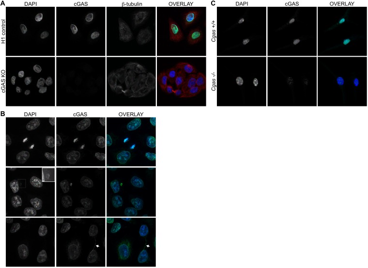
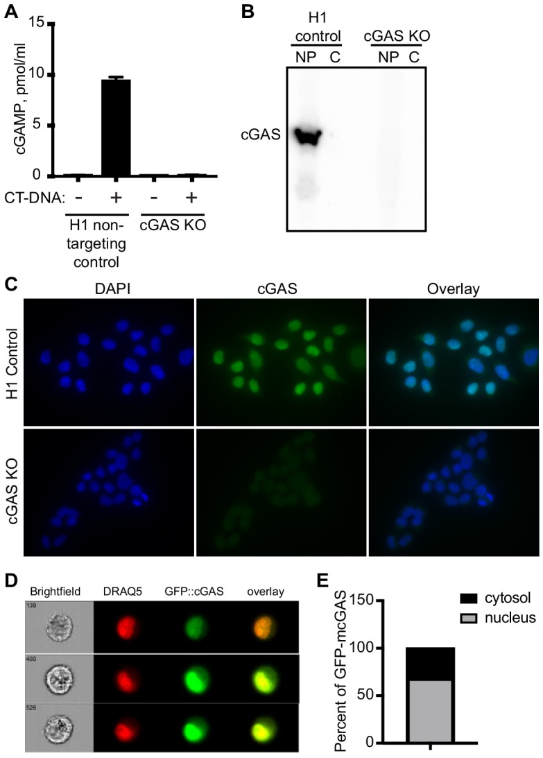
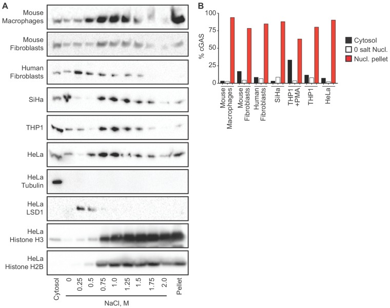
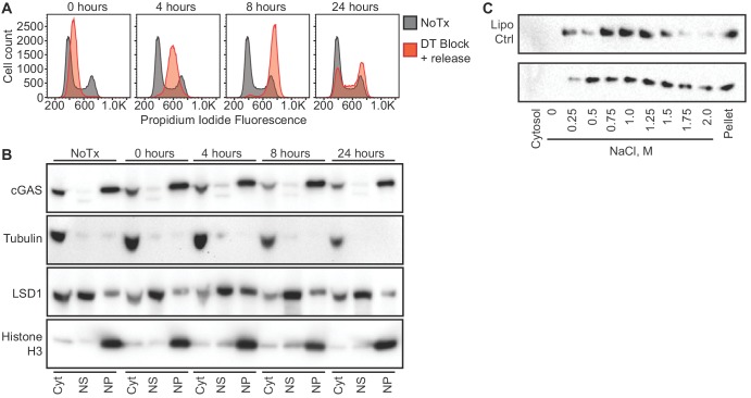
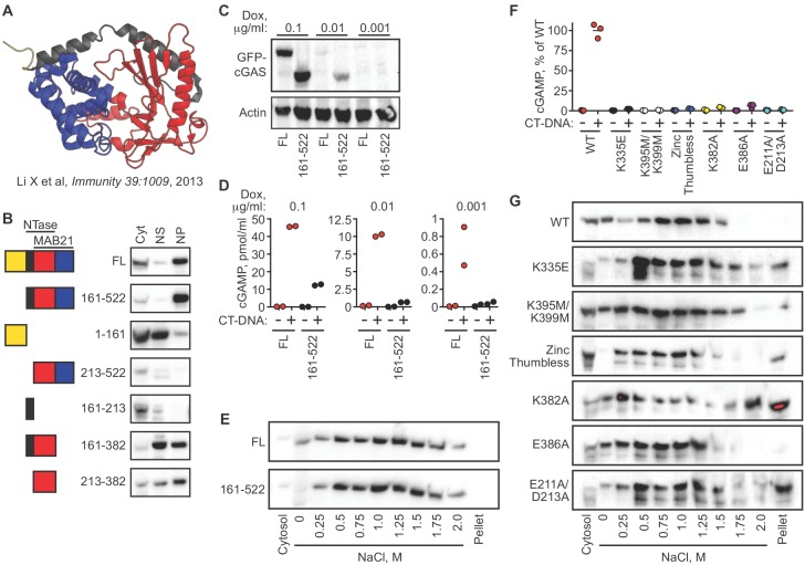
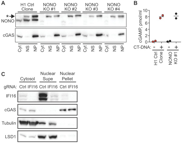
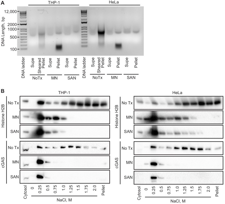
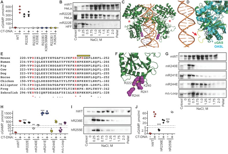
cGAS is an intracellular innate immune sensor that detects double-stranded DNA. The presence of billions of base pairs of genomic DNA in all nucleated cells raises the question of how cGAS is not constitutively activated. A widely accepted explanation for this is the sequestration of cGAS in the cytosol, which is thought to prevent cGAS from accessing nuclear DNA. Here, we demonstrate that endogenous cGAS is predominantly a nuclear protein, regardless of cell cycle phase or cGAS activation status. We show that nuclear cGAS is tethered tightly by a salt-resistant interaction. This tight tethering is independent of the domains required for cGAS activation, and it requires intact nuclear chromatin. We identify the evolutionarily conserved tethering surface on cGAS and we show that mutation of single amino acids within this surface renders cGAS massively and constitutively active against self-DNA. Thus, tight nuclear tethering maintains the resting state of cGAS and prevents autoreactivity.
Overview
The claims and statistics of this paper haven’t been checked yet.
Run the check — free, no account needed. It reads the open-access text and checks the sources it cites.
Integrity signals
Registry
Cites
Statistics
Figures
Verification
Claims the paper makes
The claims and statistics of this paper haven’t been checked yet.
Run the check — free, no account needed. It reads the open-access text and checks the sources it cites.
Claims other papers make about this one
Each is checked against this paper’s own text: does it say what the citing paper says it does?
The source backs part of the claim, not all of it — or backs it for a different population, condition or comparison.
In the citing paper
“Previously, we and others showed that cGAS is suppressed by binding to nucleosomes and that disruption of nucleosome regulation enables robust DNA-dependent activation of cGAS32–37 (Fig. 4a).”
From cGAS–STING drives ageing-related inflammation and neurodegeneration (2023) · cites this paper as [37]
Experimental — accuracy not yet measured
Self-check
Percentages, sums and reported differences re-computed from the paper’s own numbers.
The self-check needs the paper’s full text.
Citations
Cites
Improved vectors and genome-wide libraries for CRISPR screening
Sanjana, N. E., Shalem, O., & Zhang, F. (2014) · Nature Methods · doi:10.1038/nmeth.3047 · Cited by 5,239
Cyclic GMP-AMP Synthase Is a Cytosolic DNA Sensor That Activates the Type I Interferon Pathway
Sun, L., Wu, J., Du, F., Chen, X., & Chen, Z. J. (2013) · Science · doi:10.1126/science.1232458 · Cited by 4,996
STING is an endoplasmic reticulum adaptor that facilitates innate immune signalling
Ishikawa, H., & Barber, G. N. (2008) · Nature · doi:10.1038/nature07317 · Cited by 3,576
STING regulates intracellular DNA-mediated, type I interferon-dependent innate immunity
Ishikawa, H., Ma, Z., & Barber, G. N. (2009) · Nature · doi:10.1038/nature08476 · Cited by 2,759 · cited 7×
“The cGAS-STING DNA sensing pathway has emerged as a key innate immune response that is important for antiviral immunity (Goubau et al., 2013), contributes to specific autoimmune diseases (Crowl et al., 2017), and mediates important aspects of antitumor immunity (Li and Chen, 2018). cGAS binds to double-stranded DNA an…”
BackgroundInfluentialCyclic GMP-AMP Is an Endogenous Second Messenger in Innate Immune Signaling by Cytosolic DNA
Wu, J., Sun, L., Chen, X., Du, F., Shi, H., Chen, C., & Chen, Z. J. (2012) · Science · doi:10.1126/science.1229963 · Cited by 2,628
Phosphorylation of innate immune adaptor proteins MAVS, STING, and TRIF induces IRF3 activation
Liu, S., Cai, X., Wu, J., Cong, Q., Chen, X., Li, T., Du, F., Ren, J., Wu, Y.-T., Grishin, N. V., et al. (2015) · Science · doi:10.1126/science.aaa2630 · Cited by 1,927
cGAS surveillance of micronuclei links genome instability to innate immunity
Mackenzie, K. J., Carroll, P., Martin, C.-A., Murina, O., Fluteau, A., Simpson, D. J., Olova, N., Sutcliffe, H., Rainger, J. K., Leitch, A., et al. (2017) · Nature · doi:10.1038/nature23449 · Cited by 1,752
Mitotic progression following DNA damage enables pattern recognition within micronuclei
Harding, S. M., Benci, J. L., Irianto, J., Discher, D. E., Minn, A. J., & Greenberg, R. A. (2017) · Nature · doi:10.1038/nature23470 · Cited by 1,508
Cytoplasmic chromatin triggers inflammation in senescence and cancer
Dou, Z., Ghosh, K., Vizioli, M. G., Zhu, J., Sen, P., Wangensteen, K. J., Simithy, J., Lan, Y., Lin, Y., Zhou, Z., et al. (2017) · Nature · doi:10.1038/nature24050 · Cited by 1,335
The cGAS–cGAMP–STING pathway connects DNA damage to inflammation, senescence, and cancer
Li, T., & Chen, Z. J. (2018) · Journal of Experimental Medicine · doi:10.1084/jem.20180139 · Cited by 1,293
Cited by
Sorted by citations
The cGAS–STING pathway as a therapeutic target in inflammatory diseases
Decout, A., Katz, J. D., Venkatraman, S., & Ablasser, A. (2021) · Nature Reviews Immunology · doi:10.1038/s41577-021-00524-z · Cited by 2,309
Molecular mechanisms and cellular functions of cGAS–STING signalling
Hopfner, K.-P., & Hornung, V. (2020) · Nature Reviews Molecular Cell Biology · doi:10.1038/s41580-020-0244-x · Cited by 2,052
cGAS–STING drives ageing-related inflammation and neurodegeneration
Gulen, M. F., Samson, N., Keller, A., Schwabenland, M., Liu, C., Glück, S., Thacker, V. V., Favre, L., Mangeat, B., Kroese, L. J., et al. (2023) · Nature · doi:10.1038/s41586-023-06373-1 · Cited by 1,104
Mitochondrial DNA in inflammation and immunity
Riley, J. S., & Tait, S. W. G. (2020) · EMBO Reports · doi:10.15252/embr.201949799 · Cited by 910
Structures and Mechanisms in the cGAS-STING Innate Immunity Pathway
Zhang, X., Bai, X., & Chen, Z. J. (2020) · Immunity · doi:10.1016/j.immuni.2020.05.013 · Cited by 859
The cGAS–STING pathway and cancer
Samson, N., & Ablasser, A. (2022) · Nature Cancer · doi:10.1038/s43018-022-00468-w · Cited by 740
The type I interferonopathies: 10 years on
Crow, Y. J., & Stetson, D. B. (2022) · Nature Reviews Immunology · doi:10.1038/s41577-021-00633-9 · Cited by 489
Self-citationChemical and Biomolecular Strategies for STING Pathway Activation in Cancer Immunotherapy
Garland, K. M., Sheehy, T. L., & Wilson, J. T. (2022) · Chemical Reviews · doi:10.1021/acs.chemrev.1c00750 · Cited by 372
New frontiers in the cGAS-STING intracellular DNA-sensing pathway
Dvorkin, S., Cambier, S., Volkman, H. E., & Stetson, D. B. (2024) · Immunity · doi:10.1016/j.immuni.2024.02.019 · Cited by 390
Self-citationOduro, P. K., Zheng, X., Wei, J., Yang, Y., Wang, Y., Zhang, H., Liu, E., Gao, X., Du, M., & Wang, Q. (2021) · Acta Pharmaceutica Sinica B · doi:10.1016/j.apsb.2021.05.011 · Cited by 349
Causes and consequences of micronuclei
Krupina, K., Goginashvili, A., & Cleveland, D. W. (2021) · Current Opinion in Cell Biology · doi:10.1016/j.ceb.2021.01.004 · Cited by 344
DNA Damage and Cancer Immunotherapy: A STING in the Tale
Reisländer, T., Groelly, F. J., & Tarsounas, M. (2020) · Molecular Cell · doi:10.1016/j.molcel.2020.07.026 · Cited by 325
Role of the cGAS–STING pathway in systemic and organ-specific diseases
Skopelja‐Gardner, S., An, J., & Elkon, K. B. (2022) · Nature Reviews Nephrology · doi:10.1038/s41581-022-00589-6 · Cited by 318
Cooperative sensing of mitochondrial DNA by ZBP1 and cGAS promotes cardiotoxicity
Lei, Y., VanPortfliet, J. J., Chen, Y., Bryant, J. D., Li, Y., Fails, D. M., Torres-Odio, S., Ragan, K. B., Deng, J., Mohan, A., et al. (2023) · Cell · doi:10.1016/j.cell.2023.05.039 · Cited by 299
Cytoplasmic DNA: sources, sensing, and role in aging and disease
Miller, K. N., Victorelli, S. G., Salmonowicz, H., Dasgupta, N., Liu, T., Passos, J. F., & Adams, P. D. (2021) · Cell · doi:10.1016/j.cell.2021.09.034 · Cited by 288
Cytosolic DNA sensing by cGAS: regulation, function, and human diseases
Yu, L., & Liu, P. (2021) · Signal Transduction and Targeted Therapy · doi:10.1038/s41392-021-00554-y · Cited by 295
Structural mechanism of cGAS inhibition by the nucleosome
Pathare, G. R., Decout, A., Glück, S., Cavadini, S., Makasheva, K., Hovius, R., Kempf, G., Weiss, J., Kozicka, Z., Guey, B., et al. (2020) · Nature · doi:10.1038/s41586-020-2750-6 · Cited by 273
The molecular basis of tight nuclear tethering and inactivation of cGAS
Zhao, B., Xu, P., Rowlett, C. M., Jing, T., Shinde, O., Lei, Y., West, A. P., Liu, W. R., & Li, P. (2020) · Nature · doi:10.1038/s41586-020-2749-z · Cited by 261
Structural basis for sequestration and autoinhibition of cGAS by chromatin
Michalski, S., de Oliveira Mann, C. C., Stafford, C. A., Witte, G., Bartho, J., Lammens, K., Hornung, V., & Hopfner, K.-P. (2020) · Nature · doi:10.1038/s41586-020-2748-0 · Cited by 256
Structural basis for the inhibition of cGAS by nucleosomes
Kujirai, T., Zierhut, C., Takizawa, Y., Kim, R., Negishi, L., Uruma, N., Hirai, S., Funabiki, H., & Kurumizaka, H. (2020) · Science · doi:10.1126/science.abd0237 · Cited by 243
Phosphorylation and chromatin tethering prevent cGAS activation during mitosis
Li, T., Huang, T., Du, M., Chen, X., Du, F., Ren, J., & Chen, Z. J. (2021) · Science · doi:10.1126/science.abc5386 · Cited by 247
Structural basis of nucleosome-dependent cGAS inhibition
Boyer, J. A., Spangler, C. J., Strauss, J. D., Cesmat, A. P., Liu, P., McGinty, R. K., & Zhang, Q. (2020) · Science · doi:10.1126/science.abd0609 · Cited by 238
cGAS phase separation inhibits TREX1-mediated DNA degradation and enhances cytosolic DNA sensing
Zhou, W., Mohr, L., Maciejowski, J., & Kranzusch, P. J. (2021) · Molecular Cell · doi:10.1016/j.molcel.2021.01.024 · Cited by 219
BAF restricts cGAS on nuclear DNA to prevent innate immune activation
Guey, B., Wischnewski, M., Decout, A. C., Makasheva, K. A., Kaynak, M., Sakar, M. S., Fierz, B., & Ablasser, A. (2020) · Science · doi:10.1126/science.aaw6421 · Cited by 210
ER-directed TREX1 limits cGAS activation at micronuclei
Mohr, L., Toufektchan, E., von Morgen, P., Chu, K., Kapoor, A. R., & Maciejowski, J. (2021) · Molecular Cell · doi:10.1016/j.molcel.2020.12.037 · Cited by 211
cGAS-mediated induction of type I interferon due to inborn errors of histone pre-mRNA processing
Uggenti, C., Lepelley, A., Depp, M., Badrock, A. P., Rodero, M. P., El-Daher, M.-T., Rice, G. I., Dhir, S., Wheeler, A. P., Dhir, A., et al. (2020) · Nature Genetics · doi:10.1038/s41588-020-00737-3 · Cited by 192
Immune Sensing Mechanisms that Discriminate Self from Altered Self and Foreign Nucleic Acids
Bartok, E., & Hartmann, G. (2020) · Immunity · doi:10.1016/j.immuni.2020.06.014 · Cited by 190
DAMP sensing and sterile inflammation: intracellular, intercellular and inter-organ pathways
Huang, Y., Jiang, W., & Zhou, R. (2024) · Nature reviews. Immunology · doi:10.1038/s41577-024-01027-3 · Cited by 208
Mitochondria-localized cGAS suppresses ferroptosis to promote cancer progression
Qiu, S., Zhong, X., Meng, X. L., Li, S., Qian, X., Lu, H., Cai, J., Zhang, Y., Wang, M., Ye, Z., et al. (2023) · Cell Research · doi:10.1038/s41422-023-00788-1 · Cited by 186
Nuclear Membrane Rupture and Its Consequences
Maciejowski, J., & Hatch, E. M. (2020) · Annual Review of Cell and Developmental Biology · doi:10.1146/annurev-cellbio-020520-120627 · Cited by 170
The balance of STING signaling orchestrates immunity in cancer
Lanng, K. R. B., Lauridsen, E. L., & Jakobsen, M. R. (2024) · Nature Immunology · doi:10.1038/s41590-024-01872-3 · Cited by 179
Cyclic GMP-AMP synthase promotes the inflammatory and autophagy responses in Huntington disease
Sharma, M., Rajendrarao, S., Shahani, N., Ramírez-Jarquín, U. N., & Subramaniam, S. (2020) · Proceedings of the National Academy of Sciences · doi:10.1073/pnas.2002144117 · Cited by 155
Regulation of the cGAS-STING Pathway
Zhang, B., Xu, P., & Ablasser, A. (2025) · Annual Review of Immunology · doi:10.1146/annurev-immunol-101721-032910 · Cited by 164
Phosphorylation of cGAS by CDK1 impairs self-DNA sensing in mitosis
Zhong, L., Hu, M., Bian, L., Liu, Y., Chen, Q., & Shu, H. (2020) · Cell Discovery · doi:10.1038/s41421-020-0162-2 · Cited by 139
cGAS suppresses genomic instability as a decelerator of replication forks
Chen, H., Chen, H., Zhang, J., Wang, Y., Simoneau, A., Yang, H., Levine, A. S., Zou, L., Chen, Z. J., & Lan, L. (2020) · Science Advances · doi:10.1126/sciadv.abb8941 · Cited by 146
MRE11 liberates cGAS from nucleosome sequestration during tumorigenesis
Cho, M.-G., Kumar, R. J., Lin, C.-C., Boyer, J. A., Shahir, J. A., Fagan‐Solis, K. D., Simpson, D. A., Fan, C., Foster, C. E., Goddard, A. M., et al. (2024) · Nature · doi:10.1038/s41586-023-06889-6 · Cited by 156
The cGAS‐STING pathway: The role of self‐DNA sensing in inflammatory lung disease
Ma, R., Serrano, T. P. O., Davis, J., Prigge, A. D., & Ridge, K. M. (2020) · The FASEB Journal · doi:10.1096/fj.202001607r · Cited by 138
ABCC1 transporter exports the immunostimulatory cyclic dinucleotide cGAMP
Maltbaek, J. H., Cambier, S., Snyder, J. M., & Stetson, D. B. (2022) · Immunity · doi:10.1016/j.immuni.2022.08.006 · Cited by 132
Self-citationPan, J., 宁波大学农产品质量安全危害因子与风险防控国家重点实验室, 浙. 宁. 3., Fei, C.-J., Hu, Y., Wu, X., Nie, L., Chen, J., 宁波大学梅山校区海洋学院生物化学与分子生物学实验室, 浙. 宁. 3., & 浙江省海洋生物工程重点实验室, 浙. 宁. 3. (2023) · 动物学研究 · doi:10.24272/j.issn.2095-8137.2022.464 · Cited by 138
Fang, L., Hao, Y., Yu, H., Gu, X., Peng, Q., Zhuo, H., Li, Y., Liu, Z., Wang, J., Chen, Y., et al. (2023) · Cancer Cell · doi:10.1016/j.ccell.2023.05.005 · Cited by 132
RetractedStructural basis for nucleosome-mediated inhibition of cGAS activity
Cao, D., Han, X., Fan, X., Xu, R.-M., & Zhang, X. (2020) · Cell Research · doi:10.1038/s41422-020-00422-4 · Cited by 130
Biochemistry, Cell Biology, and Pathophysiology of the Innate Immune cGAS–cGAMP–STING Pathway
Ritchie, C., Carozza, J. A., & Li, L. (2022) · Annual Review of Biochemistry · doi:10.1146/annurev-biochem-040320-101629 · Cited by 123
Significance of the cGAS-STING Pathway in Health and Disease
Zhou, J., Zhuang, Z., Li, J., & Feng, Z. (2023) · International Journal of Molecular Sciences · doi:10.3390/ijms241713316 · Cited by 129
Excessive R-loops trigger an inflammatory cascade leading to increased HSPC production
Weinreb, J. T., Ghazale, N., Pradhan, K., Gupta, V., Potts, K. S., Tricomi, B. J., Daniels, N. J., Padgett, R. A., de Oliveira, S., Verma, A. K., et al. (2021) · Developmental Cell · doi:10.1016/j.devcel.2021.02.006 · Cited by 120
STING, the Endoplasmic Reticulum, and Mitochondria: Is Three a Crowd or a Conversation?
Smith, J. A. (2021) · Frontiers in Immunology · doi:10.3389/fimmu.2020.611347 · Cited by 116
Sun, Z., & Hornung, V. (2022) · Current Biology · doi:10.1016/j.cub.2022.05.027 · Cited by 106
A Nuclear Export Signal Is Required for cGAS to Sense Cytosolic DNA
Sun, H., Huang, Y., Mei, S., Xu, F., Liu, X., Zhao, F., Yin, L., Zhang, D., Wei, L., Wu, C., et al. (2021) · Cell Reports · doi:10.1016/j.celrep.2020.108586 · Cited by 105
cGAS‒STING signaling and function in metabolism and kidney diseases
Bai, J., & Liu, F. (2021) · Journal of Molecular Cell Biology · doi:10.1093/jmcb/mjab066 · Cited by 100
Quan, S., Fu, X., Cai, H., Ren, Z., Xu, Y., & Jia, L. (2025) · Molecular Neurodegeneration · doi:10.1186/s13024-025-00815-2 · Cited by 110
Molecular and Structural Basis of DNA Sensors in Antiviral Innate Immunity
Zahid, A., Ismail, H., Li, B., & Jin, T. (2020) · Frontiers in Immunology · doi:10.3389/fimmu.2020.613039 · Cited by 100
STING-Mediated Lung Inflammation and Beyond
Frémond, M., & Crow, Y. J. (2021) · Journal of Clinical Immunology · doi:10.1007/s10875-021-00974-z · Cited by 100
Translation stress and collided ribosomes are co-activators of cGAS
Wan, L., Juszkiewicz, S., Blears, D., Bajpe, P. K., Han, Z., Faull, P. A., Mitter, R., Stewart, A., Snijders, A. P., Hegde, R. S., et al. (2021) · Molecular Cell · doi:10.1016/j.molcel.2021.05.018 · Cited by 94
Sensing of transposable elements by the antiviral innate immune system
Gázquez-Gutiérrez, A., Witteveldt, J., Heras, S. R., & Macías, S. (2021) · RNA · doi:10.1261/rna.078721.121 · Cited by 101
Regulation and Consequences of cGAS Activation by Self-DNA
Zierhut, C., & Funabiki, H. (2020) · Trends in Cell Biology · doi:10.1016/j.tcb.2020.05.006 · Cited by 91
The Trinity of cGAS, TLR9, and ALRs Guardians of the Cellular Galaxy Against Host-Derived Self-DNA
Kumar, V. (2021) · Frontiers in Immunology · doi:10.3389/fimmu.2020.624597 · Cited by 95
Takaki, T., Millar, R., Hiley, C. T., & Boulton, S. J. (2024) · Molecular Cell · doi:10.1016/j.molcel.2024.04.017 · Cited by 101
Nucleic Acid Sensors as Therapeutic Targets for Human Disease
McWhirter, S. M., & Jefferies, C. A. (2020) · Immunity · doi:10.1016/j.immuni.2020.04.004 · Cited by 91
CGAS is a micronucleophagy receptor for the clearance of micronuclei
Zhao, M., Wang, F., Wu, J., Cheng, Y., Cao, Y., Wu, X., Ma, M., Tang, F., Liu, Z., Liu, H., et al. (2021) · Autophagy · doi:10.1080/15548627.2021.1899440 · Cited by 89
Mitotic disassembly and reassembly of nuclear pore complexes
Kutay, U., Jühlen, R., & Antonin, W. (2021) · Trends in Cell Biology · doi:10.1016/j.tcb.2021.06.011 · Cited by 85
Banerjee, D., Langberg, K. A., Abbas, S., Odermatt, E., Yerramothu, P., Volaric, M. P., Reidenbach, M. A., Krentz, K. J., Rubinstein, C. D., Brautigan, D. L., et al. (2021) · Nature Communications · doi:10.1038/s41467-021-26240-9 · Cited by 88
cGAS‐STING pathway agonists are promising vaccine adjuvants
Tian, X., Ai, J., Tian, X., & Wei, X. (2024) · Medicinal Research Reviews · doi:10.1002/med.22016 · Cited by 86
cGAS restricts colon cancer development by protecting intestinal barrier integrity
Hu, S., Fang, Y., Chen, X., Cheng, T., Zhao, M., Du, M., Li, T., Li, M., Zeng, Z., Wei, Y., et al. (2021) · Proceedings of the National Academy of Sciences · doi:10.1073/pnas.2105747118 · Cited by 84
DDX41 is required for cGAS-STING activation against DNA virus infection
Singh, R. S., Vidhyasagar, V., Yang, S., Arna, A. B., Yadav, M., Aggarwal, A., Aguilera, A. N., Shinriki, S., Bhanumathy, K. K., Pandey, K., et al. (2022) · Cell Reports · doi:10.1016/j.celrep.2022.110856 · Cited by 85
The cGAS-STING Pathway: Novel Perspectives in Liver Diseases
Xu, D., Tian, Y., Xia, Q., & Ke, B. (2021) · Frontiers in Immunology · doi:10.3389/fimmu.2021.682736 · Cited by 87
Wang, F., Zhao, M., Chang, B., Zhou, Y., Wu, X., Ma, M., Liu, S., Cao, Y., Zheng, M., Dang, Y., et al. (2022) · Molecular Cell · doi:10.1016/j.molcel.2022.03.034 · Cited by 79
Candida albicans extracellular vesicles trigger type I IFN signalling via cGAS and STING
Brown Harding, H., Kwaku, G. N., Reardon, C. M., Khan, N. S., Zamith-Miranda, D., Zarnowski, R., Tam, J. M., Bohaen, C. K., Richey, L., Mosallanejad, K., et al. (2024) · Nature Microbiology · doi:10.1038/s41564-023-01546-0 · Cited by 82
DNA Damage and Activation of cGAS/STING Pathway Induce Tumor Microenvironment Remodeling
Shen, R., Liu, D., Wang, X., Guo, Z., Sun, H., Song, Y., & Wang, D. (2022) · Frontiers in Cell and Developmental Biology · doi:10.3389/fcell.2021.828657 · Cited by 77
The CRL5–SPSB3 ubiquitin ligase targets nuclear cGAS for degradation
Xu, P., Liu, Y., Liu, C., Guey, B., Li, L., Mélénec, P., Ricci, J., & Ablasser, A. (2024) · Nature · doi:10.1038/s41586-024-07112-w · Cited by 78
cGAS–STING, an important signaling pathway in diseases and their therapy
Li, Q., Wu, P., Du, Q., Hanif, U., Hu, H., & Li, K. (2024) · MedComm · doi:10.1002/mco2.511 · Cited by 83
cGAS/STING cross-talks with cell cycle and potentiates cancer immunotherapy
Long, Z., Wang, J., Xu, J., Lei, X., & Liu, Q. (2022) · Molecular Therapy · doi:10.1016/j.ymthe.2022.01.044 · Cited by 76
Mitochondrial DNA signals driving immune responses: Why, How, Where?
Giordano, L., Ware, S. A., Lagranha, C. J., & Kaufman, B. A. (2025) · Cell Communication and Signaling · doi:10.1186/s12964-025-02042-0 · Cited by 82
cGAMP-activated cGAS–STING signaling: its bacterial origins and evolutionary adaptation by metazoans
Patel, D. J., Yu, Y., & Xie, W. (2023) · Nature Structural & Molecular Biology · doi:10.1038/s41594-023-00933-9 · Cited by 79
Nuclear cGAS: guard or prisoner?
Mann, C. C. D. O., & Hopfner, K. (2021) · The EMBO Journal · doi:10.15252/embj.2021108293 · Cited by 65
Nuclear cGAS: sequestration and beyond
Bai, J., & Liu, F. (2021) · Protein & Cell · doi:10.1007/s13238-021-00869-0 · Cited by 65
Torres-Odio, S., Lei, Y., Gispert, S., Maletzko, A., Key, J., Menissy, S. S., Wittig, I., Auburger, G. W. J., & West, A. P. (2021) · The Journal of Immunology · doi:10.4049/jimmunol.2001016 · Cited by 61
cGAS/STING signalling pathway in senescence and oncogenesis
Yu, L., & Liu, P. (2024) · Seminars in Cancer Biology · doi:10.1016/j.semcancer.2024.08.007 · Cited by 67
Opportunities and challenges of targeting cGAS–STING in cancer
Lu, C., Wang, W., & Fu, Y. (2026) · Nature reviews. Cancer · doi:10.1038/s41568-025-00894-9 · Cited by 73
STING Agonists and How to Reach Their Full Potential in Cancer Immunotherapy
Gehrcken, L., Deben, C., Smits, E., & Audenaerde, J. R. M. V. (2025) · Advanced Science · doi:10.1002/advs.202500296 · Cited by 66
ISG15 conjugation to proteins on nascent DNA mitigates DNA replication stress
Wardlaw, C. P., & Petrini, J. H. J. (2022) · Nature Communications · doi:10.1038/s41467-022-33535-y · Cited by 57
Nuclear and genome dynamics underlying DNA double-strand break repair
Chiolo, I., Altmeyer, M., Legube, G., & Mekhail, K. (2025) · Nature Reviews Molecular Cell Biology · doi:10.1038/s41580-025-00828-1 · Cited by 78
STING Signaling and Sterile Inflammation
Couillin, I., & Riteau, N. (2021) · Frontiers in Immunology · doi:10.3389/fimmu.2021.753789 · Cited by 60
Targeting STING in cancer: Challenges and emerging opportunities
Zhao, K., Huang, J., Zhao, Y., Wang, S., Xu, J., & Yin, K. (2023) · Biochimica et Biophysica Acta (BBA) - Reviews on Cancer · doi:10.1016/j.bbcan.2023.188983 · Cited by 57
Micronucleus is not a potent inducer of the cGAS/STING pathway
Sato, Y., & Hayashi, M. (2024) · Life Science Alliance · doi:10.26508/lsa.202302424 · Cited by 58
Mitochondrial DNA in cell death and inflammation
Heilig, R., Lee, J., & Tait, S. W. G. (2023) · Biochemical Society Transactions · doi:10.1042/bst20221525 · Cited by 56
Regulation and inhibition of the DNA sensor cGAS
Hertzog, J., & Rehwinkel, J. (2020) · EMBO Reports · doi:10.15252/embr.202051345 · Cited by 52
Lactylation orchestrates ubiquitin-independent degradation of cGAS and promotes tumor growth
Rao, K., Zhang, X., Luo, Y., Xia, Q., Jin, Y., & He, J. (2025) · Cell Reports · doi:10.1016/j.celrep.2025.115441 · Cited by 54
Tsuji, N., & Agbor-Enoh, S. T. (2021) · The Journal of Heart and Lung Transplantation · doi:10.1016/j.healun.2021.03.007 · Cited by 50
Herpes Simplex Virus and Pattern Recognition Receptors: An Arms Race
Zhao, J., Qin, C., Liu, Y., Rao, Y., & Feng, P. (2021) · Frontiers in Immunology · doi:10.3389/fimmu.2020.613799 · Cited by 52
cGAS‐STING pathway: post‐translational modifications and functions in sterile inflammatory diseases
Hong, Z., Ma, T., Liu, X., & Wang, C. (2021) · FEBS Journal · doi:10.1111/febs.16137 · Cited by 52
Barrier-to-Autointegration Factor 1 Protects against a Basal cGAS-STING Response
Ma, H., Qian, W. P., Bambousková, M., Collins, P. L., Porter, S. I., Byrum, A. K., Zhang, R., Artyomov, M. N., Oltz, E. M., Mosammaparast, N., et al. (2020) · mBio · doi:10.1128/mbio.00136-20 · Cited by 51
Ribonucleotide incorporation into mitochondrial DNA drives inflammation
Bahat, A., Milenkovic, D., Cors, E., Barnett, M., Niftullayev, S., Katsalifis, A., Schwill, M., Kirschner, P., MacVicar, T., Giavalisco, P., et al. (2025) · Nature · doi:10.1038/s41586-025-09541-7 · Cited by 53
Control of innate immunity by the cGAS‐STING pathway
Mosallanejad, K., & Kagan, J. C. (2022) · Immunology and Cell Biology · doi:10.1111/imcb.12555 · Cited by 52
Song, B., Greco, T. M., Lum, K. K., Taber, C. E., & Cristea, I. M. (2020) · Molecular & Cellular Proteomics · doi:10.1074/mcp.ra120.001981 · Cited by 46
Role of Post-Translational Modifications of cGAS in Innate Immunity
Wu, Y., & Li, S. (2020) · International Journal of Molecular Sciences · doi:10.3390/ijms21217842 · Cited by 47
STING nuclear partners contribute to innate immune signaling responses
Dixon, C. R., Malik, P. R., Heras, J. I. D. L., Saiz-Ros, N., Alves, F. D. L., Tingey, M., Gaunt, E. R., Richardson, C. A., Kelly, D. A., Goldberg, M. W., et al. (2021) · iScience · doi:10.1016/j.isci.2021.103055 · Cited by 46
Medicinal chemistry perspective on cGAS-STING signaling pathway with small molecule inhibitors
Tian, X., Xu, F., Zhu, Q., Feng, Z., Dai, W., Zhou, Y.-L., You, Q., & Xu, X. (2022) · European Journal of Medicinal Chemistry · doi:10.1016/j.ejmech.2022.114791 · Cited by 44
Molecular mechanisms of nonself nucleic acid recognition by the innate immune system
Mann, C. C. D. O., & Hornung, V. (2021) · European Journal of Immunology · doi:10.1002/eji.202049116 · Cited by 45
DNA-sensing pathways in health, autoinflammatory and autoimmune diseases
Dong, M., & Fitzgerald, K. A. (2024) · Nature Immunology · doi:10.1038/s41590-024-01966-y · Cited by 46
Crosstalk Between Autophagy and the cGAS–STING Signaling Pathway in Type I Interferon Production
Zhang, K., Wang, S., Gou, H., Zhang, J., & Li, C. (2021) · Frontiers in Cell and Developmental Biology · doi:10.3389/fcell.2021.748485 · Cited by 45
cGAS-STING DNA-sensing in inflammatory bowel diseases
Dimitrov, G., Ryffel, B., Togbe, D., & Quesniaux, V. F. J. (2024) · Trends in Molecular Medicine · doi:10.1016/j.molmed.2024.10.002 · Cited by 44
Metadata
- Authors
- Volkman, H. E., Cambier, S., Gray, E. E., & Stetson, D. B.
- Affiliations
- University of Washington (US)
- Corresponding author
- Stetson, Daniel B
- Venue
- eLife
- Publisher
- eLife Sciences Publications, Ltd
- Published
- 2019-12-06
- Volume
- 8
- ISSN
- 2050-084X (ISSN-L)
- PMC
- PMC6927687
- OpenAlex
- W2994585472
- Cited by
- 303
- References
- 42
- Citations by year
- 2026: 38 · 2025: 42 · 2024: 55 · 2023: 36 · 2022: 37 · 2021: 59 · 2020: 36
- FWCI
- 13.19
- Type
- journal article
- Language
- en
- Open access
- gold
- doi.org
- Licence
- Locations
- eLife (publishedVersion, cc-by)
- Europe PMC (PubMed Central) (publishedVersion, cc-by)
- PubMed Central (publishedVersion, cc-by)
- PubMed (publishedVersion)
- DOAJ (DOAJ: Directory of Open Access Journals) (submittedVersion, cc-by-sa)
- Funders
- National Institutes of Health (AI084914)
- Jane Coffin Childs Memorial Fund for Medical Research
- Burroughs Wellcome Fund (1013540)
- Howard Hughes Medical Institute (55108572)
- Bill and Melinda Gates Foundation (OPP1156262)
- Cancer Research Institute (A84161)
- Topics
- Life Sciences › Immunology and Microbiology › Immunology › interferon and immune responses
- Life Sciences › Immunology and Microbiology › Immunology › Immune Response and Inflammation
- Life Sciences › Biochemistry, Genetics and Molecular Biology › Molecular Biology › DNA and Nucleic Acid Chemistry
- Keywords
- cGAS, innate immunity, double-stranded DNA sensing, self-DNA, autoreactivity, chromatin, cytosolic DNA sensing, constitutive activation, nucleic acid sensors
- MeSH terms
- Cyclic Guanosine Monophosphate-Adenosine Monophosphate Synthase, Animals, Cell Nucleus, Cells, Cultured, Cytosol, DNA, DNA Damage, HeLa Cells, Humans, Mice, Inbred C57BL, Nuclear Proteins, Nucleotidyltransferases, Protein Binding, Cell Line, Tumor, DNA Cleavage
About these checks
Registry, retraction, Cites and statistics results are computed, not judged, with one exception: a printed reference that no index lists is read once by a model before it counts as a citation error. Claims are extracted by a model and kept only when their quote appears verbatim in the text; each cited source is then checked, with a model’s help, for whether it says what the claim relies on. Those verdicts can be wrong — read the claim’s quote and the cited source before relying on one.
Statistics and arithmetic are recomputed, not judged.
Confidence bands are shown only for fields calibrated on our annotated test set. None is calibrated yet, so no band is shown.
What counts as a claim ›How we measure confidence ›How we check this ›
Are you an author, or do you see a problem with this page? Request a review
