

The DOI matches its Crossref record; 85 of the 85 references it cites were found in a registry or an index.
Remyelination alters the pattern of myelin in the cerebral cortex
Open accessPublished version of a preprint
Abstract
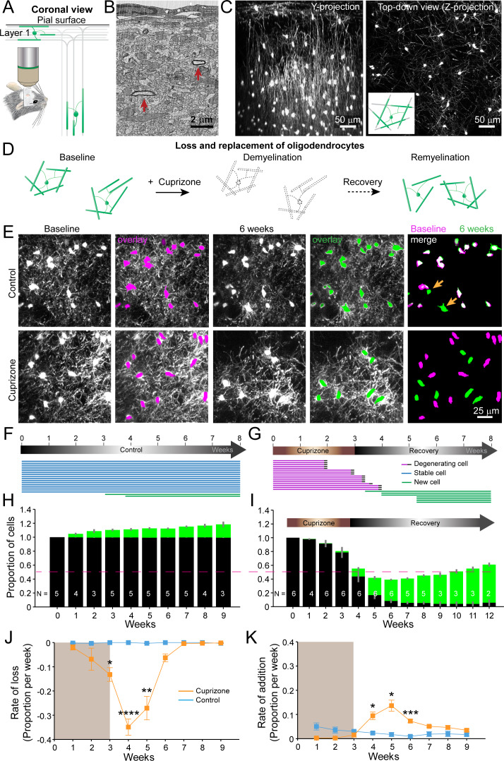
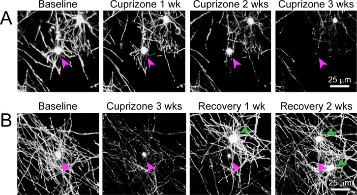
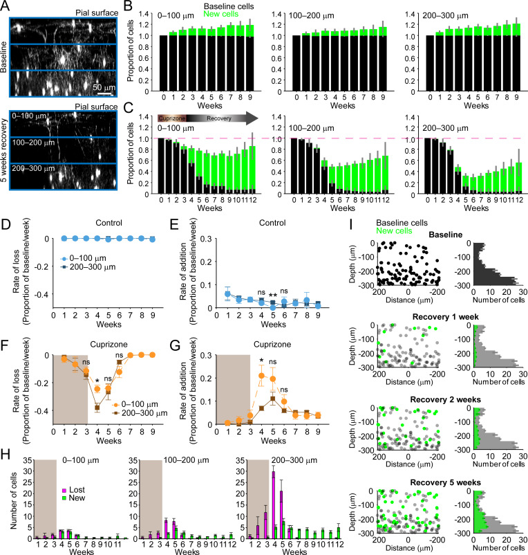
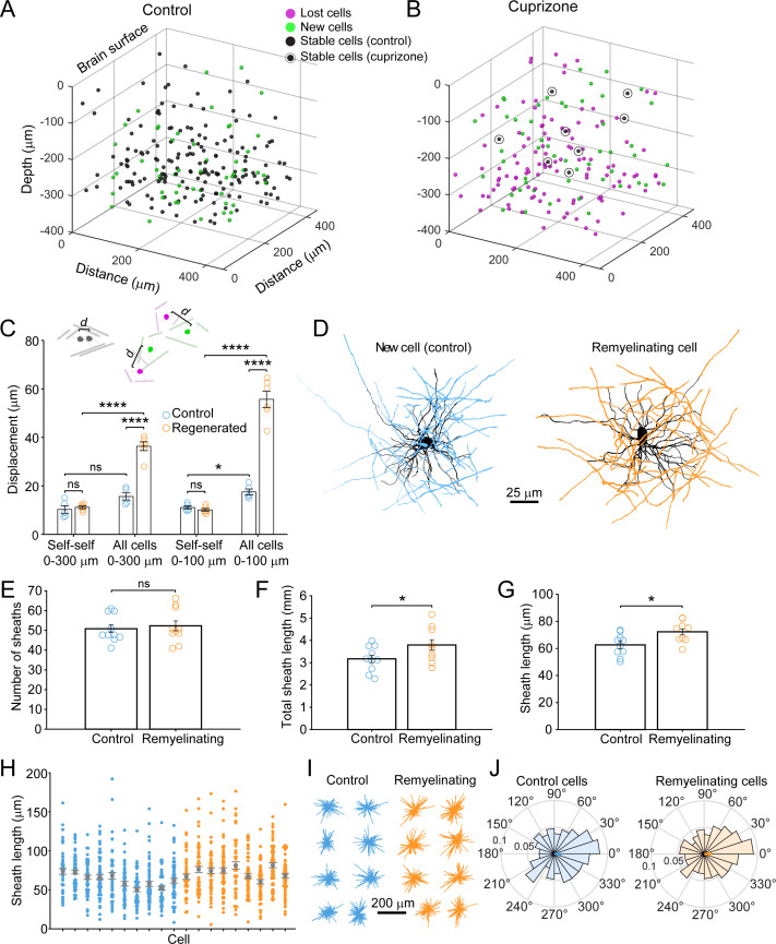
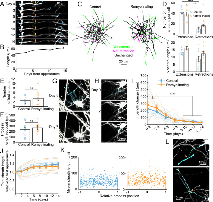
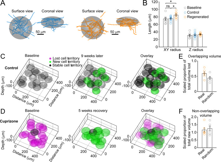
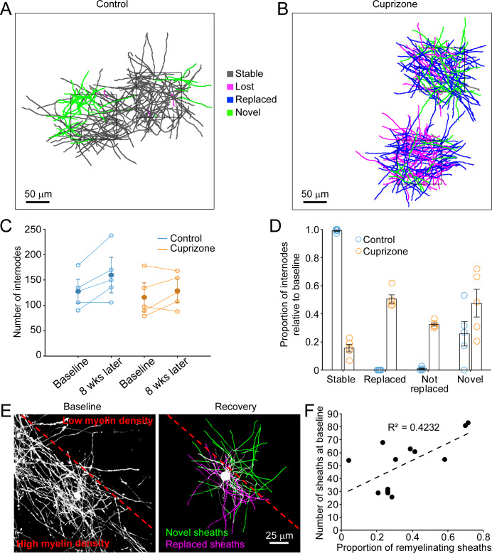
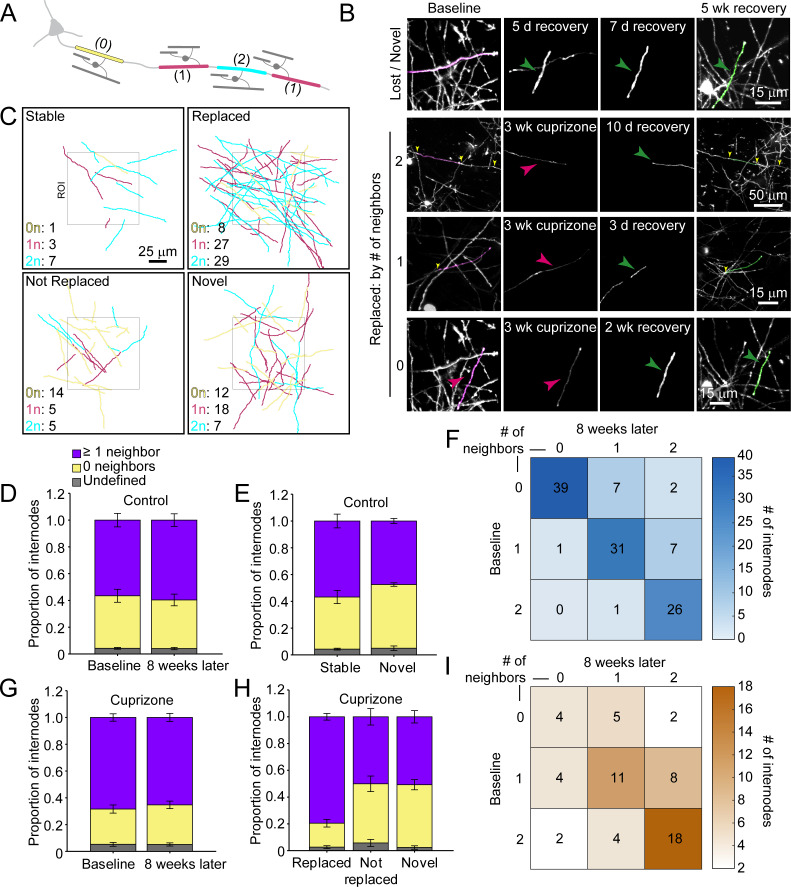
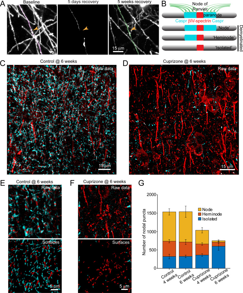
Destruction of oligodendrocytes and myelin sheaths in cortical gray matter profoundly alters neural activity and is associated with cognitive disability in multiple sclerosis (MS). Myelin can be restored by regenerating oligodendrocytes from resident progenitors; however, it is not known whether regeneration restores the complex myelination patterns in cortical circuits. Here, we performed time lapse in vivo two photon imaging in somatosensory cortex of adult mice to define the kinetics and specificity of myelin regeneration after acute oligodendrocyte ablation. These longitudinal studies revealed that the pattern of myelination in cortex changed dramatically after regeneration, as new oligodendrocytes were formed in different locations and new sheaths were often established along axon segments previously lacking myelin. Despite the dramatic increase in axonal territory available, oligodendrogenesis was persistently impaired in deeper cortical layers that experienced higher gliosis. Repeated reorganization of myelin patterns in MS may alter circuit function and contribute to cognitive decline.
Overview
The claims and statistics of this paper haven’t been checked yet.
Run the check — free, no account needed. It reads the open-access text and checks the sources it cites.
Integrity signals
Registry
Cites
Statistics
Figures
Verification
Claims the paper makes
The claims and statistics of this paper haven’t been checked yet.
Run the check — free, no account needed. It reads the open-access text and checks the sources it cites.
Claims other papers make about this one
Each is checked against this paper’s own text: does it say what the citing paper says it does?
The source backs part of the claim, not all of it — or backs it for a different population, condition or comparison.
In the citing paper
“The efficacy of remyelination has been shown to depend on multiple factors such as, age, disease duration and lesion location.”
From BCAS1-positive oligodendrocytes enable efficient cortical remyelination in multiple sclerosis (2025) · cites this paper as [8]
Experimental — accuracy not yet measuredThe source backs part of the claim, not all of it — or backs it for a different population, condition or comparison.
In the citing paper
“Similar to our prior study (Orthmann‐Murphy et al. 2020), we found no significant changes in OPC number in the deep cortex (Figure 1G, also see Figure S1C).”
From Depletion of Microglia Increases Cortical Oligodendrocyte Density During Remyelination (2026) · cites this paper as Orthmann‐Murphy et al. 2020
Experimental — accuracy not yet measuredThe source backs part of the claim, not all of it — or backs it for a different population, condition or comparison.
In the citing paper
“BCAS1 + GFP+ oligodendrocytes should represent a relatively more mature differentiating oligodendrocyte state than BCAS1 + GFP‐ cells (Chapman et al. 2024; Hughes et al. 2018; Orthmann‐Murphy et al. 2020).”
From Depletion of Microglia Increases Cortical Oligodendrocyte Density During Remyelination (2026) · cites this paper as Orthmann‐Murphy et al. 2020
Experimental — accuracy not yet measuredThe source backs part of the claim, not all of it — or backs it for a different population, condition or comparison.
In the citing paper
“We found that new oligodendrocytes formed after cessation of the cuprizone‐supplemented diet, with a peak rate of formation at 2 weeks post‐cuprizone (“early” recovery).”
From Depletion of Microglia Increases Cortical Oligodendrocyte Density During Remyelination (2026) · cites this paper as Orthmann‐Murphy et al. 2020
Experimental — accuracy not yet measuredThe source backs part of the claim, not all of it — or backs it for a different population, condition or comparison.
In the citing paper
“For cell counts, a blinded observer quantified the number of NG2+, GFAP+, Iba1+, ASPA+, BCAS1+, and GFP+ cells within each 500 μm × 750 μm region using a custom counting macro in FIJI/ImageJ (Orthmann‐Murphy et al. 2020).”
From Depletion of Microglia Increases Cortical Oligodendrocyte Density During Remyelination (2026) · cites this paper as Orthmann‐Murphy et al. 2020
Experimental — accuracy not yet measured
The source backs part of the claim, not all of it — or backs it for a different population, condition or comparison.
In the citing paper
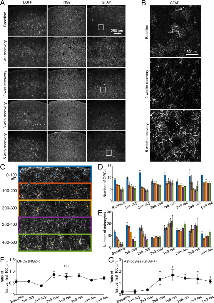
“Similar to our previous findings (Orthmann‐Murphy et al. 2020), even in the presence of Csf1r inhibitor, deep cortical reactive astrocytes persistently upregulate GFAP (Figure S5B).”
From Depletion of Microglia Increases Cortical Oligodendrocyte Density During Remyelination (2026) · cites this paper as Orthmann‐Murphy et al. 2020
Experimental — accuracy not yet measuredThe cited source says what the claim attributes to it.
In the citing paper
“In our prior in vivo imaging study, however, we showed that deep cortical oligodendrogenesis was selectively impaired, with only ~55% of the baseline oligodendrocyte population replaced after 9 weeks of recovery in the deepest cortical zone examined (200–300 μm; Orthmann‐Murphy et al. 2020).”
From Depletion of Microglia Increases Cortical Oligodendrocyte Density During Remyelination (2026) · cites this paper as Orthmann‐Murphy et al. 2020
The cited source says what the claim attributes to it.
In the citing paper
“We found that oligodendrocyte regeneration in the somatosensory cortex is incomplete, and this is driven primarily by inefficient oligodendrocyte replacement in deep cortical regions.”
From Depletion of Microglia Increases Cortical Oligodendrocyte Density During Remyelination (2026) · cites this paper as Orthmann‐Murphy et al. 2020
The cited source says what the claim attributes to it.
In the citing paper
“Nearly all oligodendrocytes present at 5 weeks (“late”) recovery were newly formed.”
From Depletion of Microglia Increases Cortical Oligodendrocyte Density During Remyelination (2026) · cites this paper as Orthmann‐Murphy et al. 2020
The cited source says what the claim attributes to it.
In the citing paper
“We previously showed that deep cortical astrocytes persistently upregulate GFAP following cuprizone administration (Orthmann‐Murphy et al. 2020);”
From Depletion of Microglia Increases Cortical Oligodendrocyte Density During Remyelination (2026) · cites this paper as Orthmann‐Murphy et al. 2020
The cited source says what the claim attributes to it.
In the citing paper
“Due to the exquisite spatial and temporal resolution of in vivo imaging, we also showed that the loss and formation of oligodendrocytes overlapped in early recovery, a finding that is difficult to appreciate by immunostaining alone.”
From Depletion of Microglia Increases Cortical Oligodendrocyte Density During Remyelination (2026) · cites this paper as Orthmann‐Murphy et al. 2020
The cited source says what the claim attributes to it.
In the citing paper
“Given that deep cortical ASPA+ oligodendrocytes recovered to baseline levels at late recovery only in PLX‐treated mice, but not cuprizone‐only treated mice (Figure 5), and late recovery oligodendrocytes are predominantly represented by newly formed cells (Orthmann‐Murphy et al. 2020), we suspect that many of these early recovery BCAS1+ cells formed in PLX‐treated mice become stably incorporated in”
From Depletion of Microglia Increases Cortical Oligodendrocyte Density During Remyelination (2026) · cites this paper as Orthmann‐Murphy et al. 2020
The cited source says what the claim attributes to it.
In the citing paper
“In addition, deep cortical astrocytes also become persistently reactive (Figure S4; Orthmann‐Murphy et al. 2020) and likely influence deep cortical reactive microglia function (Liddelow and Barres 2017).”
From Depletion of Microglia Increases Cortical Oligodendrocyte Density During Remyelination (2026) · cites this paper as Orthmann‐Murphy et al. 2020
The check wasn’t confident either way, so it gives no verdict — never disputed.
In the citing paper
“In fact, there are likely overlapping pathogenic roles of cortical reactive microglia, especially at early recovery (Orthmann‐Murphy et al. 2020).”
From Depletion of Microglia Increases Cortical Oligodendrocyte Density During Remyelination (2026) · cites this paper as Orthmann‐Murphy et al. 2020
Experimental — accuracy not yet measured
Self-check
Percentages, sums and reported differences re-computed from the paper’s own numbers.
The self-check needs the paper’s full text.
Citations
Cites
Cortical demyelination and diffuse white matter injury in multiple sclerosis
Kutzelnigg, A., Lucchinetti, C. F., Stadelmann, C., Brück, W., Rauschka, H., Bergmann, M., Schmidbauer, M., Parisi, J. E., & Lassmann, H. (2005) · Brain · doi:10.1093/brain/awh641 · Cited by 1,836
M2 microglia and macrophages drive oligodendrocyte differentiation during CNS remyelination
Miron, V. E., Boyd, A., Zhao, J.-W., Yuen, T. J., Ruckh, J. M., Shadrach, J. L., van Wijngaarden, P., Wagers, A. J., Williams, A., Franklin, R. J. M., et al. (2013) · Nature Neuroscience · doi:10.1038/nn.3469 · Cited by 1,777
Magliozzi, R., Howell, O. W., Vora, A., Serafini, B., Nicholas, R. S., Puopolo, M., Reynolds, R., & Aloisi, F. (2006) · Brain · doi:10.1093/brain/awm038 · Cited by 1,486
Neuronal activity promotes oligodendrogenesis and adaptive myelination in the mammalian brain
Gibson, E. M., Purger, D., Mount, C. W., Goldstein, A. K., Lin, G. L., Wood, L. S., Inema, I., Miller, S. E., Bieri, G., Zuchero, J. B., et al. (2014) · Science · doi:10.1126/science.1252304 · Cited by 1,472
Peterson, J. W., Bö, L., Mörk, S., Chang, A., & Trapp, B. D. (2001) · Annals of Neurology · doi:10.1002/ana.1123 · Cited by 1,403
Motor skill learning requires active central myelination
McKenzie, I. A., Ohayon, D., Li, H., Paes de Faria, J., Emery, B., Tohyama, K., & Richardson, W. D. (2014) · Science · doi:10.1126/science.1254960 · Cited by 1,177
Inflammatory cortical demyelination in early multiple sclerosis.
Lucchinetti, C. F., Popescu, B. F. G., Bunyan, R. F., Moll, N. M., Roemer, S. F., Lassmann, H., Brück, W., Parisi, J. E., Scheithauer, B. W., Giannini, C., et al. (2011) · New England Journal of Medicine · doi:10.1056/nejmoa1100648 · Cited by 1,103
Matsushima, G. K., & Morell, P. (2001) · Brain Pathology · doi:10.1111/j.1750-3639.2001.tb00385.x · Cited by 984
Longair, M., Baker, D. A., & Armstrong, J. D. (2011) · Bioinformatics · doi:10.1093/bioinformatics/btr390 · Cited by 939
Oligodendrocyte Dynamics in the Healthy Adult CNS: Evidence for Myelin Remodeling
Young, K. M., Psachoulia, K., Tripathi, R. B., Dunn, S.-J., Cossell, L., Attwell, D., Tohyama, K., & Richardson, W. D. (2013) · Neuron · doi:10.1016/j.neuron.2013.01.006 · Cited by 919
Cited by
Sorted by citations
Items 101–111 of 111
Microglia-nodes of Ranvier interaction during repair
Roux, T. (2020) · theses.fr (ABES) · doi:10.70675/a1319259z2b39z4d4ez9c05z261e4533646b
PIEZOs regulate oligodendrocyte sheath formation, expansion, and myelination potentialPreprint
Coombs, A. M., Heo, D., Orlin, D. J., Call, C. L., Bechler, M. E., Murthy, S. E., Emery, B., & Monk, K. R. (2026) · bioRxiv (Cold Spring Harbor Laboratory) · doi:10.64898/2026.04.23.720488
Self-citationCortical and white matter myelination proceed in concert during early infancy
Zika, S., Chang, K., Orhon, A., Kruper, J., Tyagi, C., Yan, X., Tung, S. S., Kalanit Grill-spector, A., Rokem, A., & Grotheer, M. (2026) · Nature Communications · doi:10.1038/s41467-026-73366-9 · Cited by 2
Tsai, H., Piya, S., Wang, J., Zhu, J., Hu, W., Gehrke, A. R., Cao, S., Guise, A. J., Chan, S. J., Sheehan, M. J., et al. (2026) · Frontiers in Bioinformatics · doi:10.3389/fbinf.2026.1832826 · Cited by 2
Chaudhary, R., Singh, A., Gupta, A., Kumar, A., & Agarwal, V. (2026) · Disease-a-Month · doi:10.1016/j.disamonth.2026.102210
Mechanisms and therapeutic strategies for central and peripheral remyelination
Zhan, J., Jo, J., Cristobal, C. D., Jeong, D. J., Park, H. T., & Lee, H. K. (2026) · Progress in Neurobiology · doi:10.1016/j.pneurobio.2026.102964
Metadata
- Authors
- Orthmann-Murphy, J., Call, C. L., Molina-Castro, G. C., Hsieh, Y. C., Rasband, M. N., Calabresi, P. A., & Bergles, D. E.
- Affiliations
- The Solomon Snyder Department of Neuroscience, Johns Hopkins University, Baltimore, United States
- Johns Hopkins University (US)
- Baylor College of Medicine (US)
- Corresponding author
- Bergles, Dwight E
- Venue
- eLife
- Publisher
- eLife Sciences Publications, Ltd
- Published
- 2020-05-27
- Volume
- 9
- ISSN
- 2050-084X (ISSN-L)
- PMC
- PMC7292648
- OpenAlex
- W3029244827
- Cited by
- 111
- References
- 85
- Citations by year
- 2026: 13 · 2025: 15 · 2024: 20 · 2023: 22 · 2022: 15 · 2021: 20 · 2020: 6
- FWCI
- 6.20
- Type
- journal article
- Language
- en
- Open access
- gold
- doi.org
- Licence
- Locations
- eLife (publishedVersion, cc-by)
- bioRxiv (Cold Spring Harbor Laboratory) (acceptedVersion, cc-by)
- PubMed Central (publishedVersion, cc-by)
- PubMed (publishedVersion)
- DOAJ (DOAJ: Directory of Open Access Journals) (submittedVersion, cc-by-sa)
- Funders
- Conrad N. Hilton Foundation
- National Multiple Sclerosis Society
- National Science Foundation (Graduate Research Fellowship)
- National Institutes of Health (NS051509, NS050274, NS080153)
- Dr. Miriam and Sheldon G Adelson Medical Research Foundation
- Topics
- Life Sciences › Neuroscience › Developmental Neuroscience › Neurogenesis and neuroplasticity mechanisms
- Health Sciences › Medicine › Clinical Neurology › Multiple Sclerosis Research Studies
- Life Sciences › Neuroscience › Neurology › Neuroinflammation and Neurodegeneration Mechanisms
- Keywords
- remyelination, oligodendrogenesis, cerebral cortex, somatosensory cortex, multiple sclerosis, gliosis, cortical layers, cognitive decline, in vivo two-photon imaging
- MeSH terms
- Remyelination, Animals, Axons, Female, Humans, Kinetics, Male, Mice, Inbred C57BL, Multiple Sclerosis, Myelin Sheath, Oligodendroglia, Somatosensory Cortex, Mice
About these checks
Registry, retraction, Cites and statistics results are computed, not judged, with one exception: a printed reference that no index lists is read once by a model before it counts as a citation error. Claims are extracted by a model and kept only when their quote appears verbatim in the text; each cited source is then checked, with a model’s help, for whether it says what the claim relies on. Those verdicts can be wrong — read the claim’s quote and the cited source before relying on one.
Statistics and arithmetic are recomputed, not judged.
Confidence bands are shown only for fields calibrated on our annotated test set. None is calibrated yet, so no band is shown.
What counts as a claim ›How we measure confidence ›How we check this ›
Are you an author, or do you see a problem with this page? Request a review
