

The DOI matches its Crossref record; 85 of the 85 references it cites were found in a registry or an index.
Remyelination alters the pattern of myelin in the cerebral cortex
Open accessPublished version of a preprint
Abstract
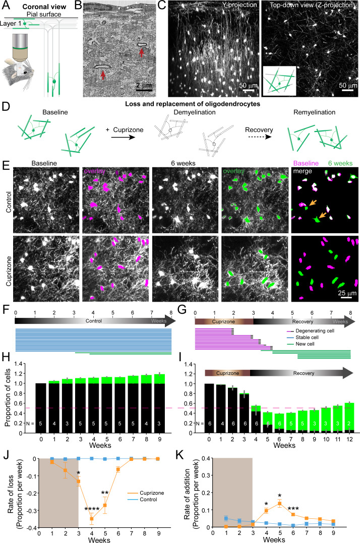
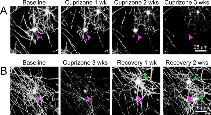
Destruction of oligodendrocytes and myelin sheaths in cortical gray matter profoundly alters neural activity and is associated with cognitive disability in multiple sclerosis (MS). Myelin can be restored by regenerating oligodendrocytes from resident progenitors; however, it is not known whether regeneration restores the complex myelination patterns in cortical circuits. Here, we performed time lapse in vivo two photon imaging in somatosensory cortex of adult mice to define the kinetics and specificity of myelin regeneration after acute oligodendrocyte ablation. These longitudinal studies revealed that the pattern of myelination in cortex changed dramatically after regeneration, as new oligodendrocytes were formed in different locations and new sheaths were often established along axon segments previously lacking myelin. Despite the dramatic increase in axonal territory available, oligodendrogenesis was persistently impaired in deeper cortical layers that experienced higher gliosis. Repeated reorganization of myelin patterns in MS may alter circuit function and contribute to cognitive decline.
Overview
The claims and statistics of this paper haven’t been checked yet.
Run the check — free, no account needed. It reads the open-access text and checks the sources it cites.
Integrity signals
Registry
Cites
Statistics
Figures
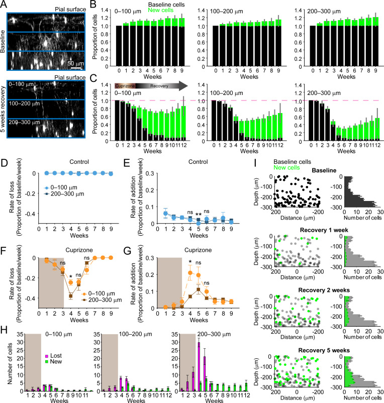
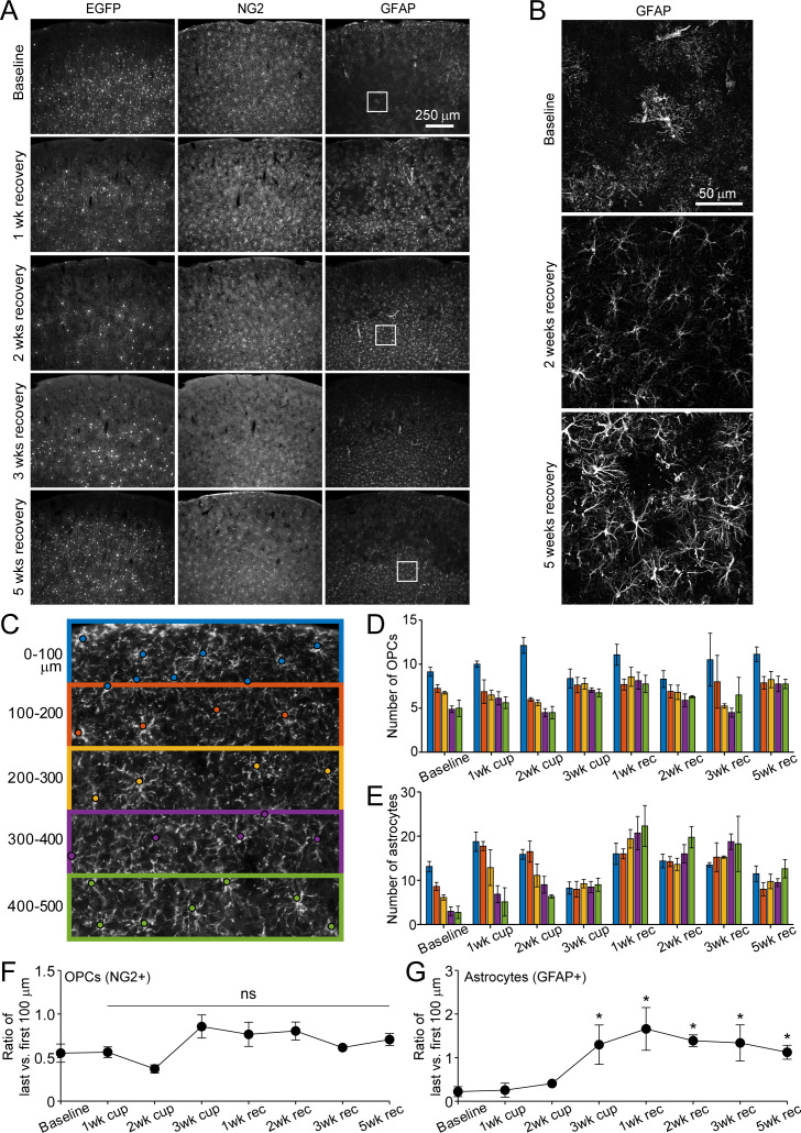
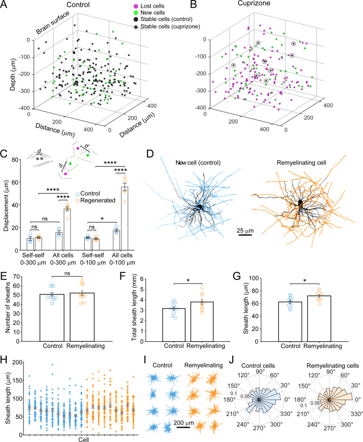
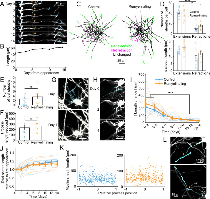
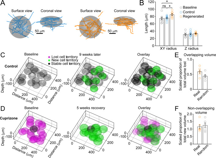
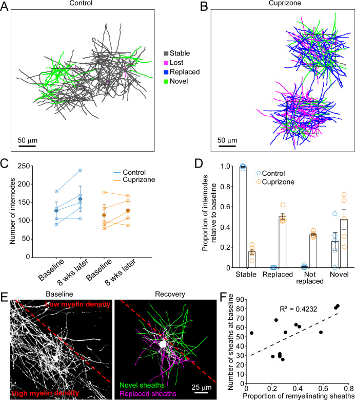
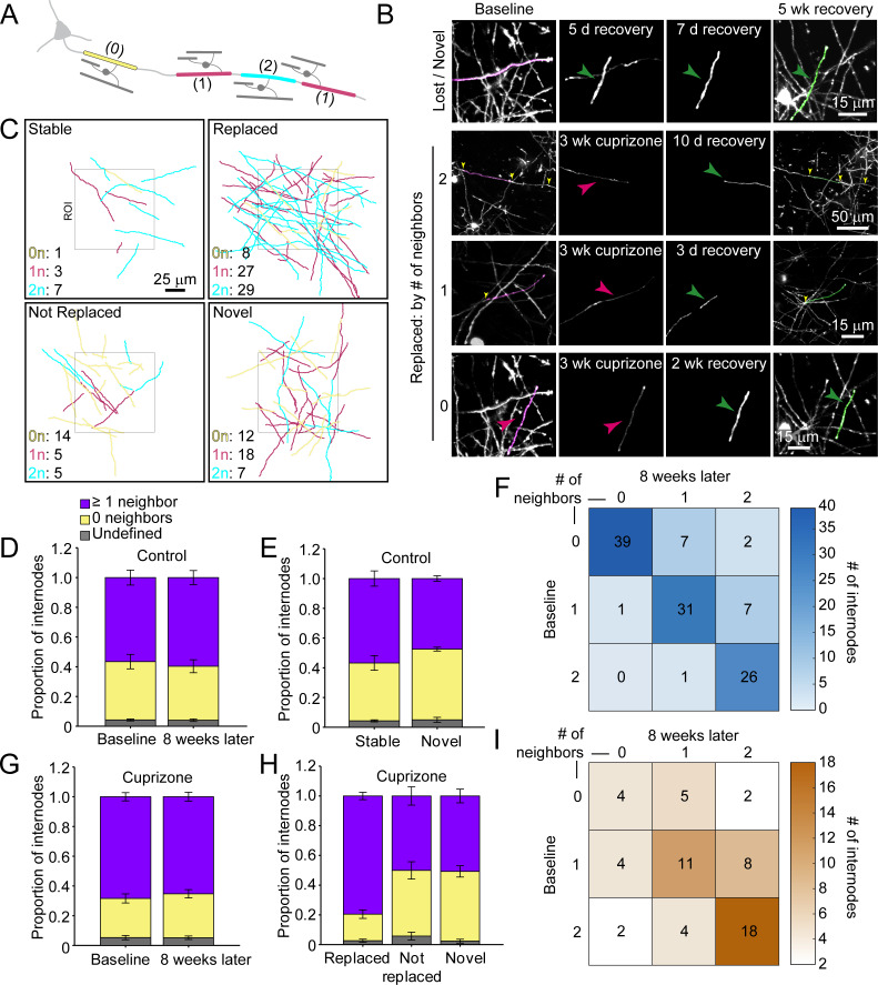
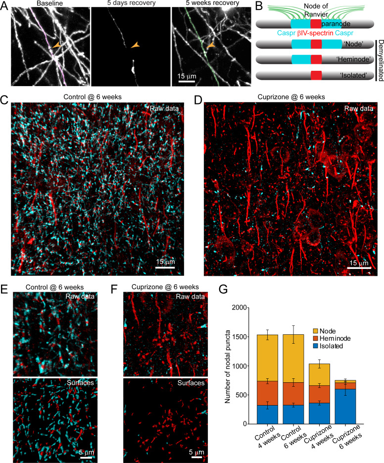
Verification
Claims the paper makes
The claims and statistics of this paper haven’t been checked yet.
Run the check — free, no account needed. It reads the open-access text and checks the sources it cites.
Claims other papers make about this one
Each is checked against this paper’s own text: does it say what the citing paper says it does?
The source backs part of the claim, not all of it — or backs it for a different population, condition or comparison.
In the citing paper
“The efficacy of remyelination has been shown to depend on multiple factors such as, age, disease duration and lesion location.”
From BCAS1-positive oligodendrocytes enable efficient cortical remyelination in multiple sclerosis (2025) · cites this paper as [8]
Experimental — accuracy not yet measuredThe source backs part of the claim, not all of it — or backs it for a different population, condition or comparison.
In the citing paper
“Similar to our prior study (Orthmann‐Murphy et al. 2020), we found no significant changes in OPC number in the deep cortex (Figure 1G, also see Figure S1C).”
From Depletion of Microglia Increases Cortical Oligodendrocyte Density During Remyelination (2026) · cites this paper as Orthmann‐Murphy et al. 2020
Experimental — accuracy not yet measuredThe source backs part of the claim, not all of it — or backs it for a different population, condition or comparison.
In the citing paper
“BCAS1 + GFP+ oligodendrocytes should represent a relatively more mature differentiating oligodendrocyte state than BCAS1 + GFP‐ cells (Chapman et al. 2024; Hughes et al. 2018; Orthmann‐Murphy et al. 2020).”
From Depletion of Microglia Increases Cortical Oligodendrocyte Density During Remyelination (2026) · cites this paper as Orthmann‐Murphy et al. 2020
Experimental — accuracy not yet measuredThe source backs part of the claim, not all of it — or backs it for a different population, condition or comparison.
In the citing paper
“We found that new oligodendrocytes formed after cessation of the cuprizone‐supplemented diet, with a peak rate of formation at 2 weeks post‐cuprizone (“early” recovery).”
From Depletion of Microglia Increases Cortical Oligodendrocyte Density During Remyelination (2026) · cites this paper as Orthmann‐Murphy et al. 2020
Experimental — accuracy not yet measuredThe source backs part of the claim, not all of it — or backs it for a different population, condition or comparison.
In the citing paper
“For cell counts, a blinded observer quantified the number of NG2+, GFAP+, Iba1+, ASPA+, BCAS1+, and GFP+ cells within each 500 μm × 750 μm region using a custom counting macro in FIJI/ImageJ (Orthmann‐Murphy et al. 2020).”
From Depletion of Microglia Increases Cortical Oligodendrocyte Density During Remyelination (2026) · cites this paper as Orthmann‐Murphy et al. 2020
Experimental — accuracy not yet measured
The source backs part of the claim, not all of it — or backs it for a different population, condition or comparison.
In the citing paper
“Similar to our previous findings (Orthmann‐Murphy et al. 2020), even in the presence of Csf1r inhibitor, deep cortical reactive astrocytes persistently upregulate GFAP (Figure S5B).”
From Depletion of Microglia Increases Cortical Oligodendrocyte Density During Remyelination (2026) · cites this paper as Orthmann‐Murphy et al. 2020
Experimental — accuracy not yet measuredThe cited source says what the claim attributes to it.
In the citing paper
“In our prior in vivo imaging study, however, we showed that deep cortical oligodendrogenesis was selectively impaired, with only ~55% of the baseline oligodendrocyte population replaced after 9 weeks of recovery in the deepest cortical zone examined (200–300 μm; Orthmann‐Murphy et al. 2020).”
From Depletion of Microglia Increases Cortical Oligodendrocyte Density During Remyelination (2026) · cites this paper as Orthmann‐Murphy et al. 2020
The cited source says what the claim attributes to it.
In the citing paper
“We found that oligodendrocyte regeneration in the somatosensory cortex is incomplete, and this is driven primarily by inefficient oligodendrocyte replacement in deep cortical regions.”
From Depletion of Microglia Increases Cortical Oligodendrocyte Density During Remyelination (2026) · cites this paper as Orthmann‐Murphy et al. 2020
The cited source says what the claim attributes to it.
In the citing paper
“Nearly all oligodendrocytes present at 5 weeks (“late”) recovery were newly formed.”
From Depletion of Microglia Increases Cortical Oligodendrocyte Density During Remyelination (2026) · cites this paper as Orthmann‐Murphy et al. 2020
The cited source says what the claim attributes to it.
In the citing paper
“We previously showed that deep cortical astrocytes persistently upregulate GFAP following cuprizone administration (Orthmann‐Murphy et al. 2020);”
From Depletion of Microglia Increases Cortical Oligodendrocyte Density During Remyelination (2026) · cites this paper as Orthmann‐Murphy et al. 2020
The cited source says what the claim attributes to it.
In the citing paper
“Due to the exquisite spatial and temporal resolution of in vivo imaging, we also showed that the loss and formation of oligodendrocytes overlapped in early recovery, a finding that is difficult to appreciate by immunostaining alone.”
From Depletion of Microglia Increases Cortical Oligodendrocyte Density During Remyelination (2026) · cites this paper as Orthmann‐Murphy et al. 2020
The cited source says what the claim attributes to it.
In the citing paper
“Given that deep cortical ASPA+ oligodendrocytes recovered to baseline levels at late recovery only in PLX‐treated mice, but not cuprizone‐only treated mice (Figure 5), and late recovery oligodendrocytes are predominantly represented by newly formed cells (Orthmann‐Murphy et al. 2020), we suspect that many of these early recovery BCAS1+ cells formed in PLX‐treated mice become stably incorporated in”
From Depletion of Microglia Increases Cortical Oligodendrocyte Density During Remyelination (2026) · cites this paper as Orthmann‐Murphy et al. 2020
The cited source says what the claim attributes to it.
In the citing paper
“In addition, deep cortical astrocytes also become persistently reactive (Figure S4; Orthmann‐Murphy et al. 2020) and likely influence deep cortical reactive microglia function (Liddelow and Barres 2017).”
From Depletion of Microglia Increases Cortical Oligodendrocyte Density During Remyelination (2026) · cites this paper as Orthmann‐Murphy et al. 2020
The check wasn’t confident either way, so it gives no verdict — never disputed.
In the citing paper
“In fact, there are likely overlapping pathogenic roles of cortical reactive microglia, especially at early recovery (Orthmann‐Murphy et al. 2020).”
From Depletion of Microglia Increases Cortical Oligodendrocyte Density During Remyelination (2026) · cites this paper as Orthmann‐Murphy et al. 2020
Experimental — accuracy not yet measured
Self-check
Percentages, sums and reported differences re-computed from the paper’s own numbers.
The self-check needs the paper’s full text.
Citations
Cites
Cortical demyelination and diffuse white matter injury in multiple sclerosis
Kutzelnigg, A., Lucchinetti, C. F., Stadelmann, C., Brück, W., Rauschka, H., Bergmann, M., Schmidbauer, M., Parisi, J. E., & Lassmann, H. (2005) · Brain · doi:10.1093/brain/awh641 · Cited by 1,836
M2 microglia and macrophages drive oligodendrocyte differentiation during CNS remyelination
Miron, V. E., Boyd, A., Zhao, J.-W., Yuen, T. J., Ruckh, J. M., Shadrach, J. L., van Wijngaarden, P., Wagers, A. J., Williams, A., Franklin, R. J. M., et al. (2013) · Nature Neuroscience · doi:10.1038/nn.3469 · Cited by 1,777
Magliozzi, R., Howell, O. W., Vora, A., Serafini, B., Nicholas, R. S., Puopolo, M., Reynolds, R., & Aloisi, F. (2006) · Brain · doi:10.1093/brain/awm038 · Cited by 1,486
Neuronal activity promotes oligodendrogenesis and adaptive myelination in the mammalian brain
Gibson, E. M., Purger, D., Mount, C. W., Goldstein, A. K., Lin, G. L., Wood, L. S., Inema, I., Miller, S. E., Bieri, G., Zuchero, J. B., et al. (2014) · Science · doi:10.1126/science.1252304 · Cited by 1,472
Peterson, J. W., Bö, L., Mörk, S., Chang, A., & Trapp, B. D. (2001) · Annals of Neurology · doi:10.1002/ana.1123 · Cited by 1,403
Motor skill learning requires active central myelination
McKenzie, I. A., Ohayon, D., Li, H., Paes de Faria, J., Emery, B., Tohyama, K., & Richardson, W. D. (2014) · Science · doi:10.1126/science.1254960 · Cited by 1,177
Inflammatory cortical demyelination in early multiple sclerosis.
Lucchinetti, C. F., Popescu, B. F. G., Bunyan, R. F., Moll, N. M., Roemer, S. F., Lassmann, H., Brück, W., Parisi, J. E., Scheithauer, B. W., Giannini, C., et al. (2011) · New England Journal of Medicine · doi:10.1056/nejmoa1100648 · Cited by 1,103
Matsushima, G. K., & Morell, P. (2001) · Brain Pathology · doi:10.1111/j.1750-3639.2001.tb00385.x · Cited by 984
Longair, M., Baker, D. A., & Armstrong, J. D. (2011) · Bioinformatics · doi:10.1093/bioinformatics/btr390 · Cited by 939
Oligodendrocyte Dynamics in the Healthy Adult CNS: Evidence for Myelin Remodeling
Young, K. M., Psachoulia, K., Tripathi, R. B., Dunn, S.-J., Cossell, L., Attwell, D., Tohyama, K., & Richardson, W. D. (2013) · Neuron · doi:10.1016/j.neuron.2013.01.006 · Cited by 919
Cited by
Sorted by citations
Periods of synchronized myelin changes shape brain function and plasticity
de Faria, O., Pivoňková, H., Varga, B. V., Timmler, S., Evans, K. A., & Káradóttir, R. T. (2021) · Nature Neuroscience · doi:10.1038/s41593-021-00917-2 · Cited by 202
Multiphoton fluorescence microscopy for in vivo imaging
Xu, C., Nedergaard, M., Fowell, D. J., Friedl, P., & Ji, N. (2024) · Cell · doi:10.1016/j.cell.2024.07.036 · Cited by 177
Mechanisms of node of Ranvier assembly
Rasband, M. N., & Peles, E. (2020) · Nature reviews. Neuroscience · doi:10.1038/s41583-020-00406-8 · Cited by 180
Self-citationRevisiting remyelination: Towards a consensus on the regeneration of CNS myelin
Franklin, R. J. M., Frisén, J., & Lyons, D. A. (2021) · Seminars in Cell & Developmental Biology · doi:10.1016/j.semcdb.2020.09.009 · Cited by 154
Neely, S. A., Williamson, J. M., Klingseisen, A., Zoupi, L., Early, J. J., Williams, A., & Lyons, D. A. (2022) · Nature Neuroscience · doi:10.1038/s41593-021-01009-x · Cited by 132
Neuron-Oligodendrocyte Interactions in the Structure and Integrity of Axons
Duncan, G. J., Simkins, T., & Emery, B. (2021) · Frontiers in Cell and Developmental Biology · doi:10.3389/fcell.2021.653101 · Cited by 129
Ronzano, R., Roux, T., Thétiot, M., Aigrot, M., Richard, L., Lejeune, F., Mazuir, E., Vallat, J., Lubetzki, C., & Desmazières, A. (2021) · Nature Communications · doi:10.1038/s41467-021-25486-7 · Cited by 122
Neurodegeneration and demyelination in multiple sclerosis
Garton, T. P., Gadani, S. P., Gill, A. J., & Calabresi, P. A. (2024) · Neuron · doi:10.1016/j.neuron.2024.05.025 · Cited by 128
Self-citationMotor learning drives dynamic patterns of intermittent myelination on learning-activated axons
Bacmeister, C. M., Huang, R., Osso, L. A., Thornton, M. A., Conant, L., Chavez, A. R., Poleg-Polsky, A., & Hughes, E. G. (2022) · Nature Neuroscience · doi:10.1038/s41593-022-01169-4 · Cited by 113
Call, C. L., & Bergles, D. E. (2021) · Nature Communications · doi:10.1038/s41467-021-25035-2 · Cited by 95
Self-citationMyelination generates aberrant ultrastructure that is resolved by microglia
Djannatian, M., Radha, S., Weikert, U., Safaiyan, S., Wrede, C., Deichsel, C., Kislinger, G., Rhomberg, A., Ruhwedel, T., Campbell, D. S., et al. (2022) · The Journal of Cell Biology · doi:10.1083/jcb.202204010 · Cited by 102
Oligodendrogenesis and myelination regulate cortical development, plasticity and circuit function
Fletcher, J. L., Makowiecki, K., Cullen, C. L., & Young, K. M. (2021) · Seminars in Cell and Developmental Biology · doi:10.1016/j.semcdb.2021.03.017 · Cited by 82
Osso, L. A., & Hughes, E. G. (2024) · Nature Neuroscience · doi:10.1038/s41593-024-01642-2 · Cited by 83
Chapman, T. W., Olveda, G. E., Bame, X., Pereira, E., & Hill, R. (2023) · Nature Neuroscience · doi:10.1038/s41593-023-01271-1 · Cited by 65
Myelin replacement triggered by single-cell demyelination in mouse cortex
Snaidero, N., Schifferer, M., Mezydlo, A., Zalc, B., Kerschensteiner, M., & Misgeld, T. (2020) · Nature Communications · doi:10.1038/s41467-020-18632-0 · Cited by 63
Moura, D. M. S., Brennan, E. J., Brock, R., & Cocas, L. A. (2022) · Frontiers in Neuroscience · doi:10.3389/fnins.2021.779125 · Cited by 70
Remyelination by surviving oligodendrocytes is inefficient in the inflamed mammalian cortex
Mezydlo, A., Treiber, N., Ullrich Gavilanes, E. M., Eichenseer, K., Ancău, M., Wens, A., Ares Carral, C., Schifferer, M., Snaidero, N., Misgeld, T., et al. (2023) · Neuron · doi:10.1016/j.neuron.2023.03.031 · Cited by 63
Stage-specific control of oligodendrocyte survival and morphogenesis by TDP-43
Heo, D., Ling, J. P., Molina-Castro, G. C., Langseth, A. J., Waisman, A., Nave, K., Möbius, W., Wong, P. C., & Bergles, D. E. (2022) · eLife · doi:10.7554/elife.75230 · Cited by 49
Self-citationHeterogeneity and regulation of oligodendrocyte morphology
Osanai, Y., Yamazaki, R., Shinohara, Y., & Ohno, N. (2022) · Frontiers in Cell and Developmental Biology · doi:10.3389/fcell.2022.1030486 · Cited by 48
Myelin Repair: From Animal Models to Humans
Cayre, M., Falque, M., Mercier, O., Magalon, K., & Durbec, P. (2021) · Frontiers in Cellular Neuroscience · doi:10.3389/fncel.2021.604865 · Cited by 43
Thornton, M. A., Futia, G. L., Stockton, M. E., Budoff, S. A., Ramirez, A. N., Ozbay, B. N., Tzang, O., Kilborn, K., Poleg-Polsky, A., Restrepo, D., et al. (2024) · Nature Neuroscience · doi:10.1038/s41593-024-01613-7 · Cited by 44
Remyelination in the central nervous system
Franklin, R. J. M., Bodini, B., & Goldman, S. A. (2024) · Cold Spring Harbor Perspectives in Biology · doi:10.1101/cshperspect.a041371 · Cited by 40
Feng, X., Li, M., Lin, Z., Lu, Y., Zhuang, Y., Lei, J., Liu, X., & Zhao, H. (2023) · Neurochemistry International · doi:10.1016/j.neuint.2023.105607 · Cited by 38
Lu, T.-Y., Hanumaihgari, P., Hsu, E. T., Agarwal, A., Kawaguchi, R., Calabresi, P. A., & Bergles, D. E. (2023) · Nature Neuroscience · doi:10.1038/s41593-023-01426-0 · Cited by 40
Self-citationOligodendrocyte Maturation Alters the Cell Death Mechanisms That Cause Demyelination
Chapman, T. W., Kamen, Y., Piedra, E. T., & Hill, R. A. (2024) · The Journal of Neuroscience · doi:10.1523/jneurosci.1794-23.2024 · Cited by 33
Mild Traumatic Brain Injury Results in Significant and Lasting Cortical Demyelination
Mahoney, S., Chowdhury, N. F., Ngo, V., Imms, P. E., & Andrei Irimia, A. (2022) · Frontiers in Neurology · doi:10.3389/fneur.2022.854396 · Cited by 34
Breaking the barriers to remyelination in multiple sclerosis
Gharagozloo, M., Bannon, R., & Calabresi, P. A. (2022) · Current Opinion in Pharmacology · doi:10.1016/j.coph.2022.102194 · Cited by 33
Self-citationBaranger, D. A. A., Halchenko, Y. O., Satz, S., Ragozzino, R., Iyengar, S., Swartz, H. A., & Manelis, A. (2021) · NeuroImage Clinical · doi:10.1016/j.nicl.2021.102790 · Cited by 31
Palhol, J. S. C., Balia, M., Terán, F. S.-R., Labarchède, M., Gontier, É., & Battefeld, A. (2023) · Fluids and Barriers of the CNS · doi:10.1186/s12987-023-00425-4 · Cited by 30
Pathways Involved in Remyelination after Cerebral Ischemia
Garcia‐Martin, G., Alcover‐Sanchez, B., Wandosell, F. G., & Cubelos, B. (2021) · Current Neuropharmacology · doi:10.2174/1570159x19666210610093658 · Cited by 27
Mercier, O., Quilichini, P., Magalon, K., Gil, F., Ghestem, A., Richard, F., Boudier, T., Cayre, M., & Durbec, P. (2024) · Glia · doi:10.1002/glia.24513 · Cited by 24
Myelin is repaired by constitutive differentiation of oligodendrocyte progenitors
Mironova, Y. A., Dang, B., Heo, D., Xu, Y. K. T., Hsu, A. T.-W., von Bernhardi, J. E., Molina-Castro, G. C., Kim, A. A., LIN, J., Reich, D. S., et al. (2026) · Science · doi:10.1126/science.adu2896 · Cited by 28
Self-citationLynch, C. M. K., Nagpal, J. K., Clarke, G. A., & Cryan, J. F. (2021) · Current Opinion in Physiology · doi:10.1016/j.cophys.2021.100468 · Cited by 25
The Current Challenges for Drug Discovery in CNS Remyelination
Balestri, S., Giovane, A. D., Sposato, C., Ferrarelli, M., & Ragnini‐Wilson, A. (2021) · International Journal of Molecular Sciences · doi:10.3390/ijms22062891 · Cited by 23
Roufagalas, I., Avloniti, M., Fortosi, A., Xingi, E., Thomaidou, D., Probert, L., & Kyrargyri, V. (2021) · Neurobiology of Disease · doi:10.1016/j.nbd.2021.105449 · Cited by 23
Xing, J., Rheaume, B. A., Kim, J., Łukomska, A., Sajid, M. S., Damania, A., & Trakhtenberg, E. F. (2023) · Development · doi:10.1242/dev.201311 · Cited by 22
Remyelination in animal models of multiple sclerosis: finding the elusive grail of regeneration
Packer, D., Fresenko, E. E., & Harrington, E. P. (2023) · Frontiers in Molecular Neuroscience · doi:10.3389/fnmol.2023.1207007 · Cited by 21
The role of TSC1 and TSC2 proteins in neuronal axons
Karalis, V., Wood, D., Teaney, N. A., & Sahin, M. (2024) · Molecular Psychiatry · doi:10.1038/s41380-023-02402-7 · Cited by 21
Live imaging of remyelination in the adult mouse corpus callosum
Bottes, S., & Jessberger, S. (2021) · Proceedings of the National Academy of Sciences · doi:10.1073/pnas.2025795118 · Cited by 22
Microglia regulate cortical remyelination via TNFR1-dependent phenotypic polarization
Boutou, A., Roufagalas, I., Politopoulou, K., Tastsoglou, S., Abouzeid, M., Skoufos, G., de Juan, L. V., Ko, J. H., Kyrargyri, V., Hatzigeorgiou, A. G., et al. (2024) · Cell Reports · doi:10.1016/j.celrep.2024.114894 · Cited by 21
Della-Flora Nunes, G., Osso, L. A., Haynes, J. A., Conant, L., Thornton, M. A., Stockton, M. E., Brassell, K. A., Morris, A., Mancha Corchado, Y. I., Gaynes, J. A., et al. (2025) · Nature Communications · doi:10.1038/s41467-025-56092-6 · Cited by 19
Djannatian, M., Weikert, U., Safaiyan, S., Wrede, C., Deichsel, C., Kislinger, G., Ruhwedel, T., Campbell, D. S., van Ham, T. J., Schmid, B., et al. (2021) · bioRxiv (Cold Spring Harbor Laboratory) · doi:10.1101/2021.02.02.429485 · Cited by 17
BCAS1-positive oligodendrocytes enable efficient cortical remyelination in multiple sclerosis
Bergner, C. G., van der Meer, F., Franz, J., Vakrakou, A., Würfel, T., Nessler, S., Schäfer, L., Nau-Gietz, C., Winkler, A., Lagumersindez-Denis, N., et al. (2025) · Brain · doi:10.1093/brain/awae293 · Cited by 16
Myelination Deficits in the Auditory Brainstem of a Mouse Model of Fragile X Syndrome
Lucas, A., Poleg, S., Klug, A., & McCullagh, E. (2021) · bioRxiv · doi:10.3389/fnins.2021.772943 · Cited by 12
Brain region dependent molecular signatures and myelin repair following chronic demyelination
Samtani, G., Kim, S.-J., Michaud, D., Hillhouse, A., Szule, J. A., Konganti, K., & Li, J. (2023) · Frontiers in Cellular Neuroscience · doi:10.3389/fncel.2023.1169786 · Cited by 16
Oliveira, M. S., Schira‐Heinen, J., Reiche, L., Han, S. K., de Amorim, V. C. M., Lewen, I., Gruchot, J., Göttle, P., Akkermann, R., Azim, K., et al. (2022) · EBioMedicine · doi:10.1016/j.ebiom.2022.104204 · Cited by 15
Development of myelination and axon diameter for fast and precise action potential conductance
Nabel, A. L., Teich, L., Wohlfrom, H., Alexandrova, O., Heß, M., Pecka, M., & Grothe, B. (2024) · Glia · doi:10.1002/glia.24504 · Cited by 13
Automated in vivo Tracking of Cortical Oligodendrocytes
Xu, Y. K. T., Call, C. L., Sulam, J., & Bergles, D. E. (2021) · Frontiers in Cellular Neuroscience · doi:10.3389/fncel.2021.667595 · Cited by 13
Self-citationKaplanis, S. I., Kaffe, D., Ktena, N., Lygeraki, A., Kolliniati, O., Savvaki, M., & Karagogeos, D. (2023) · Frontiers in Cellular Neuroscience · doi:10.3389/fncel.2023.1201317 · Cited by 13
Brain Stiffness Follows Cuprizone-Induced Variations in Local Myelin Content
Zhang, X., & Weickenmeier, J. (2023) · Acta Biomaterialia · doi:10.1016/j.actbio.2023.08.033 · Cited by 11
Astrocyte signaling and interactions in Multiple Sclerosis
Ortiz, C. K. C., & Eroğlu, Ç. (2023) · Current Opinion in Cell Biology · doi:10.1016/j.ceb.2023.102307 · Cited by 10
Antiretroviral treatment reveals a novel role for lysosomes in oligodendrocyte maturation
Festa, L. K., Clyde, A. E., Long, C. C., Roth, L. M., Grinspan, J. B., & Jordan‐Sciutto, K. L. (2023) · Journal of Neurochemistry · doi:10.1111/jnc.15773 · Cited by 10
Xiong, G., Metheny, H. E., Hood, K., Jean, I., Farrugia, A. M., Johnson, B. N., Tummala, S. R., Cohen, N. A., & Cohen, A. S. (2023) · Brain Pathology · doi:10.1111/bpa.13163 · Cited by 10
Layer 5 myelination gates corticothalamic coincidence detection
Jamann, N., Montijn, J. S., Petersen, N., Lokhorst, R. E., van den Burg, D., Balemans, M., Driessens, S. L. W., Heimel, J. A., & Kole, M. H. P. (2025) · Nature Communications · doi:10.1038/s41467-025-66157-1 · Cited by 9
Paired vagus nerve stimulation drives precise remyelination and motor recovery after myelin loss
Huang, R., Carter, E., Hughes, E. G., & Welle, C. G. (2024) · Neuromodec journal. · doi:10.31641/nmj-fdgu6868 · Cited by 9
Brain-wide mapping of oligodendrocyte organization and oligodendrogenesis across the murine lifespanPreprint
Xu, Y. K. T., Bush, A., Musheyev, E., Kim, A. A., Zhang, S., von Bernhardi, J. E., Sulam, J., & Bergles, D. E. (2024) · bioRxiv (Cold Spring Harbor Laboratory) · doi:10.1101/2024.09.06.611254 · Cited by 8
Self-citationCollins, H. Y., Doan, R. A., Li, J., Early, J. J., Madden, M., Simkins, T., Lyons, D. A., Monk, K. R., & Emery, B. (2025) · Nature Communications · doi:10.1038/s41467-025-62715-9 · Cited by 11
Characterization of a new mouse line triggering transient oligodendrocyte progenitor depletion
Brousse, B., Mercier, O., Magalon, K., Gubellini, P., Malapert, P., Cayre, M., & Durbec, P. L. (2023) · Scientific Reports · doi:10.1038/s41598-023-48926-4 · Cited by 8
Fernandez-Alvarez, M., Atienza, M., Zallo, F., Matute, C., Capetillo‐Zarate, E., & Cantero, J. L. (2022) · Frontiers in Aging Neuroscience · doi:10.3389/fnagi.2022.896848 · Cited by 8
Flower, G., Vorthmann, S., Fulton, D. J., & Hamilton, N. B. (2025) · Advances in neurobiology · doi:10.1007/978-3-031-87919-7_8 · Cited by 9
Temporal static and dynamic imaging of skeletal muscle in vivo
Evano, B., Sarde, L., & Tajbakhsh, S. (2023) · Experimental Cell Research · doi:10.1016/j.yexcr.2023.113484 · Cited by 7
Remyelinating Drugs at a Crossroad: How to Improve Clinical Efficacy and Drug Screenings
Jaf, A. I. A. A., Peria, S., Fabiano, T., & Ragnini‐Wilson, A. (2024) · Cells · doi:10.3390/cells13161326 · Cited by 7
Promoting remyelination: A case study in regenerative medicine
Beyer, B. A., & Lairson, L. L. (2022) · Current Opinion in Chemical Biology · doi:10.1016/j.cbpa.2022.102201 · Cited by 8
Remyelination by preexisting oligodendrocytes: Glass half full or half empty?
Nocera, S., & Chan, J. R. (2023) · Neuron · doi:10.1016/j.neuron.2023.05.001 · Cited by 7
Rocha, D. N., Carvalho, E. D., Pires, L. R., Gardin, C., Zanolla, I., Szewczyk, P. K., Ribeiro-Machado, C., Fernandes, R., Stachewicz, U., Zavan, B., et al. (2023) · Biomaterials Advances · doi:10.1016/j.bioadv.2023.213429 · Cited by 6
How do different cell populations orchestrate myelin regeneration?
Grassi, S., & Prinetti, A. (2025) · Biochemical Society Transactions · doi:10.1042/bst20231085 · Cited by 5
Pantazou, V., Roux, T., Moreira, V. O., Lubetzki, C., & Desmazières, A. (2021) · Life · doi:10.3390/life11030231 · Cited by 6
Spatial Heterogeneity in Myelin Sheathing Impacts Signaling Reliability and Susceptibility to Injury
Talidou, A., & Lefebvre, J. (2025) · eNeuro · doi:10.1523/eneuro.0402-24.2025 · Cited by 5
Label-free and fluorescence imaging of oligodendrocytes and myelin
Kamen, Y., Doty, M. E., Ciolkowski, M. E., Bame, X., Pietramale, A. N., Schain, A. J., Grutzendler, J., & Hill, R. (2025) · npj Imaging · doi:10.1038/s44303-025-00098-8 · Cited by 6
Brain-wide mapping of oligodendrocyte organization, oligodendrogenesis, and myelin injury
Xu, Y. K. T., Bush, A., Musheyev, E., Umans, J., Zhang, L., Kim, A. A., Zhang, S., von Bernhardi, J. E., Yan, Y., Sulam, J., et al. (2026) · Cell · doi:10.1016/j.cell.2026.01.025 · Cited by 5
Self-citationFlexible ensheathment of axons enables myelination of complex CNS networks
Call, C. L., Neely, S. A., Early, J. J., James, O. G., Zoupi, L., Williams, A., Xu, Y. K. T., Chandran, S., Lyons, D. A., Monk, K. R., et al. (2026) · Nature · doi:10.1038/s41586-026-10312-1 · Cited by 4
Self-citationGronseth, J. R., Nelson, H. N., Johnson, T. L., Mallon, T. A., Martell, M. R., Pfaffenbach, K. A., Duxbury, B. B., Henke, J. T., Treichel, A. J., & Hines, J. H. (2024) · Frontiers in Cellular Neuroscience · doi:10.3389/fncel.2024.1386352 · Cited by 4
Baranger, D. A. A., Halchenko, Y. O., Satz, S., Ragozzino, R., Iyengar, S., Swartz, H. A., & Manelis, A. (2021) · medRxiv · doi:10.1101/2021.02.25.21252472 · Cited by 4
Age-related myelin deficits in the auditory brain stem contribute to cocktail-party deficitsPreprint
Poleg, S., Li, B., Sergison, M. D., Ridenour, M., Hughes, E. G., Tollin, D. J., & Klug, A. (2024) · bioRxiv (Cold Spring Harbor Laboratory) · doi:10.1101/2024.07.29.605710 · Cited by 4
Hijazi, S., Pascual-Garcia, M., Nabawi, Y., & Kushner, S. A. (2025) · PLoS Biology · doi:10.1371/journal.pbio.3003421 · Cited by 8
Remyelination restores myelin content on distinct neuronal subtypes in the cerebral cortexPreprint
Call, C. L., & Bergles, D. E. (2021) · bioRxiv (Cold Spring Harbor Laboratory) · doi:10.1101/2021.02.17.431685 · Cited by 3
Self-citationDepletion of Microglia Increases Cortical Oligodendrocyte Density During Remyelination
Loo, H. K., Gallegos, J., Mialki, C., Perrin, G. E., Malloy, T., & Orthmann‐Murphy, J. L. (2026) · Glia · doi:10.1002/glia.70120 · Cited by 2
“BCAS1 + GFP+ oligodendrocytes should represent a relatively more mature differentiating oligodendrocyte state than BCAS1 + GFP-cells (Chapman et al. 2024; Hughes et al. 2018; Orthmann-Murphy et al. 2020).”
InfluentialSelf-citationUchewa, O. O., Alobu, E. J., Favour, I. C., Jennifer, U. C., Chinwendu, J. C., Maria-Goretti, I. C., Favour, I. C., Chikwado, I. P., Precious, I. N., Chukwuemeka, I., et al. (2025) · Neuroscience · doi:10.1016/j.neuroscience.2025.10.015 · Cited by 3
Ferreira, C., Carvalho, F. S., Vieira, P., Alves, A. D. C., Palavra, F., Almeida, J., Alves, V., Coscueta, E. R., Dias‐Pereira, P., Pintado, M., et al. (2025) · Frontiers in Immunology · doi:10.3389/fimmu.2025.1682183 · Cited by 2
Wang, Y., Yan, W., Yao, L., Liu, W., Yin, Z., Xiao, L., & Niu, J. (2026) · Journal of Translational Medicine · doi:10.1186/s12967-026-08101-5 · Cited by 2
Chauca, J. M. N., VanDyck, M. V., Oseguera, A. L. G., Ramírez, C. M., Escobar, A. D. G., Orozco, I. P., & Sanchez, M. B. R. (2026) · International Journal of Molecular Sciences · doi:10.3390/ijms27083603 · Cited by 3
Glucose, glycolysis, and neurodegenerative disorders
Coelho, P., & Rego, A. C. (2023) · Elsevier eBooks · doi:10.1016/b978-0-323-91704-9.00012-4 · Cited by 2
Intracortical myelin across laminae in adult individuals with 47,XXX: a 7 Tesla MRI study
Serrarens, C., Ruiz-Fernandez, J., Otter, M., Campforts, B. C. M., Schrander-Stumpel, C. T. R. M., Linden, D. E. J., van Amelsvoort, T., Kashyap, S., & Vingerhoets, C. (2024) · Cerebral Cortex · doi:10.1093/cercor/bhae343 · Cited by 3
FBXW7 regulates MYRF levels to control myelin capacity and homeostasis in the adult CNSPreprint
Collins, H. Y., Doan, R. A., Li, J., Early, J. J., Madden, M., Simkins, T., Lyons, D. A., Monk, K. R., & Emery, B. (2024) · bioRxiv (Cold Spring Harbor Laboratory) · doi:10.1101/2024.10.15.618515 · Cited by 2
Tsai, H., Piya, S., Wang, J., Zhu, J. K., Hu, W., Gehrke, A. R., Cao, S., Guise, A. J., Chan, S. J., Sheehan, M. J., et al. (2025) · bioRxiv (Cold Spring Harbor Laboratory) · doi:10.1101/2025.02.07.637125 · Cited by 2
Cortical and white matter myelination proceed in concert during early infancyPreprint
Zika, S., Chang, K., Orhon, A., Kruper, J., Tyagi, C., Yan, X., Tung, S. S., Kalanit Grill-spector, A., Rokem, A., & Grotheer, M. (2025) · bioRxiv (Cold Spring Harbor Laboratory) · doi:10.1101/2025.07.07.663449 · Cited by 2
Louie, A. Y., Drnevich, J. M., Johnson, J. L., Woodard, M., Kukekova, A. V., Johnson, R. W., & Steelman, A. J. (2024) · iScience · doi:10.1016/j.isci.2024.110464 · Cited by 2
FAST: Fast, Free, Consistent, and Unsupervised Oligodendrocyte Segmentation and Tracking System
Bae, E., Perrin, G. E., Gonzenbach, V., Orthmann‐Murphy, J. L., & Shinohara, R. T. (2025) · eNeuro · doi:10.1523/eneuro.0025-24.2024 · Cited by 1
Self-citationDynamic glia in the living brain: insights into glial physiology and function from in vivo imaging
Liu, Y., Eyo, U. B., & Lu, T.-Y. (2025) · Journal of Neurophysiology · doi:10.1152/jn.00292.2025 · Cited by 1
Oligodendroglial primary cilium heterogeneity during development and demyelination/remyelination
Delfino, G., Bénardais, K., Graff, J., Samama, B., Antal, M. C., Ghandour, M. S., & Boehm, N. (2022) · Frontiers in Cellular Neuroscience · doi:10.3389/fncel.2022.1049468 · Cited by 1
Cheng, G. W., Ma, I. W.-T., Huang, J., Yeung, S. H., Ho, P., Chen, Z., Mak, H. K., Herrup, K., Chan, K. W. Y., & Tse, K. (2023) · bioRxiv (Cold Spring Harbor Laboratory) · doi:10.1101/2023.07.24.547147 · Cited by 1
Age and axon-specific forms of cortical remyelination by divergent populations of NG2-gliaPreprint
Chapman, T. W., Olveda, G. E., Pereira, E., & Hill, R. (2020) · bioRxiv (Cold Spring Harbor Laboratory) · doi:10.1101/2020.12.09.414755
BOSS: Beta-mixture Unsupervised Oligodendrocytes Segmentation SystemPreprint
Bae, E., Gonzenbach, V., Orthmann‐Murphy, J. L., & Shinohara, R. T. (2022) · bioRxiv (Cold Spring Harbor Laboratory) · doi:10.1101/2022.06.17.495689
Self-citationFeng, X., Li, M., Lin, Z., Lu, Y., Zhuang, Y., Lei, J., Wang, L., & Zhao, H. (2023) · Research Square · doi:10.21203/rs.3.rs-2725089/v1
Matrosova, M. S., Bryukhov, V. V., Popova, E. V., Belskaya, G. N., & Кротенкова, М. В. (2023) · Neurology neuropsychiatry Psychosomatics · doi:10.14412/2074-2711-2023-6-48-55
Ouali, I. E., Naggar, A., Berrada, K. R., Jiddane, M., & Touarsa, F. (2024) · SAGE Open Medical Case Reports · doi:10.1177/2050313x241261021
Longitudinal changes in cortical cell body and neurite density in people with multiple sclerosis
Krijnen, E. A., Lee, H., Ma, Y., Lee, H., Schoonheim, M. M., Klawiter, E. C., & Huang, S. Y. (2025) · Neuroscience · doi:10.1016/j.neuroscience.2025.07.027
Local demyelination rekindles oligodendrocyte precursor dynamics and remyelination in old agePreprint
Eichenseer, K., Askari, S., Shkurti, L., Misgeld, T., & Snaidero, N. (2026) · bioRxiv (Cold Spring Harbor Laboratory) · doi:10.64898/2026.02.15.705983
Premyelinating Oligodendrocyte Survival Governs CNS RemyelinationPreprint
Stockton, M. E., Wentling, M., Corchado, Y. I. M., Kucera, A. J., Karas, S., Seabrook, T. A., Mutschler, C., Osso, L. A., Schuster, K. H., Nunes, G. D. F., et al. (2026) · bioRxiv (Cold Spring Harbor Laboratory) · doi:10.64898/2026.02.23.707571 · Cited by 1
Le cil primaire : une nouvelle cible pour la remyélinisation ?
Delfino, G. (2020) · theses.fr (ABES) · doi:10.70675/a62c179az64bfz4247zb041zc7cfc961677c
Metadata
- Authors
- Orthmann-Murphy, J., Call, C. L., Molina-Castro, G. C., Hsieh, Y. C., Rasband, M. N., Calabresi, P. A., & Bergles, D. E.
- Affiliations
- The Solomon Snyder Department of Neuroscience, Johns Hopkins University, Baltimore, United States
- Johns Hopkins University (US)
- Baylor College of Medicine (US)
- Corresponding author
- Bergles, Dwight E
- Venue
- eLife
- Publisher
- eLife Sciences Publications, Ltd
- Published
- 2020-05-27
- Volume
- 9
- ISSN
- 2050-084X (ISSN-L)
- PMC
- PMC7292648
- OpenAlex
- W3029244827
- Cited by
- 111
- References
- 85
- Citations by year
- 2026: 13 · 2025: 15 · 2024: 20 · 2023: 22 · 2022: 15 · 2021: 20 · 2020: 6
- FWCI
- 6.20
- Type
- journal article
- Language
- en
- Open access
- gold
- doi.org
- Licence
- Locations
- eLife (publishedVersion, cc-by)
- bioRxiv (Cold Spring Harbor Laboratory) (acceptedVersion, cc-by)
- PubMed Central (publishedVersion, cc-by)
- PubMed (publishedVersion)
- DOAJ (DOAJ: Directory of Open Access Journals) (submittedVersion, cc-by-sa)
- Funders
- Conrad N. Hilton Foundation
- National Multiple Sclerosis Society
- National Science Foundation (Graduate Research Fellowship)
- National Institutes of Health (NS051509, NS050274, NS080153)
- Dr. Miriam and Sheldon G Adelson Medical Research Foundation
- Topics
- Life Sciences › Neuroscience › Developmental Neuroscience › Neurogenesis and neuroplasticity mechanisms
- Health Sciences › Medicine › Clinical Neurology › Multiple Sclerosis Research Studies
- Life Sciences › Neuroscience › Neurology › Neuroinflammation and Neurodegeneration Mechanisms
- Keywords
- remyelination, oligodendrogenesis, cerebral cortex, somatosensory cortex, multiple sclerosis, gliosis, cortical layers, cognitive decline, in vivo two-photon imaging
- MeSH terms
- Remyelination, Animals, Axons, Female, Humans, Kinetics, Male, Mice, Inbred C57BL, Multiple Sclerosis, Myelin Sheath, Oligodendroglia, Somatosensory Cortex, Mice
About these checks
Registry, retraction, Cites and statistics results are computed, not judged, with one exception: a printed reference that no index lists is read once by a model before it counts as a citation error. Claims are extracted by a model and kept only when their quote appears verbatim in the text; each cited source is then checked, with a model’s help, for whether it says what the claim relies on. Those verdicts can be wrong — read the claim’s quote and the cited source before relying on one.
Statistics and arithmetic are recomputed, not judged.
Confidence bands are shown only for fields calibrated on our annotated test set. None is calibrated yet, so no band is shown.
What counts as a claim ›How we measure confidence ›How we check this ›
Are you an author, or do you see a problem with this page? Request a review
