

The DOI matches its Crossref record; 85 of the 85 references it cites were found in a registry or an index.
Remyelination alters the pattern of myelin in the cerebral cortex
Open accessPublished version of a preprint
Abstract
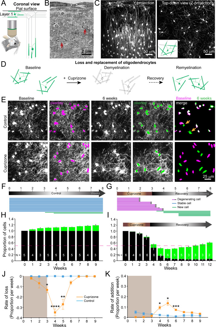
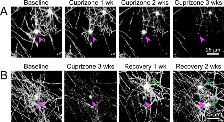
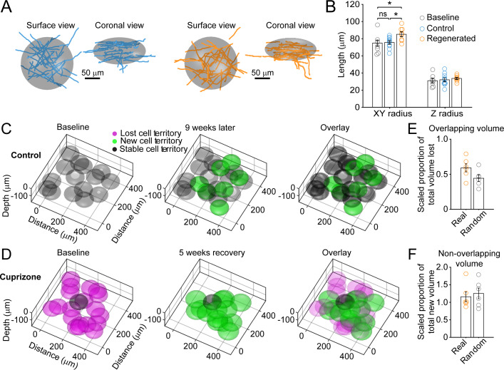
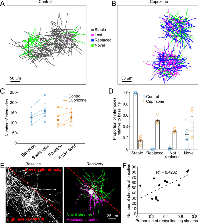
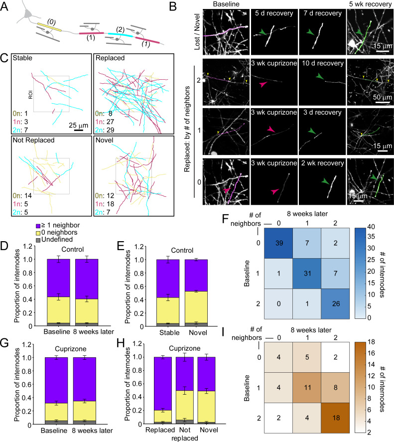
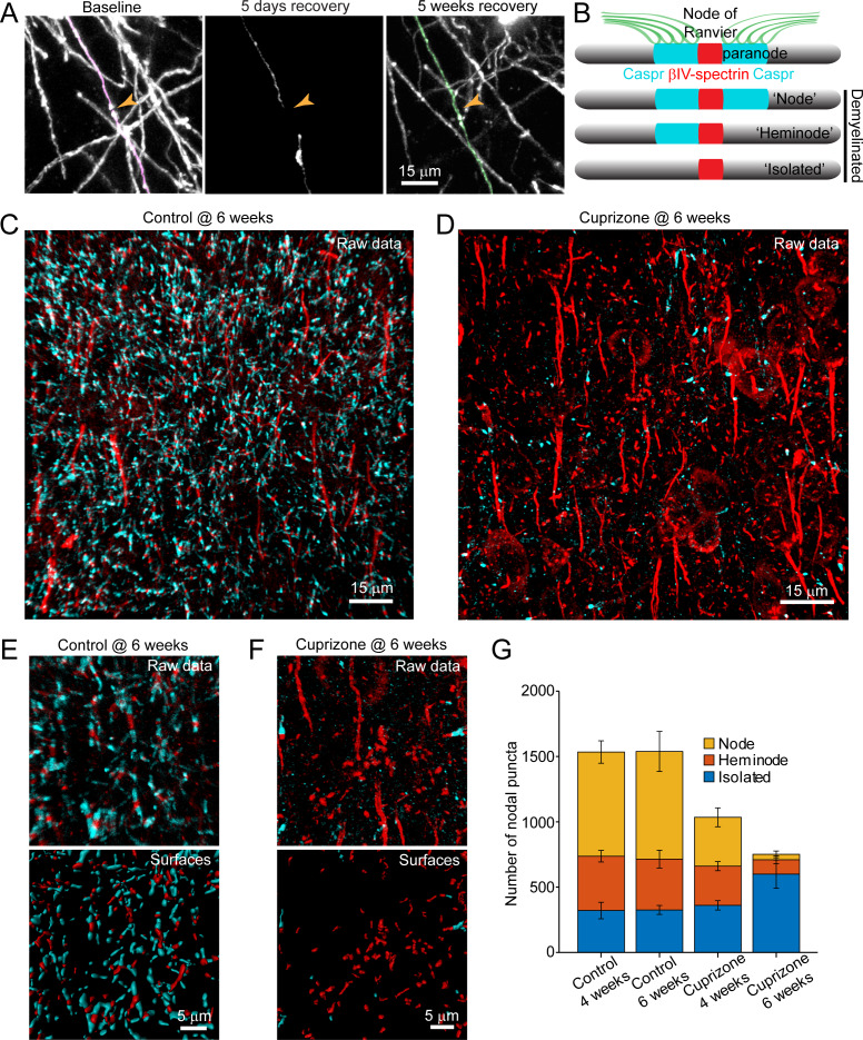
Destruction of oligodendrocytes and myelin sheaths in cortical gray matter profoundly alters neural activity and is associated with cognitive disability in multiple sclerosis (MS). Myelin can be restored by regenerating oligodendrocytes from resident progenitors; however, it is not known whether regeneration restores the complex myelination patterns in cortical circuits. Here, we performed time lapse in vivo two photon imaging in somatosensory cortex of adult mice to define the kinetics and specificity of myelin regeneration after acute oligodendrocyte ablation. These longitudinal studies revealed that the pattern of myelination in cortex changed dramatically after regeneration, as new oligodendrocytes were formed in different locations and new sheaths were often established along axon segments previously lacking myelin. Despite the dramatic increase in axonal territory available, oligodendrogenesis was persistently impaired in deeper cortical layers that experienced higher gliosis. Repeated reorganization of myelin patterns in MS may alter circuit function and contribute to cognitive decline.
Overview
The claims and statistics of this paper haven’t been checked yet.
Run the check — free, no account needed. It reads the open-access text and checks the sources it cites.
Integrity signals
Registry
Cites
Statistics
Figures
Verification
Claims the paper makes
The claims and statistics of this paper haven’t been checked yet.
Run the check — free, no account needed. It reads the open-access text and checks the sources it cites.
Claims other papers make about this one
Each is checked against this paper’s own text: does it say what the citing paper says it does?
The source backs part of the claim, not all of it — or backs it for a different population, condition or comparison.
In the citing paper
“The efficacy of remyelination has been shown to depend on multiple factors such as, age, disease duration and lesion location.”
From BCAS1-positive oligodendrocytes enable efficient cortical remyelination in multiple sclerosis (2025) · cites this paper as [8]
Experimental — accuracy not yet measuredThe source backs part of the claim, not all of it — or backs it for a different population, condition or comparison.
In the citing paper
“Similar to our prior study (Orthmann‐Murphy et al. 2020), we found no significant changes in OPC number in the deep cortex (Figure 1G, also see Figure S1C).”
From Depletion of Microglia Increases Cortical Oligodendrocyte Density During Remyelination (2026) · cites this paper as Orthmann‐Murphy et al. 2020
Experimental — accuracy not yet measuredThe source backs part of the claim, not all of it — or backs it for a different population, condition or comparison.
In the citing paper
“BCAS1 + GFP+ oligodendrocytes should represent a relatively more mature differentiating oligodendrocyte state than BCAS1 + GFP‐ cells (Chapman et al. 2024; Hughes et al. 2018; Orthmann‐Murphy et al. 2020).”
From Depletion of Microglia Increases Cortical Oligodendrocyte Density During Remyelination (2026) · cites this paper as Orthmann‐Murphy et al. 2020
Experimental — accuracy not yet measuredThe source backs part of the claim, not all of it — or backs it for a different population, condition or comparison.
In the citing paper
“We found that new oligodendrocytes formed after cessation of the cuprizone‐supplemented diet, with a peak rate of formation at 2 weeks post‐cuprizone (“early” recovery).”
From Depletion of Microglia Increases Cortical Oligodendrocyte Density During Remyelination (2026) · cites this paper as Orthmann‐Murphy et al. 2020
Experimental — accuracy not yet measuredThe source backs part of the claim, not all of it — or backs it for a different population, condition or comparison.
In the citing paper
“For cell counts, a blinded observer quantified the number of NG2+, GFAP+, Iba1+, ASPA+, BCAS1+, and GFP+ cells within each 500 μm × 750 μm region using a custom counting macro in FIJI/ImageJ (Orthmann‐Murphy et al. 2020).”
From Depletion of Microglia Increases Cortical Oligodendrocyte Density During Remyelination (2026) · cites this paper as Orthmann‐Murphy et al. 2020
Experimental — accuracy not yet measured
The source backs part of the claim, not all of it — or backs it for a different population, condition or comparison.
In the citing paper
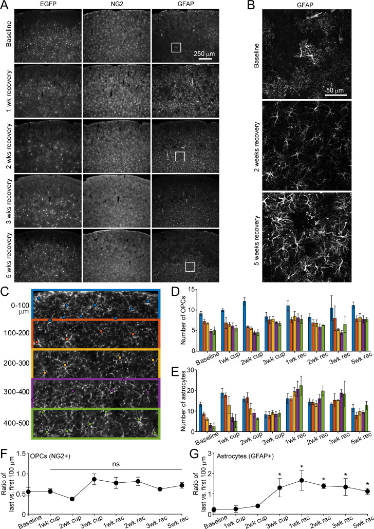
“Similar to our previous findings (Orthmann‐Murphy et al. 2020), even in the presence of Csf1r inhibitor, deep cortical reactive astrocytes persistently upregulate GFAP (Figure S5B).”
From Depletion of Microglia Increases Cortical Oligodendrocyte Density During Remyelination (2026) · cites this paper as Orthmann‐Murphy et al. 2020
Experimental — accuracy not yet measuredThe cited source says what the claim attributes to it.
In the citing paper
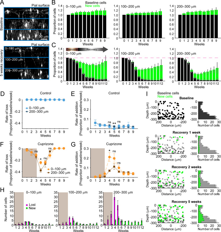
“In our prior in vivo imaging study, however, we showed that deep cortical oligodendrogenesis was selectively impaired, with only ~55% of the baseline oligodendrocyte population replaced after 9 weeks of recovery in the deepest cortical zone examined (200–300 μm; Orthmann‐Murphy et al. 2020).”
From Depletion of Microglia Increases Cortical Oligodendrocyte Density During Remyelination (2026) · cites this paper as Orthmann‐Murphy et al. 2020
The cited source says what the claim attributes to it.
In the citing paper
“We found that oligodendrocyte regeneration in the somatosensory cortex is incomplete, and this is driven primarily by inefficient oligodendrocyte replacement in deep cortical regions.”
From Depletion of Microglia Increases Cortical Oligodendrocyte Density During Remyelination (2026) · cites this paper as Orthmann‐Murphy et al. 2020
The cited source says what the claim attributes to it.
In the citing paper
“Nearly all oligodendrocytes present at 5 weeks (“late”) recovery were newly formed.”
From Depletion of Microglia Increases Cortical Oligodendrocyte Density During Remyelination (2026) · cites this paper as Orthmann‐Murphy et al. 2020
The cited source says what the claim attributes to it.
In the citing paper
“We previously showed that deep cortical astrocytes persistently upregulate GFAP following cuprizone administration (Orthmann‐Murphy et al. 2020);”
From Depletion of Microglia Increases Cortical Oligodendrocyte Density During Remyelination (2026) · cites this paper as Orthmann‐Murphy et al. 2020
The cited source says what the claim attributes to it.
In the citing paper
“Due to the exquisite spatial and temporal resolution of in vivo imaging, we also showed that the loss and formation of oligodendrocytes overlapped in early recovery, a finding that is difficult to appreciate by immunostaining alone.”
From Depletion of Microglia Increases Cortical Oligodendrocyte Density During Remyelination (2026) · cites this paper as Orthmann‐Murphy et al. 2020
The cited source says what the claim attributes to it.
In the citing paper
“Given that deep cortical ASPA+ oligodendrocytes recovered to baseline levels at late recovery only in PLX‐treated mice, but not cuprizone‐only treated mice (Figure 5), and late recovery oligodendrocytes are predominantly represented by newly formed cells (Orthmann‐Murphy et al. 2020), we suspect that many of these early recovery BCAS1+ cells formed in PLX‐treated mice become stably incorporated in”
From Depletion of Microglia Increases Cortical Oligodendrocyte Density During Remyelination (2026) · cites this paper as Orthmann‐Murphy et al. 2020
The cited source says what the claim attributes to it.
In the citing paper
“In addition, deep cortical astrocytes also become persistently reactive (Figure S4; Orthmann‐Murphy et al. 2020) and likely influence deep cortical reactive microglia function (Liddelow and Barres 2017).”
From Depletion of Microglia Increases Cortical Oligodendrocyte Density During Remyelination (2026) · cites this paper as Orthmann‐Murphy et al. 2020
The check wasn’t confident either way, so it gives no verdict — never disputed.
In the citing paper
“In fact, there are likely overlapping pathogenic roles of cortical reactive microglia, especially at early recovery (Orthmann‐Murphy et al. 2020).”
From Depletion of Microglia Increases Cortical Oligodendrocyte Density During Remyelination (2026) · cites this paper as Orthmann‐Murphy et al. 2020
Experimental — accuracy not yet measured
Self-check
Percentages, sums and reported differences re-computed from the paper’s own numbers.
The self-check needs the paper’s full text.
Citations
Cites
Cortical demyelination and diffuse white matter injury in multiple sclerosis
Kutzelnigg, A., Lucchinetti, C. F., Stadelmann, C., Brück, W., Rauschka, H., Bergmann, M., Schmidbauer, M., Parisi, J. E., & Lassmann, H. (2005) · Brain · doi:10.1093/brain/awh641 · Cited by 1,836
M2 microglia and macrophages drive oligodendrocyte differentiation during CNS remyelination
Miron, V. E., Boyd, A., Zhao, J.-W., Yuen, T. J., Ruckh, J. M., Shadrach, J. L., van Wijngaarden, P., Wagers, A. J., Williams, A., Franklin, R. J. M., et al. (2013) · Nature Neuroscience · doi:10.1038/nn.3469 · Cited by 1,777
Magliozzi, R., Howell, O. W., Vora, A., Serafini, B., Nicholas, R. S., Puopolo, M., Reynolds, R., & Aloisi, F. (2006) · Brain · doi:10.1093/brain/awm038 · Cited by 1,486
Neuronal activity promotes oligodendrogenesis and adaptive myelination in the mammalian brain
Gibson, E. M., Purger, D., Mount, C. W., Goldstein, A. K., Lin, G. L., Wood, L. S., Inema, I., Miller, S. E., Bieri, G., Zuchero, J. B., et al. (2014) · Science · doi:10.1126/science.1252304 · Cited by 1,472
Peterson, J. W., Bö, L., Mörk, S., Chang, A., & Trapp, B. D. (2001) · Annals of Neurology · doi:10.1002/ana.1123 · Cited by 1,403
Motor skill learning requires active central myelination
McKenzie, I. A., Ohayon, D., Li, H., Paes de Faria, J., Emery, B., Tohyama, K., & Richardson, W. D. (2014) · Science · doi:10.1126/science.1254960 · Cited by 1,177
Inflammatory cortical demyelination in early multiple sclerosis.
Lucchinetti, C. F., Popescu, B. F. G., Bunyan, R. F., Moll, N. M., Roemer, S. F., Lassmann, H., Brück, W., Parisi, J. E., Scheithauer, B. W., Giannini, C., et al. (2011) · New England Journal of Medicine · doi:10.1056/nejmoa1100648 · Cited by 1,103
Matsushima, G. K., & Morell, P. (2001) · Brain Pathology · doi:10.1111/j.1750-3639.2001.tb00385.x · Cited by 984
Longair, M., Baker, D. A., & Armstrong, J. D. (2011) · Bioinformatics · doi:10.1093/bioinformatics/btr390 · Cited by 939
Oligodendrocyte Dynamics in the Healthy Adult CNS: Evidence for Myelin Remodeling
Young, K. M., Psachoulia, K., Tripathi, R. B., Dunn, S.-J., Cossell, L., Attwell, D., Tohyama, K., & Richardson, W. D. (2013) · Neuron · doi:10.1016/j.neuron.2013.01.006 · Cited by 919
Network anatomy and in vivo physiology of visual cortical neurons
Bock, D. D., Lee, W.-C. A., Kerlin, A., Andermann, M. L., Hood, G., Wetzel, A. W., Yurgenson, S., Soucy, E. R., Kim, H. S., & Reid, R. C. (2011) · Nature · doi:10.1038/nature09802 · Cited by 867
Meningeal inflammation is widespread and linked to cortical pathology in multiple sclerosis
Howell, O. W., Reeves, C. A., Nicholas, R., Carassiti, D., Radotra, B., Gentleman, S. M., Serafini, B., Aloisi, F., Roncaroli, F., Magliozzi, R., et al. (2011) · Brain · doi:10.1093/brain/awr182 · Cited by 858
Kang, S. H., Fukaya, M., Yang, J. K., Rothstein, J. D., & Bergles, D. E. (2010) · Neuron · doi:10.1016/j.neuron.2010.09.009 · Cited by 815
Self-citationHughes, E. G., Kang, S. H., Fukaya, M., & Bergles, D. E. (2013) · Nature Neuroscience · doi:10.1038/nn.3390 · Cited by 808
Self-citationMyelin Impairs CNS Remyelination by Inhibiting Oligodendrocyte Precursor Cell Differentiation
Kotter, M. R., Li, W.-W., Zhao, C., & Franklin, R. J. M. (2006) · The Journal of Neuroscience · doi:10.1523/jneurosci.2615-05.2006 · Cited by 768
Cortical lesions in multiple sclerosis
Kidd, D., Barkhof, F., McConnell, R., Algra, P. R., Allen, I. V., & Revesz, T. (1999) · Brain · doi:10.1093/brain/122.1.17 · Cited by 766
NG2-Positive Oligodendrocyte Progenitor Cells in Adult Human Brain and Multiple Sclerosis Lesions
Chang, A., Nishiyama, A., Peterson, J. W., Prineas, J. W., & Trapp, B. D. (2000) · Journal of Neuroscience · doi:10.1523/jneurosci.20-17-06404.2000 · Cited by 711
Rejuvenation of Regeneration in the Aging Central Nervous System
Ruckh, J. M., Zhao, J.-W., Shadrach, J. L., van Wijngaarden, P., Rao, T. N., Wagers, A. J., & Franklin, R. J. M. (2012) · Cell Stem Cell · doi:10.1016/j.stem.2011.11.019 · Cited by 649
LINGO-1 negatively regulates myelination by oligodendrocytes
Mi, S., Miller, R. H., Lee, X., Scott, M. L., Shulag-Morskaya, S., Shao, Z., Chang, J., Thill, G., Levesque, M., Zhang, M., et al. (2005) · Nature Neuroscience · doi:10.1038/nn1460 · Cited by 628
Hyaluronan accumulates in demyelinated lesions and inhibits oligodendrocyte progenitor maturation
Back, S. A., Tuohy, T. M. F., Chen, H., Wallingford, N., Craig, A., Struve, J., Luo, N. L., Banine, F., Liu, Y., Chang, A., et al. (2005) · Nature Medicine · doi:10.1038/nm1279 · Cited by 612
Distinct Profiles of Myelin Distribution Along Single Axons of Pyramidal Neurons in the Neocortex
Tomassy, G. S., Berger, D. R., Chen, H.-H., Kasthuri, N., Hayworth, K. J., Vercelli, A., Seung, H. S., Lichtman, J. W., & Arlotta, P. (2014) · Science · doi:10.1126/science.1249766 · Cited by 600
Kang, S. H., Li, Y., Fukaya, M., Lorenzini, I., Cleveland, D. W., Ostrow, L. W., Rothstein, J. D., & Bergles, D. E. (2013) · Nature Neuroscience · doi:10.1038/nn.3357 · Cited by 599
Self-citationMicropillar arrays as a high-throughput screening platform for therapeutics in multiple sclerosis
Mei, F., Fancy, S. P. J., Shen, Y. A., Niu, J., Zhao, C., Presley, B., Miao, E., Lee, S., Mayoral, S. R., Redmond, S., et al. (2014) · Nature Medicine · doi:10.1038/nm.3618 · Cited by 595
Dimou, L., Simon, C., Kirchhoff, F., Takebayashi, H., & Götz, M. (2008) · The Journal of Neuroscience · doi:10.1523/jneurosci.2831-08.2008 · Cited by 551
Dynamics of Oligodendrocyte Generation and Myelination in the Human Brain
Yeung, M. S. Y., Zdunek, S., Bergmann, O., Bernard, S., Salehpour, M., Alkass, K., Perl, S., Tisdale, J., Possnert, G., Brundin, L., et al. (2014) · Cell · doi:10.1016/j.cell.2014.10.011 · Cited by 541
Myelin remodeling through experience-dependent oligodendrogenesis in the adult somatosensory cortex
Hughes, E. G., Orthmann-Murphy, J. L., Langseth, A. J., & Bergles, D. E. (2018) · Nature Neuroscience · doi:10.1038/s41593-018-0121-5 · Cited by 540
Self-citationA regenerative approach to the treatment of multiple sclerosis
Deshmukh, V. A., Tardif, V., Lyssiotis, C. A., Green, C. C., Kerman, B., Kim, H. J., Padmanabhan, K., Swoboda, J. G., Ahmad, I., Kondo, T., et al. (2013) · Nature · doi:10.1038/nature12647 · Cited by 531
Rapid production of new oligodendrocytes is required in the earliest stages of motor-skill learning
Xiao, L., Ohayon, D., McKenzie, I. A., Sinclair-Wilson, A., Wright, J. L., Fudge, A. D., Emery, B., Li, H., & Richardson, W. D. (2016) · Nature Neuroscience · doi:10.1038/nn.4351 · Cited by 507
Lifelong cortical myelin plasticity and age-related degeneration in the live mammalian brain
Hill, R. A., Li, A. M., & Grutzendler, J. (2018) · Nature Neuroscience · doi:10.1038/s41593-018-0120-6 · Cited by 467
Metformin restores CNS remyelination capacity by rejuvenating aged stem cells
Neumann, B., Baror, R., Zhao, C., Segel, M., Dietmann, S., Rawji, K. S., Foerster, S., McClain, C. R., Chalut, K., van Wijngaarden, P., et al. (2019) · Cell Stem Cell · doi:10.1016/j.stem.2019.08.015 · Cited by 449
Craner, M. J., Newcombe, J., Black, J. A., Hartle, C., Cuzner, M. L., & Waxman, S. G. (2004) · Proceedings of the National Academy of Sciences · doi:10.1073/pnas.0402765101 · Cited by 446
Werneburg, S., Jung, J., Kunjamma, R. B., Ha, S.-K., Luciano, N. J., Willis, C. M., Gao, G., Biscola, N. P., Havton, L. A., Crocker, S. J., et al. (2020) · Immunity · doi:10.1016/j.immuni.2019.12.004 · Cited by 440
Kirby, L., Jin, J., Cardona, J. G., Smith, M. D., Martin, K. A., Wang, J., Strasburger, H., Herbst, L., Alexis, M., Karnell, J., et al. (2019) · Nature Communications · doi:10.1038/s41467-019-11638-3 · Cited by 436 · cited 3×
“Like other progenitor cells, OPCs exhibit a decline in regenerative potential with age and can undergo senescence, a process that may be accelerated by exposure to inflammatory cytokines (Kirby et al., 2019; Neumann et al., 2019; Nicaise et al., 2019).”
BackgroundSelf-citationDifferentiation and death of premyelinating oligodendrocytes in developing rodent brain
Trapp, B. D., Nishiyama, A., Cheng, D., & Macklin, W. (1997) · The Journal of Cell Biology · doi:10.1083/jcb.137.2.459 · Cited by 395
Generating Neuronal Diversity in the Mammalian Cerebral Cortex
Lodato, S., & Arlotta, P. (2015) · Annual Review of Cell and Developmental Biology · doi:10.1146/annurev-cellbio-100814-125353 · Cited by 389
Dynamics of oligodendrocyte generation in multiple sclerosis
Yeung, M. S. Y., Djelloul, M., Steiner, E., Bernard, S., Salehpour, M., Possnert, G., Brundin, L., & Frisén, J. (2019) · Nature · doi:10.1038/s41586-018-0842-3 · Cited by 376
Individual Oligodendrocytes Have Only a Few Hours in which to Generate New Myelin Sheaths In Vivo
Czopka, T., ffrench‐Constant, C., & Lyons, D. A. (2013) · Developmental Cell · doi:10.1016/j.devcel.2013.05.013 · Cited by 354
Cortical lesion load associates with progression of disability in multiple sclerosis.
Calabrese, M., Poretto, V., Favaretto, A., Alessio, S., Bernardi, V., Romualdi, C., Rinaldi, F., Perini, P., & Gallo, P. (2012) · Brain : a journal of neurology · doi:10.1093/brain/aws246 · Cited by 341
Mei, F., Lehmann‐Horn, K., Shen, Y. A., Rankin, K. A., Stebbins, K. J., Lorrain, D. S., Pekarek, K. L., Sagan, S. A., Xiao, L., Teuscher, C., et al. (2016) · eLife · doi:10.7554/elife.18246 · Cited by 325
Motor learning promotes remyelination via new and surviving oligodendrocytes
Bacmeister, C. M., Barr, H. J., McClain, C. R., Thornton, M. A., Nettles, D., Welle, C. G., & Hughes, E. G. (2020) · Nature Neuroscience · doi:10.1038/s41593-020-0637-3 · Cited by 320
Maintaining Tissue Homeostasis: Dynamic Control of Somatic Stem Cell Activity
Biteau, B., Hochmuth, C. E., & Jasper, H. (2011) · Cell stem cell · doi:10.1016/j.stem.2011.10.004 · Cited by 310
A large fraction of neocortical myelin ensheathes axons of local inhibitory neurons
Micheva, K. D., Wolman, D. N., Mensh, B. D., Pax, E., Buchanan, J., Smith, S. J., & Bock, D. D. (2016) · eLife · doi:10.7554/elife.15784 · Cited by 289
Gouw, A. A., Seewann, A., Vrenken, H., van der Flier, W. M., Rozemuller, J. M., Barkhof, F., Scheltens, P., & Geurts, J. J. G. (2008) · Brain · doi:10.1093/brain/awn265 · Cited by 281
Cortical Demyelination Is Prominent in the Murine Cuprizone Model and Is Strain-Dependent
Skripuletz, T., Lindner, M., Kotsiari, A., Garde, N., Fokuhl, J., Linsmeier, F., Trebst, C., & Stangel, M. (2008) · The American Journal of Pathology · doi:10.2353/ajpath.2008.070850 · Cited by 271
Chondroitin sulfate proteoglycans in demyelinated lesions impair remyelination
Lau, L. W., Keough, M. B., Haylock‐Jacobs, S., Cua, R., Döring, A., Sloka, S., Stirling, D. P., Rivest, S., & Yong, V. W. (2012) · Annals of Neurology · doi:10.1002/ana.23599 · Cited by 255
Regional differences between grey and white matter in cuprizone induced demyelination
Gudi, V., Moharregh-Khiabani, D., Skripuletz, T., Koutsoudaki, P. N., Kotsiari, A., Skuljec, J., Trebst, C., & Stangel, M. (2009) · Brain Research · doi:10.1016/j.brainres.2009.06.005 · Cited by 252
Ihara, M., Polvikoski, T. M., Hall, R., Slade, J. Y., Perry, R. H., Oakley, A. E., Englund, E. M., O'Brien, J. T., Ince, P. G., & Kalaria, R. N. (2010) · Acta Neuropathologica · doi:10.1007/s00401-009-0635-8 · Cited by 251
Chong, S. Y. C., Rosenberg, S. S., Fancy, S. P. J., Zhao, C., Shen, Y.-A. A., Hahn, A. T., McGee, A. W., Xu, X., Zheng, B., Zhang, L. I., et al. (2012) · Proceedings of the National Academy of Sciences · doi:10.1073/pnas.1113540109 · Cited by 232
Nicaise, A. M., Wagstaff, L. J., Willis, C. M., Paisie, C. A., Chandok, H., Robson, P., Fossati, V., Williams, A., & Crocker, S. J. (2019) · Proceedings of the National Academy of Sciences · doi:10.1073/pnas.1818348116 · Cited by 228
Cortical remyelination: A new target for repair therapies in multiple sclerosis
Chang, A., Staugaitis, S. M., Dutta, R., Batt, C. E., Easley, K. E., Chomyk, A. M., Yong, V. W., Fox, R. J., Kidd, G. J., & Trapp, B. D. (2012) · Annals of Neurology · doi:10.1002/ana.23693 · Cited by 216
Coman, I., Aigrot, M., Seilhean, D., Reynolds, R., Girault, J., Zalc, B., & Lubetzki, C. (2006) · Brain · doi:10.1093/brain/awl144 · Cited by 210
Regulation of myelin structure and conduction velocity by perinodal astrocytes
Dutta, D. J., Woo, D. H., Lee, P. R., Pajevic, S., Bukalo, O., Huffman, W. C., Wake, H., Basser, P. J., SheikhBahaei, S., Lazarevic, V., et al. (2018) · Proceedings of the National Academy of Sciences · doi:10.1073/pnas.1811013115 · Cited by 203
Remyelination in multiple sclerosis
Lassmann, H., Brück, W., Lucchinetti, C., & Rodriguez, M. (1997) · Multiple Sclerosis Journal · doi:10.1177/135245859700300213 · Cited by 191
Remyelination of mouse spinal cord axons demyelinated by local injection of lysolecithin
Jeffery, N. D., & Blakemore, W. F. (1995) · Journal of Neurocytology · doi:10.1007/bf01191213 · Cited by 189
Five Decades of Cuprizone, an Updated Model to Replicate Demyelinating Diseases
Vega-Riquer, J. M., Mendez-Victoriano, G., Morales-Luckie, R. A., & Gonzalez-Perez, O. (2019) · Current Neuropharmacology · doi:10.2174/1570159x15666170717120343 · Cited by 188
Sample Drift Correction Following 4D Confocal Time-lapse Imaging
Parslow, A. C., Cardona, A., & Bryson‐Richardson, R. J. (2014) · Journal of Visualized Experiments · doi:10.3791/51086 · Cited by 187
Inhibition of Gli1 mobilizes endogenous neural stem cells for remyelination
Samanta, J., Grund, E. M., Silva, H. M., Lafaille, J. J., Fishell, G., & Salzer, J. L. (2015) · Nature · doi:10.1038/nature14957 · Cited by 170
Stedehouder, J., Couey, J. J., Brizee, D., Hosseini, B., Slotman, J. A., Dirven, C. M. F., Shpak, G., Houtsmuller, A. B., & Kushner, S. A. (2017) · Cerebral Cortex · doi:10.1093/cercor/bhx203 · Cited by 166
Walhovd, K. B., Johansen-Berg, H., & Káradóttir, R. T. (2014) · Neuroscience · doi:10.1016/j.neuroscience.2014.06.058 · Cited by 156
Contribution of cortical lesion subtypes at 7T MRI to physical and cognitive performance in MS
Nielsen, A. S., Kinkel, R. P., Madigan, N. K., Tinelli, E., Benner, T., & Mainero, C. (2013) · Neurology · doi:10.1212/wnl.0b013e3182a08ce8 · Cited by 133
A neuronal PI(3,4,5)P3-dependent program of oligodendrocyte precursor recruitment and myelination
Goebbels, S., Wieser, G. L., Pieper, A., Spitzer, S. O., Weege, B., Yan, K., Edgar, J. M., Yagensky, O., Wichert, S. P., Agarwal, A., et al. (2016) · Nature Neuroscience · doi:10.1038/nn.4425 · Cited by 125
Evidence for Myelin Sheath Remodeling in the CNS Revealed by In Vivo Imaging
Auer, F., Vagionitis, S., & Czopka, T. (2018) · Current Biology · doi:10.1016/j.cub.2018.01.017 · Cited by 123
Su, Z., Yuan, Y., Chen, J., Zhu, Y., Qiu, Y., Zhu, F., Huang, A., & He, C. (2011) · Journal of Neurotrauma · doi:10.1089/neu.2010.1597 · Cited by 123
Stedehouder, J., & Kushner, S. A. (2016) · Molecular Psychiatry · doi:10.1038/mp.2016.147 · Cited by 120
Baxi, E. G., DeBruin, J., Tosi, D. M., Grishkan, I. V., Smith, M. D., Kirby, L. A., Strasburger, H. J., Fairchild, A. N., Calabresi, P. A., & Gocke, A. R. (2015) · The Journal of Neuroscience · doi:10.1523/jneurosci.3817-14.2015 · Cited by 103
Self-citationChanged distribution of sodium channels along demyelinated axons.
England, J. D., Gamboni, F., Levinson, S. R., & Finger, T. E. (1990) · Proceedings of the National Academy of Sciences · doi:10.1073/pnas.87.17.6777 · Cited by 98
Baxi, E. G., DeBruin, J., Jin, J., Strasburger, H. J., Smith, M. D., Orthmann‐Murphy, J. L., Schott, J. T., Fairchild, A. N., Bergles, D. E., & Calabresi, P. A. (2017) · Glia · doi:10.1002/glia.23229 · Cited by 97
Self-citationImproved Visualization of Cortical Lesions in Multiple Sclerosis Using 7T MP2RAGE
Beck, E., Sati, P., Sethi, V., Kober, T., Dewey, B. E., Bhargava, P., Nair, G., Cortese, I. C. M., & Reich, D. S. (2018) · American Journal of Neuroradiology · doi:10.3174/ajnr.a5534 · Cited by 95
Synaptophysin Is a Reliable Marker for Axonal Damage
Gudi, V., Gai, L., Herder, V., Tejedor, L. S., Kipp, M., Amor, S., Sühs, K.-W., Hansmann, F., Beineke, A., Baumgärtner, W., et al. (2017) · Journal of Neuropathology & Experimental Neurology · doi:10.1093/jnen/nlw114 · Cited by 87
Early, J. J., Marshall-Phelps, K. L., Williamson, J., Swire, M., Kamadurai, H., Muskavitch, M. A. T., & Lyons, D. A. (2018) · eLife · doi:10.7554/elife.35136 · Cited by 86
Imaging outcome measures of neuroprotection and repair in MS
Oh, J., Ontaneda, D., Azevedo, C. J., Klawiter, E. C., Absinta, M., Arnold, D. L., Bakshi, R., Calabresi, P. A., Crainiceanu, C., Dewey, B. E., et al. (2019) · Neurology · doi:10.1212/wnl.0000000000007099 · Cited by 76
Self-citationMorphometric analysis of oligodendrocytes in the adult mouse frontal cortex
Murtie, J. C., Macklin, W. B., & Corfas, G. (2007) · Journal of Neuroscience Research · doi:10.1002/jnr.21339 · Cited by 73
Dupree, J. L., Mason, J. L., Marcus, J., Stull, M., Levinson, R., Matsushima, G. K., & Popko, B. (2004) · Neuron Glia Biology · doi:10.1017/s1740925x04000304 · Cited by 69
Kilsdonk, I. D., de Graaf, W. L., Soriano, A. L., Zwanenburg, J. J. M., Visser, F., Kuijer, J. P. A., Geurts, J. J. G., Pouwels, P. J. W., Polman, C. H., Castelijns, J. A., et al. (2012) · American Journal of Neuroradiology · doi:10.3174/ajnr.a3289 · Cited by 68
Myelin replacement triggered by single-cell demyelination in mouse cortex
Snaidero, N., Schifferer, M., Mezydlo, A., Zalc, B., Kerschensteiner, M., & Misgeld, T. (2020) · Nature Communications · doi:10.1038/s41467-020-18632-0 · Cited by 63
Zhang, Y., Zhang, J., Navrazhina, K., Argaw, A. T., Zameer, A., Gurfein, B. T., Brosnan, C. F., & John, G. R. (2010) · Glia · doi:10.1002/glia.20978 · Cited by 62
Ultra-high-field MR imaging in multiple sclerosis
Filippi, M., Evangelou, N., Kangarlu, A., Inglese, M., Mainero, C., Horsfield, M. A., & Rocca, M. A. (2013) · Journal of Neurology Neurosurgery & Psychiatry · doi:10.1136/jnnp-2013-305246 · Cited by 55
Imaging Neocortical Neurons through a Chronic Cranial WindowPreprint
Holtmaat, A. J. D. G., Paola, V. D., Wilbrecht, L., Trachtenberg, J. T., Svoboda, K., & Portera‐Cailliau, C. (2012) · Cold Spring Harbor Protocols · doi:10.1101/pdb.prot069617 · Cited by 51
MRI of cortical lesions and its use in studying their role in MS pathogenesis and disease course
Magliozzi, R., Reynolds, R., & Calabrese, M. (2018) · Brain Pathology · doi:10.1111/bpa.12642 · Cited by 51
Klingseisen, A., Ristoiu, A.-M., Kegel, L., Sherman, D. L., Rubio-Brotons, M., Almeida, R., Koudelka, S., Benito-Kwiecinski, S. K., Poole, R. J., Brophy, P., et al. (2019) · Developmental Cell · doi:10.1016/j.devcel.2019.10.016 · Cited by 44
Inflammatory demyelination alters subcortical visual circuits
Araújo, S. E. S., Mendonça, H. R., Wheeler, N. A., Campello-Costa, P., Jacobs, K. M., Gomes, F. C. A., Fox, M. A., & Fuss, B. (2017) · Journal of Neuroinflammation · doi:10.1186/s12974-017-0936-0 · Cited by 43
Profiles of cortical inflammation in multiple sclerosis by 11 C-PBR28 MR-PET and 7 Tesla imaging
Herranz, E., Louapre, C., Treaba, C. A., Govindarajan, S. T., Ouellette, R., Mangeat, G., Loggia, M. L., Cohen‐Adad, J., Klawiter, E. C., Sloane, J. A., et al. (2019) · Multiple Sclerosis Journal · doi:10.1177/1352458519867320 · Cited by 34
Submembranous cytoskeletons stabilize nodes of Ranvier
Susuki, K., Otani, Y., & Rasband, M. N. (2016) · Experimental Neurology · doi:10.1016/j.expneurol.2015.11.012 · Cited by 31
Self-citationPrecision in a sea of profusion: Myelin replacement triggered by single-cell cortical demyelinationPreprint
Snaidero, N., Schifferer, M., Mężydło, A., Kerschensteiner, M., & Misgeld, T. (2019) · bioRxiv (Cold Spring Harbor Laboratory) · doi:10.1101/2019.12.16.877597 · Cited by 3
Rankin, K. A., Mei, F., Kim, K., Shen, Y. A., Sylvester, S., Fung, A., Gallo, V., Fancy, S. P. J., & Green, P. E. (2019) · Journal of Neuroscience · doi:10.1523/jneurosci.1475-18.2019
Cited by
Sorted by citations
Periods of synchronized myelin changes shape brain function and plasticity
de Faria, O., Pivoňková, H., Varga, B. V., Timmler, S., Evans, K. A., & Káradóttir, R. T. (2021) · Nature Neuroscience · doi:10.1038/s41593-021-00917-2 · Cited by 202
Multiphoton fluorescence microscopy for in vivo imaging
Xu, C., Nedergaard, M., Fowell, D. J., Friedl, P., & Ji, N. (2024) · Cell · doi:10.1016/j.cell.2024.07.036 · Cited by 177
Mechanisms of node of Ranvier assembly
Rasband, M. N., & Peles, E. (2020) · Nature reviews. Neuroscience · doi:10.1038/s41583-020-00406-8 · Cited by 180
Self-citationRevisiting remyelination: Towards a consensus on the regeneration of CNS myelin
Franklin, R. J. M., Frisén, J., & Lyons, D. A. (2021) · Seminars in Cell & Developmental Biology · doi:10.1016/j.semcdb.2020.09.009 · Cited by 154
Neely, S. A., Williamson, J. M., Klingseisen, A., Zoupi, L., Early, J. J., Williams, A., & Lyons, D. A. (2022) · Nature Neuroscience · doi:10.1038/s41593-021-01009-x · Cited by 132
Metadata
- Authors
- Orthmann-Murphy, J., Call, C. L., Molina-Castro, G. C., Hsieh, Y. C., Rasband, M. N., Calabresi, P. A., & Bergles, D. E.
- Affiliations
- The Solomon Snyder Department of Neuroscience, Johns Hopkins University, Baltimore, United States
- Johns Hopkins University (US)
- Baylor College of Medicine (US)
- Corresponding author
- Bergles, Dwight E
- Venue
- eLife
- Publisher
- eLife Sciences Publications, Ltd
- Published
- 2020-05-27
- Volume
- 9
- ISSN
- 2050-084X (ISSN-L)
- PMC
- PMC7292648
- OpenAlex
- W3029244827
- Cited by
- 111
- References
- 85
- Citations by year
- 2026: 13 · 2025: 15 · 2024: 20 · 2023: 22 · 2022: 15 · 2021: 20 · 2020: 6
- FWCI
- 6.20
- Type
- journal article
- Language
- en
- Open access
- gold
- doi.org
- Licence
- Locations
- eLife (publishedVersion, cc-by)
- bioRxiv (Cold Spring Harbor Laboratory) (acceptedVersion, cc-by)
- PubMed Central (publishedVersion, cc-by)
- PubMed (publishedVersion)
- DOAJ (DOAJ: Directory of Open Access Journals) (submittedVersion, cc-by-sa)
- Funders
- Conrad N. Hilton Foundation
- National Multiple Sclerosis Society
- National Science Foundation (Graduate Research Fellowship)
- National Institutes of Health (NS051509, NS050274, NS080153)
- Dr. Miriam and Sheldon G Adelson Medical Research Foundation
- Topics
- Life Sciences › Neuroscience › Developmental Neuroscience › Neurogenesis and neuroplasticity mechanisms
- Health Sciences › Medicine › Clinical Neurology › Multiple Sclerosis Research Studies
- Life Sciences › Neuroscience › Neurology › Neuroinflammation and Neurodegeneration Mechanisms
- Keywords
- remyelination, oligodendrogenesis, cerebral cortex, somatosensory cortex, multiple sclerosis, gliosis, cortical layers, cognitive decline, in vivo two-photon imaging
- MeSH terms
- Remyelination, Animals, Axons, Female, Humans, Kinetics, Male, Mice, Inbred C57BL, Multiple Sclerosis, Myelin Sheath, Oligodendroglia, Somatosensory Cortex, Mice
About these checks
Registry, retraction, Cites and statistics results are computed, not judged, with one exception: a printed reference that no index lists is read once by a model before it counts as a citation error. Claims are extracted by a model and kept only when their quote appears verbatim in the text; each cited source is then checked, with a model’s help, for whether it says what the claim relies on. Those verdicts can be wrong — read the claim’s quote and the cited source before relying on one.
Statistics and arithmetic are recomputed, not judged.
Confidence bands are shown only for fields calibrated on our annotated test set. None is calibrated yet, so no band is shown.
What counts as a claim ›How we measure confidence ›How we check this ›
Are you an author, or do you see a problem with this page? Request a review
