

The DOI matches its Crossref record; 9 of the 9 references it cites were found in a registry or an index.
Recent progress of magnetic nanoparticles in biomedical applications: A review
Abstract
Magnetic nanoparticles (MNPs) offer tremendous potentialities in biomedical applications for a long while. Since these materials' interactions in biological media largely rely on their crystal structures, sizes, and shapes, detailed studies on their synthesis mechanism for medicinal aspects are crucial. Despite many review reports that have already been published on MNPs, they mainly have focused either on their perspective in biomedical applications or their synthesis and characterization along with functionalization mechanisms as individual entities. For this reason, this review uncovers a comprehensive insight into the ongoing improvement of fabrication processes, surface functionalization of MNPs for biomedical applications together. Besides, various magnetic nanocomposite (MNCs) for smart drug delivery, recent hyperthermia treatment, lab‐on‐a‐chip, and magnetic bio‐separation, and some of the recent emerging imaging techniques using MNPs are discussed. A detailed analysis of toxicity, challenges, and recent progress of clinical trials of MNPs is sketched out to open numerous entryways for advanced research on MNPs for biomedical applications.
Overview
The claims and statistics of this paper haven’t been checked yet.
Run the check — free, no account needed. It reads the open-access text and checks the sources it cites.
Integrity signals
Registry
Cites
Statistics
Figures
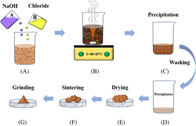
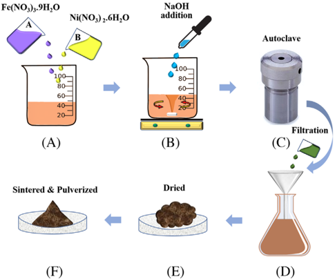

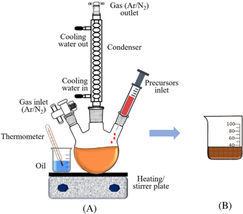
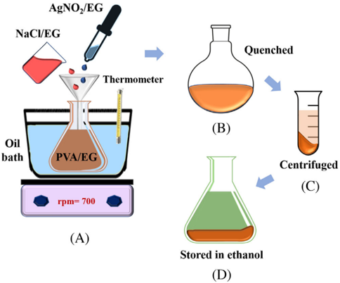






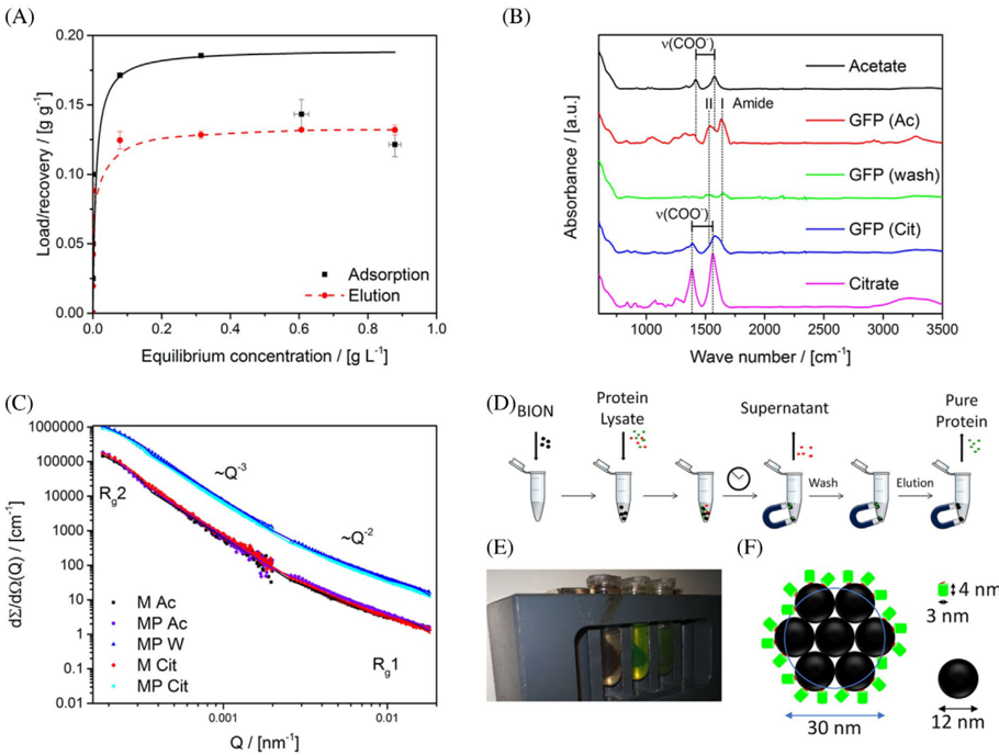

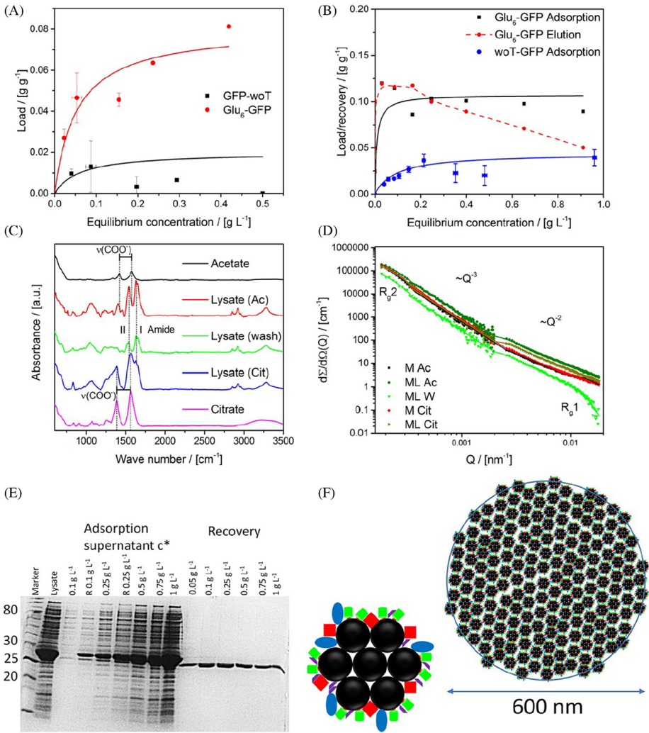

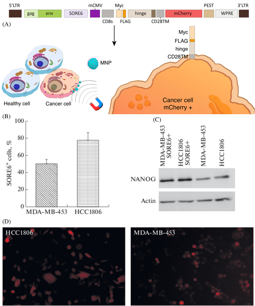

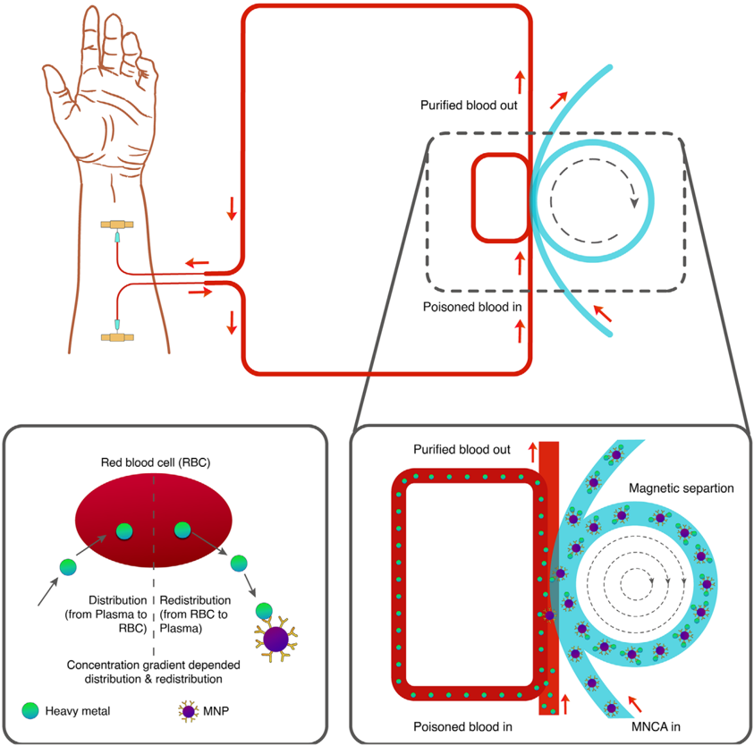









Verification
Claims the paper makes
The claims and statistics of this paper haven’t been checked yet.
Run the check — free, no account needed. It reads the open-access text and checks the sources it cites.
Claims other papers make about this one
Each is checked against this paper’s own text: does it say what the citing paper says it does?
In the citing paper
“These properties are affected by many factors, including particle size, shape, crystallinity, and types of iron oxides used [63].”
From Advances in green-synthesized magnetic nanoparticles for targeted cancer therapy: mechanisms, applications, and future perspectives (2026) · cites this paper as [63]
Self-check
Percentages, sums and reported differences re-computed from the paper’s own numbers.
The self-check needs the paper’s full text.
Citations
Cites
Laurent, S., Forge, D., Port, M., Roch, A., Robic, C., Vander Elst, L., & Muller, R. N. (2008) · Chemical Reviews · doi:10.1021/cr068445e · Cited by 6,852
Nanoparticle-Based Medicines: A Review of FDA-Approved Materials and Clinical Trials to Date
Bobo, D., Robinson, K. J., Islam, J., Thurecht, K. J., & Corrie, S. R. (2016) · Pharmaceutical Research · doi:10.1007/s11095-016-1958-5 · Cited by 2,586
Magnetic iron oxide nanoparticles: synthesis and surface functionalization strategies
Wu, W., He, Q., & Jiang, C. (2008) · Nanoscale Research Letters · doi:10.1007/s11671-008-9174-9 · Cited by 2,300
Wu, W., Wu, Z., Yu, T., Jiang, C., & Kim, W.-S. (2015) · Science and Technology of Advanced Materials · doi:10.1088/1468-6996/16/2/023501 · Cited by 1,574
Block copolymer micelles in nanomedicine applications
Cabral, H., Miyata, K., Osada, K., & Kataoka, K. (2018) · Chemical Reviews · doi:10.1021/acs.chemrev.8b00199 · Cited by 1,309
Surface modification of magnetic iron oxide nanoparticles
Zhu, N., Ji, H., Yu, P., Niu, J., Farooq, M. U., Akram, M. W., Udego, I. O., Li, H., & Niu, X. (2018) · Nanomaterials · doi:10.3390/nano8100810 · Cited by 569
Tay, Z. W., Chandrasekharan, P., Chiu-Lam, A., Hensley, D. W., Dhavalikar, R., Zhou, X. Y., Yu, E. Y., Goodwill, P. W., Zheng, B., Rinaldi, C., et al. (2018) · ACS Nano · doi:10.1021/acsnano.8b00893 · Cited by 459
Systematic review of the preparation techniques of iron oxide magnetic nanoparticles
F. Hasany, S., Ahmed, I., J, R., & Rehman, A. (2013) · Nanoscience and Nanotechnology · doi:10.5923/j.nn.20120206.01 · Cited by 318
Dendrimer-Based Drug Delivery Systems for Brain Targeting
Zhu, Y., Liu, C., & Pang, Z. (2019) · Biomolecules · doi:10.3390/biom9120790 · Cited by 220
Research works printed but not linked: 316
- 10.1080/10717544.2019.1667450 · DOI
- 10.1096/fj.201900716RR · DOI
- 10.3390/pharmaceutics12090841 · DOI
- 10.1016/j.jare.2018.06.005 · DOI · Paper page
- 10.1002/adhm.201700845 · DOI
- 10.1515/ntrev-2017-0193 · DOI
- Wong J., 2017, Curr. Pharm. Des., 23, 10.2174/1381612823666170215104659 · DOI
- 10.3390/magnetochemistry5040066 · DOI
- 10.1016/j.addr.2011.03.008 · DOI
- 10.1016/j.addr.2008.03.018 · DOI
- 10.1016/j.partic.2016.06.001 · DOI · Paper page
- 10.1016/j.jconrel.2015.09.039 · DOI
- Dobson, 2008, Int. J. Nanomedicine, 3, 169, 10.2147/IJN.S1608 · DOI
- 10.1016/j.jmmm.2005.02.045 · DOI
- 10.1016/j.msec.2015.04.047 · DOI
- 10.1263/jbb.100.1 · DOI
- Majidi S., 2016, Nanomed. Biotechnol., 44, 722
- 10.1080/10408347.2020.1797465 · DOI
- Mosayebi J., 2017, Mater, 6, 1700306
- 10.1039/b207789b · DOI
- 10.1007/s00216-005-0255-7 · DOI
- 10.1016/j.jmmm.2006.06.003 · DOI
- 10.1016/j.jallcom.2018.11.280 · DOI
- Calle I., 2019, Talanta, 199, 449, 10.1016/j.talanta.2019.02.085 · DOI
- 10.1016/j.solidstatesciences.2019.05.005 · DOI
- 10.1016/j.proeng.2011.12.498 · DOI
- 10.1016/j.jmmm.2018.12.090 · DOI
- 10.1016/j.jmmm.2008.08.083 · DOI
- 10.1016/j.apsusc.2012.01.127 · DOI
- 10.1016/j.jmmm.2010.08.048 · DOI
- 10.1007/s11051-010-9982-6 · DOI
- 10.1021/jp0686285 · DOI
- 10.1016/j.partic.2015.09.011 · DOI
- 10.1016/j.cej.2015.06.066 · DOI
- 10.1021/acsnano.7b00609 · DOI
- 10.1016/j.colsurfa.2009.02.003 · DOI
- 10.1016/j.jallcom.2019.152179 · DOI
- 10.1016/j.jmmm.2019.166083 · DOI
- 10.1007/s10948-018-4834-1 · DOI
- Lee Y. J., 2020, J. Nanoparticle Res., 22, 1, 10.1007/s11051-019-4718-8 · DOI
- 10.1016/j.matchemphys.2020.122950 · DOI
- Oh A. H., 2020, Ceram. Int., 46, 10723, 10.1016/j.ceramint.2020.01.080 · DOI
- 10.1016/j.optmat.2020.110381 · DOI
- 10.1186/1556-276X-7-144 · DOI
- 10.3390/ma12030465 · DOI
- Faraji M., 2010, Chem. Soc., 7, 1
- 10.1088/0022-3727/36/13/202 · DOI
- Ananjana K., 2020, New J. Chem., 44, 1962, 10.1039/C9NJ05343C · DOI
- 10.1016/j.ceramint.2019.10.005 · DOI
- 10.1080/10426914.2017.1401720 · DOI
- 10.1016/j.physe.2018.04.009 · DOI
- 10.1002/anie.200602866 · DOI
- 10.1186/1752-153X-6-23 · DOI
- 10.1088/1674-1056/23/3/037503 · DOI
- 10.1088/1361-6528/aaae59 · DOI
- 10.1039/b815548h · DOI
- 10.1088/1361-6528/aa5ab0 · DOI
- Orsini N. Jović, 2018, J. Magn. Magn. Mater., 449, 286, 10.1016/j.jmmm.2017.10.053 · DOI
- 10.1021/ja016812s · DOI
- Eatemadi A., 2016, lls Nanomedicine Biotechnol, 44, 111
- 10.1039/C5GC00943J · DOI
- 10.3390/ijms141019636 · DOI
- 10.2147/NSA.S99986 · DOI · Paper page
- A. H. Choi R. C. Conway S. Cazalbou B. Ben‐Nissan 2018 Elsevier 63 93 · DOI
- Y. Lu W. Dong J. Ding W. Wang A. Wang 2019 Elsevier 485 536 · DOI
- 10.1590/S0366-69132009000400013 · DOI
- 10.1038/s41598-018-30802-1 · DOI
- 10.1016/j.jmmm.2014.05.015 · DOI · Paper page
- 10.1007/s11051-011-0631-5 · DOI
- 10.1039/c3ra40348c · DOI
- 10.1016/B978-0-12-804609-8.00007-8 · DOI
- D'Arienzo M., 2017, Studies in Surface Science Catalysis, 477
- 10.3389/fmats.2019.00179 · DOI
- T. D. Clemons R. H. Kerr A. Joos 2019 Elsevier 193 210 · DOI
- 10.1016/B978-0-12-813731-4.00001-1 · DOI
- 10.1039/C7CS00777A · DOI
- 10.1016/B978-0-323-37829-1.00006-7 · DOI
- 10.1039/C6RA11819D · DOI
- 10.1016/j.ces.2006.02.032 · DOI
- 10.1039/c1ra00023c · DOI
- 10.1007/978-3-319-15338-4_4 · DOI
- 10.1080/00986445.2013.858039 · DOI
- 10.1007/s11051-012-0964-8 · DOI
- Wang X., 2003, Molecules, 8, 40, 10.3390/80100040 · DOI
- 10.1146/annurev-chembioeng-062011-080930 · DOI
- 10.3390/molecules24071224 · DOI
- Riaz S., 2014, IEEE Trans. Magn., 50, 1
- Ganesh Kumar C., 2017, Nanostructures Drug Deliv, 1
- Dawi E. A., 2020, Appl. Phys. A, 126, 316, 10.1007/s00339-020-03498-2 · DOI
- 10.1016/j.cis.2010.02.001 · DOI
- 10.1039/C6RA02129H · DOI
- 10.2217/nnm.10.114 · DOI
- 10.1186/s11671-017-2084-y · DOI
- 10.1016/j.msec.2017.02.143 · DOI
- 10.2147/IJN.S156528 · DOI
- 10.1039/c2jm16851k · DOI
- Tang S., 2016, Nanoscale Res. Lett., 11, 334, 10.1186/s11671-016-1536-0 · DOI
- 10.1186/1556-276X-9-304 · DOI
- 10.1186/s11671-015-0920-5 · DOI
- 10.1007/s10854-017-6957-8 · DOI
- 10.1023/B:JMSM.0000021126.32934.20 · DOI
- 10.1364/BOE.3.002500 · DOI
- 10.3390/antibiotics7020046 · DOI
- Shi D., 2016, Int. J. Nanomedicine, 11, 5371, 10.2147/IJN.S108333 · DOI
- Margel S., 2012, Int. J. Nanomedicine, 7, 5517, 10.2147/IJN.S33710 · DOI
- 10.2147/IJN.S39048 · DOI
- 10.1021/tx200369s · DOI
- 10.3390/ijms19082230 · DOI
- Mahmoud M. E., 2016, Chem. Eng. J., 293, 200, 10.1016/j.cej.2016.02.056 · DOI
- 10.1007/s11595-017-1555-4 · DOI
- 10.1080/03602532.2019.1697282 · DOI
- 10.1021/acsomega.8b03348 · DOI
- 10.1089/dna.2005.24.479 · DOI
- 10.3390/ph13060121 · DOI
- 10.3389/fphar.2015.00286 · DOI
- 10.1016/j.jconrel.2019.03.003 · DOI
- 10.1039/D0BM00532K · DOI
- 10.2147/IJN.S117495 · DOI
- 10.1021/acs.accounts.0c00280 · DOI
- 10.1039/C6PY01006G · DOI
- 10.1016/j.cclet.2018.10.038 · DOI
- Rȩkorajska A., 2019, Nanotechnology, 30, 315101, 10.1088/1361-6528/ab19d3 · DOI
- 10.1021/acsami.9b20603 · DOI
- 10.1016/j.polymdegradstab.2020.109255 · DOI
- 10.1088/1741-2552/ab4a22 · DOI
- 10.1007/s10904-019-01301-1 · DOI
- 10.1016/j.carres.2019.107889 · DOI
- 10.3390/ma13010188 · DOI
- 10.1016/j.ijbiomac.2019.04.005 · DOI
- 10.1081/DDC-43985 · DOI
- 10.1002/pi.5930 · DOI
- 10.1002/adhm.202000118 · DOI
- 10.1016/j.snb.2019.03.030 · DOI
- 10.1016/j.jddst.2019.101293 · DOI
- 10.1016/j.colsurfb.2020.110896 · DOI
- 10.1016/j.jddst.2020.101542 · DOI
- 10.1021/acs.biomac.8b00636 · DOI
- 10.1021/acsbiomaterials.8b01307 · DOI
- 10.1016/j.colsurfb.2014.07.049 · DOI
- 10.1016/j.carbpol.2010.06.033 · DOI
- 10.1016/j.ijpharm.2006.07.042 · DOI
- 10.1021/acs.biomac.5b00801 · DOI
- 10.1080/09205063.2014.956166 · DOI
- 10.1088/0964-1726/25/2/027001 · DOI
- 10.1016/j.actbio.2007.07.009 · DOI
- 10.3390/molecules23092272 · DOI
- 10.1016/j.jcis.2014.03.046 · DOI
- 10.1080/02652048.2017.1339738 · DOI
- Il Choi W., 2019, RSC Adv., 9, 15053, 10.1039/C9RA02529D · DOI
- 10.1016/j.cej.2019.05.055 · DOI
- 10.1039/C8TB02684J · DOI
- Du Nguyen V., 2017, Colloids Surfaces B Biointerfaces, 154, 104, 10.1016/j.colsurfb.2017.03.008 · DOI
- 10.1016/j.ceramint.2018.04.034 · DOI
- 10.1016/j.jmmm.2016.10.122 · DOI
- 10.1016/j.jinorgbio.2019.110778 · DOI
- 10.1038/nrn.2015.2 · DOI
- 10.1038/nbt.3659 · DOI · Paper page
- 10.1038/mt.2016.10 · DOI
- 10.1038/nrcardio.2016.139 · DOI
- 10.1146/annurev-biophys-062215-010822 · DOI · Paper page
- 10.1146/annurev-genom-083115-022258 · DOI
- 10.1038/s41551-018-0318-7 · DOI
- 10.1038/s41598-020-61465-6 · DOI
- 10.1021/la9041919 · DOI
- 10.1007/s11095-014-1448-6 · DOI
- 10.1007/s12033-018-0145-9 · DOI
- 10.1038/s41598-019-40222-4 · DOI
- 10.1021/acsnano.8b00763 · DOI
- 10.1021/acs.langmuir.9b02759 · DOI
- 10.1007/BF01490027 · DOI
- 10.1021/acs.langmuir.0c00908 · DOI
- 10.1016/j.colsurfb.2019.110461 · DOI
- 10.1016/j.jddst.2020.101563 · DOI
- 10.1016/j.xphs.2018.11.044 · DOI
- 10.1016/j.msec.2019.03.004 · DOI
- 10.1080/00914037.2018.1493687 · DOI
- 10.1039/C8BM01672K · DOI
- Rashid M., 2019, Drugs Delivery System, 297
- Taheri‐Kafrani A., 2017, Nano‐Microscale Drug Delivery System, 75, 10.1016/B978-0-323-52727-9.00005-4 · DOI
- 10.1016/j.drudis.2016.06.028 · DOI
- 10.1021/acs.langmuir.7b00742 · DOI
- 10.1109/TMAG.2016.2517602 · DOI
- 10.1016/j.colsurfb.2018.10.045 · DOI
- 10.1016/j.matchemphys.2018.01.064 · DOI
- 10.1016/j.colsurfb.2019.110531 · DOI
- 10.1016/j.biopha.2016.01.002 · DOI
- 10.1080/03639045.2018.1451881 · DOI
- 10.1002/adfm.201902634 · DOI
- 10.1016/j.molliq.2019.111367 · DOI
- 10.1039/C8NJ06105J · DOI
- 10.1016/j.nano.2019.102040 · DOI
- 10.1039/D0NJ00939C · DOI
- 10.1016/j.jcis.2019.02.086 · DOI
- 10.1016/j.jiec.2015.01.021 · DOI
- 10.1007/s12272-020-01206-5 · DOI
- 10.3390/nano5010063 · DOI
- 10.1016/j.jmmm.2013.11.006 · DOI
- 10.1016/S0304-8853(99)00065-7 · DOI
- Chang D., 2018, Front. Pharmacol., 9
- 10.1021/jacs.9b13082 · DOI
200 of 316 shown
Cited by
Sorted by citations
Magnetic nanomaterials for wireless thermal and mechanical neuromodulation
Signorelli, L., Hescham, S. A., Pralle, A., & Gregurec, D. (2022) · iScience · doi:10.1016/j.isci.2022.105401 · Cited by 32
El-khawaga, A. M., Youssef, O. I., El-dydamoni, O. A., Ragab, W. M., & Mohammed, R. A. E. (2026) · European Journal of Medical Research · doi:10.1186/s40001-025-03804-9 · Cited by 14
Metadata
- Authors
- Anik, M. I., Hossain, M. K., Hossain, I., Mahfuz, A. M. U. B., Rahman, M. T., & Ahmed, I.
- Venue
- Nano Select
- Publisher
- Wiley
- Published
- 2021-01-21
- Volume
- 2
- Issue
- 6
- Pages
- 1146-1186
- ISSN
- 2688-4011
- Cited by
- 300
- References
- 9
- Language
- en
About these checks
Registry, retraction, Cites and statistics results are computed, not judged, with one exception: a printed reference that no index lists is read once by a model before it counts as a citation error. Claims are extracted by a model and kept only when their quote appears verbatim in the text; each cited source is then checked, with a model’s help, for whether it says what the claim relies on. Those verdicts can be wrong — read the claim’s quote and the cited source before relying on one.
Statistics and arithmetic are recomputed, not judged.
Confidence bands are shown only for fields calibrated on our annotated test set. None is calibrated yet, so no band is shown.
What counts as a claim ›How we measure confidence ›How we check this ›
Are you an author, or do you see a problem with this page? Request a review
