

The DOI matches its Crossref record; 260 of the 267 references it cites were found in a registry or an index.
Mechanisms of secretion and spreading of pathological tau protein
Open accessAbstract
Accumulation of misfolded and aggregated forms of tau protein in the brain is a neuropathological hallmark of tauopathies, such as Alzheimer’s disease and frontotemporal lobar degeneration. Tau aggregates have the ability to transfer from one cell to another and to induce templated misfolding and aggregation of healthy tau molecules in previously healthy cells, thereby propagating tau pathology across different brain areas in a prion-like manner. The molecular mechanisms involved in cell-to-cell transfer of tau aggregates are diverse, not mutually exclusive and only partially understood. Intracellular accumulation of misfolded tau induces several mechanisms that aim to reduce the cellular burden of aggregated proteins and also promote secretion of tau aggregates. However, tau may also be released from cells physiologically unrelated to protein aggregation. Tau secretion involves multiple vesicular and non-vesicle-mediated pathways, including secretion directly through the plasma membrane. Consequently, extracellular tau can be found in various forms, both as a free protein and in vesicles, such as exosomes and ectosomes. Once in the extracellular space, tau aggregates can be internalized by neighboring cells, both neurons and glial cells, via endocytic, pinocytic and phagocytic mechanisms. Importantly, accumulating evidence suggests that prion-like propagation of misfolding protein pathology could provide a general mechanism for disease progression in tauopathies and other related neurodegenerative diseases. Here, we review the recent literature on cellular mechanisms involved in cell-to-cell transfer of tau, with a particular focus in tau secretion.
Overview
The claims and statistics of this paper haven’t been checked yet.
Run the check — free, no account needed. It reads the open-access text and checks the sources it cites.
Integrity signals
Registry
Cites
Statistics
Figures
Verification
Claims the paper makes
The claims and statistics of this paper haven’t been checked yet.
Run the check — free, no account needed. It reads the open-access text and checks the sources it cites.
Claims other papers make about this one
Each is checked against this paper’s own text: does it say what the citing paper says it does?
The source backs part of the claim, not all of it — or backs it for a different population, condition or comparison.
In the citing paper
“Thus, it is suggested that CSF p-tau biomarkers likely indicate an active process of tau secretion, and one that is correlated with cerebral Aβ deposition in early disease.20, 21, 22”
From Cerebrospinal fluid p-tau231 as an early indicator of emerging pathology in Alzheimer's disease (2022) · cites this paper as [22]
Experimental — accuracy not yet measured
Self-check
Percentages, sums and reported differences re-computed from the paper’s own numbers.
The self-check needs the paper’s full text.
Citations
Cites
Neuropathological stageing of Alzheimer-related changes.
Braak, H., & Braak, E. (1991) · Acta Neuropathologica · doi:10.1007/bf00308809 · Cited by 16,123 · cited 1×
“It is well established that in AD the development of tau pathology occurs in a hierarchical pattern of accumulation starting from the layer II of entorhinal cortex (EC) and spreading toward cortical regions, as originally proposed by the findings of Heiko and Eva Braak [107].”
ResultAmyloid oligomers exacerbate tau pathology in a mouse model of tauopathy
Selenica, M.-L. B., Brownlow, M., Jimenez, J. P., Lee, D. C., Pena, G., Dickey, C. A., Gordon, M. N., & Morgan, D. (2013) · Neurodegenerative Diseases · doi:10.1159/000337230 · Cited by 10,359
Iliff, J. J., Wang, M., Liao, Y., Plogg, B. A., Peng, W., Gundersen, G. A., Benveniste, H., Vates, G. E., Deane, R., Goldman, S. A., et al. (2012) · Science Translational Medicine · doi:10.1126/scitranslmed.3003748 · Cited by 5,824 · cited 1×
“One important contributor to clearance of tau aggregates from the brain is the brain glymphatic system, a brain-wide interstitial solute clearance system composed of astrocytes, paravascular routes and dural lymphatics [230, 231].”
BackgroundFrontotemporal lobar degeneration: A consensus on clinical diagnostic criteria
Neary, D., Snowden, J. S., Gustafson, L., Passant, U., Stuss, D., Black, S., Freedman, M., Kertesz, A., Robert, P. H., Albert, M., et al. (1998) · Neurology · doi:10.1212/wnl.51.6.1546 · Cited by 5,047
Association of missense and 5′-splice-site mutations in tau with the inherited dementia FTDP-17
Hutton, M., Lendon, C. L., Rizzu, P., Baker, M., Froelich, S., Houlden, H., Pickering-Brown, S., Chakraverty, S., Isaacs, A., Grover, A., et al. (1998) · Nature · doi:10.1038/31508 · Cited by 3,498 · cited 3×
“First mutations in the MAPT gene were found in 1998 [31, 32]”
BackgroundA protein factor essential for microtubule assembly
Weingarten, M. D., Lockwood, A. H., Hwo, S. Y., & Kirschner, M. W. (1975) · Proceedings of the National Academy of Sciences · doi:10.1073/pnas.72.5.1858 · Cited by 3,089
A Liquid-to-Solid Phase Transition of the ALS Protein FUS Accelerated by Disease Mutation
Patel, A., Lee, H. O., Jawerth, L. M., Maharana, S., Jahnel, M., Hein, M. Y., Stoynov, S., Mahamid, J., Saha, S., Franzmann, T. M., et al. (2015) · Cell · doi:10.1016/j.cell.2015.07.047 · Cited by 2,927
Microglia Function in the Central Nervous System During Health and Neurodegeneration
Colonna, M., & Butovsky, O. (2017) · Annual Review of Immunology · doi:10.1146/annurev-immunol-051116-052358 · Cited by 2,894
Molliex, A., Temirov, J. P., Lee, J., Coughlin, M., Kanagaraj, A., Kim, H. J., Mittag, T., & Taylor, J. P. (2015) · Cell · doi:10.1016/j.cell.2015.09.015 · Cited by 2,690
Tau in physiology and pathology
Wang, Y., & Mandelkow, E. (2016) · Nature Reviews Neuroscience · doi:10.1038/nrn.2015.1 · Cited by 2,251
Research works printed but not linked: 3
- Liu F, Gong CX (2008) Tau exon 10 alternative splicing and tauopathies. Mol Neurodegener 3:8 · DOI · Paper page
- Grundke-Iqbal I, Iqbal K, Tung YC, Quinlan M, Wisniewski HM, Binder LI (1986) Abnormal phosphorylation of the microtubule-associated protein tau (tau) in Alzheimer cytoskeletal pathology. Proc Natl Acad Sci USA 83(13):4913–4917 · DOI · Paper page
- Yamada K et al (2014) Neuronal activity regulates extracellular tau in vivo. J Exp Med 211(3):387–393 · DOI
Cited by
Sorted by citations
Items 101–200 of 272
Weismiller, H. A., Holub, T. J., Krzesinski, B. J., & Margittai, M. (2021) · Journal of Biological Chemistry · doi:10.1016/j.jbc.2021.101021 · Cited by 24
“Tau protein that is secreted into extracellular space (69) occupies an oxidizing environment (70).”
BackgroundGiaimo, R. D., Penna, E., Pizzella, A., Cirillo, R., Perrone‐Capano, C., & Crispino, M. (2020) · International Journal of Molecular Sciences · doi:10.3390/ijms21239133 · Cited by 26
Physical Exercise Counteracts Aging-Associated White Matter Demyelination Causing Cognitive Decline
Butt, T. H., Tobiume, M., Ré, D. B., & Kariya, S. (2024) · Aging and Disease · doi:10.14336/ad.2024.0216 · Cited by 25
“This phenomenon known as “cell-to-cell transfer of tau aggregates” is recognized as one of the mechanisms involved in disease progression of different tauopathies including AD [72, 73].”
BackgroundGuzman‐Hernandez, R. A., & Fossati, S. (2025) · Alzheimer s & Dementia · doi:10.1002/alz.70077 · Cited by 23
“49,50 Although both oligomeric and protofibrillar tauaggregateshavebeenshowntoinducecytotoxicityinneuronaland glial cells, the toxic effects of tau protofibrils or fibrils on cerebral ECs and the BBB remain understudied.”
Tau kinetics in Alzheimer's disease
Hier, D. B., Azizi, S., Thimgan, M. S., & Wunsch, D. C. (2022) · Frontiers in Aging Neuroscience · doi:10.3389/fnagi.2022.1055170 · Cited by 22
“Rising levels of tau could be due to increased neuronal synthesis, increased neuronal release, decreased neuronal clearance, or impaired CSF clearance of tau (de Vrij et al., 2004; Yamada et al., 2011, 2014, 2015; Chai et al., 2012; Gendreau and Hall, 2013; Tarasoff-Conway et al., 2015; Orr et al., 2017; Merezhko et a…”
Backgroundp38 Inhibition Decreases Tau Toxicity in Microglia and Improves Their Phagocytic Function
Perea, J. R., Bolós, M., Cuadros, R., García, E., García‐Escudero, V., Hernández, F., McManus, R. M., Heneka, M. T., & Ávila, J. (2022) · Molecular Neurobiology · doi:10.1007/s12035-021-02715-0 · Cited by 24
“In this context, tau can be released by neurons into the extracellular space and subsequently be internalized by neighboring cells [54].”
BackgroundHong, S., Nagayach, A., Lü, Y., Peng, H., Duong, Q.-V., Pham, N. B., Vuong, C. A., & Bazan, N. G. (2021) · CNS Neuroscience & Therapeutics · doi:10.1111/cns.13726 · Cited by 24
Cellular Biology of Tau Diversity and Pathogenic Conformers
Kang, S., Eskandari‐Sedighi, G., Hromádková, L., Safar, J., & Westaway, D. A. (2020) · Frontiers in Neurology · doi:10.3389/fneur.2020.590199 · Cited by 29
“Homologous Cells Intracellular depositions of abnormally folded proteins act as dangerous molecular signals (DAMPs, damage associated molecular patterns) causing stress conditions and provoking diverse responses which can address burdens such as accumulation of misfolded tau, α-syn, Abeta, TDP-43, and PrPSc by upregul…”
BackgroundInfluentialMicroglia internalize tau monomers and fibrils using distinct receptors but similar mechanisms
Falkon, K. F., Danford, L., Kuri, E. G., Esquinca‐Moreno, P., Señeriz, Y. L. P., Smith, S. A., Wickline, J. L., Louwrier, A., McPhail, J. A., Sayre, N. L., et al. (2024) · Alzheimer s & Dementia · doi:10.1002/alz.14418 · Cited by 26
“10 Therefore, extracellular tau, which can be detected in the extracellular space as both a free protein and within vesicles, 11 may be a key player in the progression of tau pathology.”
Unraveling the Molecular Jam: How Crowding Shapes Protein Aggregation in Neurodegenerative Disorders
Patel, S. P., Nikam, T., Sreepathi, B., Karankar, V. S., Jaiswal, A., Vardhan, S. V., Rana, A., Toga, V., Srivastava, N., Saraf, S. A., et al. (2024) · ACS Chemical Biology · doi:10.1021/acschembio.4c00365 · Cited by 22
Gut Microbiota as a Hidden Player in the Pathogenesis of Alzheimer’s Disease
Choi, H., Lee, D., & Mook‐Jung, I. (2022) · Journal of Alzheimer s Disease · doi:10.3233/jad-215235 · Cited by 24
“Thereafter, the tau aggregates propagate in a prion-like manner from the entorhinal cortex to the neocortex of AD patients, causing serial neurodegeneration [5].”
α-Linolenic acid induces clearance of Tau seeds via Actin-remodeling in Microglia
Desale, S. E., & Chinnathambi, S. (2021) · Molecular Biomedicine · doi:10.1186/s43556-021-00028-1 · Cited by 43
The role of wild-type tau in Alzheimer’s disease and related tauopathies
Lo, C. H., & Sachs, J. N. (2020) · JoLS Journal of Life Sciences · doi:10.36069/jols/20201201 · Cited by 15
Ashrafizadeh, M., Zarrabi, A., Najafi, M., Samarghandian, S., Mohammadinejad, R., & Ahn, K. S. (2020) · Phytotherapy Research · doi:10.1002/ptr.6732 · Cited by 24
Bittar, A., Al-Lahham, R., Bhatt, N., Moore, K., Montalbano, M., Jerez, C., Fung, L., McAllen, S., Ellsworth, A., & Kayed, R. (2022) · Journal of Alzheimer's Disease · doi:10.3233/jad-220518 · Cited by 18
Virus-Induced Membrane Fusion in Neurodegenerative Disorders
Osorio, C. P., Sfera, A., Anton, J. J., Thomas, K. G., Andronescu, C. V., Li, E., Yahia, R., Avalos, A. G., & Kozlakidis, Z. (2022) · Frontiers in Cellular and Infection Microbiology · doi:10.3389/fcimb.2022.845580 · Cited by 23
“Indeed, exogenous viruses were demonstrated to spread pTau, probably explaining the previously noted prion-like properties of this protein (Lasagna-Reeves et al., 2014; Brunello et al., 2020).”
BackgroundWu, C., Zhao, J., Wu, Q., Tan, Q., Liu, Q., & Xiao, S. (2021) · International Journal of Molecular Sciences · doi:10.3390/ijms22189728 · Cited by 19
“Tau protein dysfunction and accumulation is the most common pathology among over 20 degenerative brain diseases, including AD, progressive supranuclear palsy, traumatic brain injury, and others [26,27].”
BackgroundIs it accurate to classify ALS as a neuromuscular disorder?
van Es, M. A., Goedee, H. S., Westeneng, H., Nijboer, T. C. W., & van den Berg, L. H. (2020) · Expert Review of Neurotherapeutics · doi:10.1080/14737175.2020.1806061 · Cited by 24
Terron, H. M., Parikh, S. J., Abdul‐Hay, S. O., Sahara, T., Kang, D., Dickson, D. W., Säftig, P., LaFerla, F. M., Lane, S., & Leissring, M. A. (2024) · Alzheimer s Research & Therapy · doi:10.1186/s13195-024-01443-6 · Cited by 22
Insights into the Structural Conformations of the Tau Protein in Different Aggregation Status
Pinzi, L., Bisi, N., Sorbi, C., Franchini, S., Tonali, N., & Rastelli, G. (2023) · Molecules · doi:10.3390/molecules28114544 · Cited by 20
“According to recent findings, variations in the composition of the MBR region play a key role in the affinity of Tau for microtubules, the 4R isoform having the highest affinity [28].”
Heparan Sulfate Proteoglycans in Tauopathy
Zhu, Y., Gandy, L. A., Zhang, F., Liu, J., Wang, C., Blair, L. J., Linhardt, R. J., & Wang, L. (2022) · Biomolecules · doi:10.3390/biom12121792 · Cited by 27
“released from pre-synaptic neurons can be internalized by the post-synaptic neurons through multiple molecular mechanisms, showing a prion-like disease progression in the mouse brain [75].”
BackgroundZhang, J., Chi, H., Wang, T., Zhang, S., Shen, T., Leng, B., Sun, H., Li, Z., & Li, F. (2021) · Journal of Alzheimer s Disease · doi:10.3233/jad-210216 · Cited by 20
A review of the roles of pathogens in Alzheimer’s disease
Zhao, M., Wang, Y., Shen, Y., Wei, C., Zhang, G., & Sun, L. (2024) · Frontiers in Neuroscience · doi:10.3389/fnins.2024.1439055 · Cited by 18
“Brunello et al. (2020) highlighted that HSV-1 can increase tau phosphorylation and aggregation by disrupting cellular processes and activating inflammatory pathways.”
BackgroundChiu, P., Yang, F., Chiu, M., Lin, W.-C., Lu, C., & Yang, S. (2022) · Scientific Reports · doi:10.1038/s41598-022-22647-6 · Cited by 20
Brain structure and structural basis of neurodegenerative diseases
Yang, J., Sui, S., & Liu, Z. (2022) · Biophysics Reports · doi:10.52601/bpr.2022.220013 · Cited by 20
“Tau not only can aggregate themselves but also cause further misfolding in a process called templated misfolding (Brunello et al. 2020) while there is more and more evidence suggesting that inclusions of misfolded pathological proteins spread along neuronal connection areas in neurodegenerative disease (Jucker and Wal…”
BackgroundLau, S., Fu, A. K. Y., & Ip, N. Y. (2023) · Journal of Neurochemistry · doi:10.1111/jnc.15933 · Cited by 21
Tau trajectory in Alzheimer’s disease: Evidence from the connectome-based computational models
Bitra, V. R., Challa, S. R., Adiukwu, P. C., & Rapaka, D. (2023) · Brain Research Bulletin · doi:10.1016/j.brainresbull.2023.110777 · Cited by 15
Disease modification in Parkinsonism: obstacles and ways forward
Höllerhage, M., Klietz, M., & Höglinger, G. U. (2022) · Journal of Neural Transmission · doi:10.1007/s00702-022-02520-6 · Cited by 29
“2021; Brás and Outeiro 2021) and tau (Brunello et al. 2020) in a so-called “prion like manner” is considered to be the mechanism behind the progression of synucleinopathies and tauopathies throughout the brain (Uemura et al.”
BackgroundIdentification of cis-acting determinants mediating the unconventional secretion of tau
Katsinelos, T., McEwan, W. A., Jahn, T. R., & Nickel, W. (2021) · Scientific Reports · doi:10.1038/s41598-021-92433-3 · Cited by 16
Beyond microtubule regulation: the multifaceted roles of tau in neuronal function and dysfunction
Bakota, L., Tulva, K., Trushina, N. I., & Brandt, R. (2026) · Translational Psychiatry · doi:10.1038/s41398-026-03947-1 · Cited by 12
“Finally,itshould also be considered that the toxic properties of pathologically altered tau are not limited to individual neurons but can also spread between neurons via multiple, non-exclusive mechanisms (for a review, see [118]).”
Controlling interfacial protein adsorption, desorption and aggregation in biomolecular condensates
Visser, B. S., van Haren, M. H. I., Lipiński, W. P., van Leijenhorst-Groener, K. A., Claessens, M. M. A. E., Queirós, M. V. A., Ramos, C. H. I., Eeftens, J. M., & Spruijt, E. (2025) · Nature Communications · doi:10.1038/s41467-025-65030-5 · Cited by 18
Complement receptor 4 mediates the clearance of extracellular tau fibrils by microglia
Yoo, C. J., Choi, Y., Bok, E., Lin, Y., Cheon, M., Lee, Y., & Kim, J. (2024) · FEBS Journal · doi:10.1111/febs.17150 · Cited by 16
“Inhibiting CR4 leads to more seed-competent tau fibrils remaining in the culture media of microglial cells than control As already mentioned, extracellular tau fibrils can serve as seeds to induce tau aggregation through inducing templated misfolding in the recipient cells [20].”
Unconventional protein secretion (UPS): role in important diseases
Meldolesi, J. (2023) · Molecular Biomedicine · doi:10.1186/s43556-022-00113-z · Cited by 15
“Results that induce not only secretion but also spreading of tau by navigation of exosomes, are active for tauopathies and other related neurodegenerative diseases [65, 66].”
BackgroundInteractions with tau’s microtubule-binding repeats modulate amyloid-β aggregation and toxicity
Kim, M., Lin, Y., Nam, E., Kang, D. M., Lim, S., Kim, Y. K., Lee, Y., & Lim, M. H. (2025) · Nature Chemical Biology · doi:10.1038/s41589-025-01987-0 · Cited by 15
Current Progress and Future Directions in Non-Alzheimer’s Disease Tau PET Tracers
Wongso, H., Harada, R., & Furumoto, S. (2025) · ACS Chemical Neuroscience · doi:10.1021/acschemneuro.4c00319 · Cited by 13
Chen, C.-H., Liang, H.-H., Wang, C.-C., Yang, Y.-T., Lin, Y.-H., & Chen, Y.-L. (2024) · Journal of Food and Drug Analysis · doi:10.38212/2224-6614.3520 · Cited by 12
“Consequently, the Tau protein gets released into the cell [30].”
BackgroundMonoclonal antibody therapy for Alzheimer's disease focusing on intracerebral targets
Gu, X., Long, Q., Qi, Q., Zhou, J., Chen, S., & Wang, L. (2024) · BioScience Trends · doi:10.5582/bst.2023.01288 · Cited by 17
Emerging perspectives of synaptic biomarkers in ALS and FTD
Krishnamurthy, K., & Pradhan, R. K. (2024) · Frontiers in Molecular Neuroscience · doi:10.3389/fnmol.2023.1279999 · Cited by 13
“Tau is known to regulate synaptic function and plasticity (Brunello et al., 2020; Robbins et al., 2021) and Tau pathology is associated with synaptic loss (Coomans et al., 2021; Wu et al., 2021).”
BackgroundNew insights into the therapeutic approaches for the treatment of tauopathies
Singh, H., Das, A., Khan, M. M., & Pourmotabbed, T. (2023) · Neural Regeneration Research · doi:10.4103/1673-5374.385288 · Cited by 19
“Once seeded, it stimulates the aggregation of natively folded tau protein inside the naive cells, causing cellular toxicity and the spread of disease, resembling a prion-like propagation hypothesis (Duyckaerts et al., 2019; Brunello et al., 2020).”
BackgroundTau immunotherapy in Alzheimer’s disease and progressive supranuclear palsy
Karimi, N., Catak, F. B., Arslan, E. O., Saghazadeh, A., & Rezaei, N. (2022) · International Immunopharmacology · doi:10.1016/j.intimp.2022.109445 · Cited by 11
Murti, B. T., Putri, A. D., Huang, Y., Wei, S.-M., Peng, C., & Yang, P. (2021) · RSC Advances · doi:10.1039/d1ra01553b · Cited by 28
Tau Isoform-Driven CBD Pathology Transmission in Oligodendrocytes in Humanized Tau Mice
Zarȩba-Pasławska, J., Patra, K., Kluzer, L., Révész, T., & Svenningsson, P. (2021) · Frontiers in Neurology · doi:10.3389/fneur.2020.589471 · Cited by 14
“Since the first classification of tau as a phospho-protein (15), at least 85 phosphorylation sites have been identified (16).”
BackgroundBiologics as Therapeutical Agents Under Perspective Clinical Studies for Alzheimer’s Disease
Li, H., Shen, X., Zhang, B., & Zhu, Z. (2025) · Molecules · doi:10.3390/molecules30173479 · Cited by 11
“Studies have shown that pathological tau can spread across synapses (i.e., trans-synoptic propagation) from one neuron to another, acting as a template that induces misfolding and aggregation of endogenous tau in recipient cells [48].”
The Multifaceted Role of Extracellular Vesicles in Alzheimer's Disease
Conceição, A. R., Marinatto, J., Pinheiro, L. S., Rody, T., & Felice, F. G. D. (2025) · Journal of Neurochemistry · doi:10.1111/jnc.70209 · Cited by 15
“…have shown that tau is primarily secreted in its free form, with a minor fraction found in EVs, as observed in the CSF and blood of both control subjects and AD patients (Arai et al. 1998; Saman et al. 2012; Zetterberg et al. 2013; Fiandaca et al. 2015; Ruan and Ikezu 2020; Brunello et al. 2020).”
Neuron-derived extracellular vesicle-based diagnostics for Tau and Alzheimer’s disease
Chinnathambi, S., Kumarappan, M., Malik, S., Velmurugan, G., & Chandrashekar, M. (2025) · Brain Network Disorders · doi:10.1016/j.bnd.2024.12.006 · Cited by 21
Marvian, A. T., Strauß, T., Tang, Q., Tuck, B. J., Keeling, S., Rüdiger, D., Dizaji, N. M., Mohammad‐Beigi, H., Nuscher, B., Chakraborty, P., et al. (2024) · Molecular Neurodegeneration · doi:10.1186/s13024-024-00786-w · Cited by 16
“Despite the lack of direct evidence, this raises the possibility of a physiological function for eTau [25].”
Unknown roles of tau pathology in neurological disorders. Challenges and new perspectives
Olesen, M. A., Villavicencio-Tejo, F., Cuevas-Espinoza, V., & Quintanilla, R. A. (2024) · Ageing Research Reviews · doi:10.1016/j.arr.2024.102594 · Cited by 12
Hossen, F., Geng, X. H., Sun, G. Y., Yao, X., & Lee, J. C. (2024) · Molecular Neurobiology · doi:10.1007/s12035-024-04138-z · Cited by 13
Pluta, R. M., Miziak, B., & Czuczwar, S. J. (2023) · Molecules · doi:10.3390/molecules28155624 · Cited by 12
“After inoculation, it induces the aggregation of natively folded tau protein in naive cells, triggering cellular toxicity and the spread of prion-like pathology [148,149].”
BackgroundDilcher, R., Malpas, C. B., O'Brien, T. J., & Vivash, L. E. (2023) · Journal of Alzheimer s Disease · doi:10.3233/jad-221171 · Cited by 13
“In FTLD-tau, abnormal hyperphosphorylated tau aggregates in glia and neurons detach from microtubules by propagating transcellularly and extracellular in the cerebrospinal fluid (CSF) and thereby spreading tau pathology into different brain areas as the disease progresses [6].”
Apolipoprotein E isoform does not influence trans-synaptic spread of tau pathology in a mouse model
Davies, C., Tulloch, J., Yip, E., Currie, L., Colom‐Cadena, M., Wegmann, S., Hyman, B. T., Wilkins, L., Hooley, M., Tzioras, M., et al. (2023) · Brain and Neuroscience Advances · doi:10.1177/23982128231191046 · Cited by 11
“Local and circuit-based tau spread may employ different and altered balance of propagation mechanisms (Brunello et al., 2020), which in turn might be variably influenced by APOE isoforms.”
BackgroundKrishna, G., Santhoshkumar, R., Sivakumar, P. T., Alladi, S., Mahadevan, A., Dahale, A. B., Arshad, F., & Subramanian, S. (2022) · Journal of Alzheimer s Disease · doi:10.3233/jad-220829 · Cited by 14
“But the main brain regions affected in AD such as parietal, temporal cortices, amygdala, and hippocampus are the regions with highest Ng expression [33].”
BackgroundChimeric antigen receptors discriminate between tau and distinct amyloid-beta species
Siebrand, C. J., Bergo, N. J., Lee, S., Andersen, J. K., & Walton, C. C. (2025) · Journal of Translational Medicine · doi:10.1186/s12967-025-06572-6 · Cited by 14
“Aβ monomers are generated through proteolytic cleavage of the amyloid precursor protein (APP) and are released into the extracellular space [13], while tau is a cytosolic protein confined to the intracellular environment under physiological conditions [14].”
Lo, C. H., Cheong, L. Y. T., & Zeng, J. (2025) · Nanomaterials · doi:10.3390/nano15100704 · Cited by 10
“This results in an increase in the cytosolic hyperphosphorylated tau species, which becomes The adult brain expresses approximately equal amounts of 4R ‐ tau and 3R ‐ tau isoforms [31]. more vulnerable to aggregation into PHFs and NFTs [30,33].”
Targeting Tau Protein with Proximity Inducing Modulators: A New Frontier to Combat Tauopathies
Uliassi, E., Bolognesi, M. L., & Milelli, A. (2025) · ACS Pharmacology & Translational Science · doi:10.1021/acsptsci.4c00733 · Cited by 13
Structural and functional damage to neuronal nuclei caused by extracellular tau oligomers
Sun, X., Eastman, G., Shi, Y., Saibaba, S., Oliveira, A. K., Lukens, J. R., Norambuena, A., Thompson, J. A., Purdy, M. D., Dryden, K. A., et al. (2023) · Alzheimer s & Dementia · doi:10.1002/alz.13535 · Cited by 24
“Previous studies established that pathogenically mutated intracellular tau can provoke neuronal nuclear deformation, 13,15 but possible roles for xcTauOs 27 in this process have not been reported.”
BackgroundPhysiological and Pathological Roles of the Cytohesin Family in Neurons
Ito, A., Fukaya, M., Okamoto, H., & Sakagami, H. (2022) · International Journal of Molecular Sciences · doi:10.3390/ijms23095087 · Cited by 9
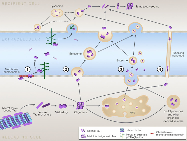
“Although pathogenic tau proteins are transferred via various mechanisms, including direct secretion through the plasma membrane, secretion in exosomes or ectosomes, and direct cell–cell transfer via tunneling nanotubes [162], our understanding of the detailed molecular mechanisms for tau secretion, which facilitates t…”
BackgroundExtracellular PHF-tau modulates astrocyte mitochondrial dynamics and mediates neuronal connectivity
Zufferey, V., Barve, A., Parietti, E., Belinga, L. N., Bringaud, A., Varisco, Y., Fabbri, K., Capotosti, F., Bezzi, P., Déglon, N., et al. (2025) · Translational Neurodegeneration · doi:10.1186/s40035-025-00474-9 · Cited by 11
“Astrocytes can capture, internalize, and some-times transmit these aggregates, thereby aggravating the pathology [14, 19, 100].”
Wang, M., Huang, W., Huang, J., Shi, J., Huang, N., & Luo, Y. (2024) · Pharmacological Research - Modern Chinese Medicine · doi:10.1016/j.prmcm.2024.100543 · Cited by 9
Molecular Insights into Tau Pathology and its Therapeutic Strategies in Alzheimer's Disease
Zhou, F., Zhao, Y., Sun, Y., & Chen, W. (2024) · Journal of Integrative Neuroscience · doi:10.31083/j.jin2311197 · Cited by 13
“Once in the extracellular space, tau species can be internalized by neighboring neurons and glia via endocytosis, pinocytosis, or phagocytosis [32].”
Glucose Metabolic Abnormality: A Crosstalk between Depression and Alzheimer’s Disease
Yang, S., Li, Y., Tang, Q., Zhang, Y., & Shao, T. (2024) · Current Neuropharmacology · doi:10.2174/011570159x343281240912190309 · Cited by 12
“Further, recent studies have suggested that Tau protein may contribute to the spread of AD within the brain [54].”
Ury‐Thiery, V., Fichou, Y., Alves, I. D., Molinari, M., Lecomte, S., & Feuillie, C. (2024) · Nanoscale · doi:10.1039/d4nr01343c · Cited by 13
Tian, S., Liu, Y., Hong, H., & Li, S. (2025) · Molecular Neurobiology · doi:10.1007/s12035-025-05280-y · Cited by 8
“As an IDP, tau exhibits high structural flexibility, enabling reversible interactions with a variety of proteins and organelles [24, 25].”
Overview of Proteomic Analysis of Amyloid Plaques and Neurofibrillary Tangles in Alzheimer’s Disease
Grewal, A., Raikundalia, S., Zaia, J., & Sethi, M. K. (2025) · Biomolecules · doi:10.3390/biom15091310 · Cited by 9
“This cell-to-cell transmission occurs through mechanisms such as exocytosis, endocytosis, and extracellular vesicles, including exosomes [24–26].”
Denus, M., Filaquier, A., Fargues, W., Néel, É., Stewart, S. E., Colladant, M., Curel, T., Mezghrani, A., Marin, P., Claeysen, S., et al. (2025) · Traffic · doi:10.1111/tra.70009 · Cited by 8
Chiang, M., Nicol, C. J. B., Yang, Y., Chiang, T., & Yen, C. (2025) · Brain Research · doi:10.1016/j.brainres.2025.149506 · Cited by 7
Korde, D. S., & Humpel, C. (2024) · Biomolecules · doi:10.3390/biom14020165 · Cited by 9
“The prion hypothesis postulates systemic aggregation and propagation of A β and tau pathologies via interconnected pathways, akin to prion protein transmission [38–42].”
BackgroundParekh, P. A., Mu, Q., Badachhape, A., Bhavane, R. C., Srivastava, M., Devkota, L., Sun, X., Bhandari, P., Eriksen, J. L., Tanifum, E. A., et al. (2022) · Theranostics · doi:10.7150/thno.72258 · Cited by 14
“healthy cell, inducing tau misfolding, aggregation, and neuro-toxicity [8,9].”
BackgroundN-Amination Converts Amyloidogenic Tau Peptides into Soluble Antagonists of Cellular Seeding
Makwana, K. M., Sarnowski, M. P., Miao, J., Lin, Y., & Valle, J. R. D. (2021) · ACS Chemical Neuroscience · doi:10.1021/acschemneuro.1c00528 · Cited by 12
Designed Cell-Penetrating Peptide Constructs for Inhibition of Pathogenic Protein Self-Assembly
Kalmouni, M., Oh, Y., Alata, W., & Magzoub, M. M. (2024) · Pharmaceutics · doi:10.3390/pharmaceutics16111443 · Cited by 7
“Eventually, the protofibrils turn into paired helical filaments or straight filaments to form NFTs that are commonly observed during the later stages of AD [25,26].”
Glucocorticoid stress hormones stimulate vesicle-free Tau secretion and spreading in the brain
Yu, Q., Du, F., Belli, I., Gomes, P., Sotiropoulos, I., & Waites, C. L. (2024) · Cell Death and Disease · doi:10.1038/s41419-024-06458-3 · Cited by 14
“[22], together with our in vitro and ex vivo fi ndings showing that NaClO 3 and methyl-β -cyclodextrin strongly attenuate DEX-mediated Tau secretion, we think it likely that type 1 UPS contributes substantially to GC-induced Tau spreading in vivo.”
Chen, M., Fu, W., Xu, H., & Liu, C.-J. (2023) · Arthritis Research & Therapy · doi:10.1186/s13075-023-03133-4 · Cited by 7
Latham, A. S., Geer, C. E., Ackart, D. F., Anderson, I. K., Vittoria, K. M., Podell, B. K., Basaraba, R. J., & Moreno, J. A. (2023) · Frontiers in Neuroscience · doi:10.3389/fnins.2023.1157652 · Cited by 9
“This spread occurs through a cycle of intracellular formation and secretion followed by transcellular uptake by nearby cells (Wang et al., 2017; Brunello et al., 2020).”
BackgroundStructural and functional damage to neuronal nuclei caused by extracellular tau oligomers
Sun, X., Eastman, G., Shi, Y., Saibaba, S., Oliveira, A. K., Lukens, J. R., Norambuena, A., Mandell, J., & Bloom, G. S. (2023) · bioRxiv · doi:10.1101/2023.05.08.539873 · Cited by 7
“Previous studies established that pathogenically mutated intracellular tau can provoke neuronal nuclear deformation [9, 11], but possible roles for xcTauOs [22] in this process have not been reported.”
Sala‐Jarque, J., Zimkowska, K., Ávila, J., Ferrer, I., & del Río, J. A. (2022) · International Journal of Molecular Sciences · doi:10.3390/ijms231911527 · Cited by 11
Eun, J. D., Jimenez, H. T., Adrien, L. R., Wolin, A., Marambaud, P., Davies, P., & Koppel, J. L. (2022) · Molecular Medicine · doi:10.1186/s10020-022-00506-4 · Cited by 7
“Meanwhile, in vitro studies have shown that cells secrete hyperphosphorylated tau more efficiently than non-phosphorylated tau (Brunello et al. 2020; Plouffe et al. 2012; Pooler et al. 2012), and elevated tau biomarkers in humans following anesthesia and surgery indicate neuronal release of hyperphosphorylated tau (Ev…”
BackgroundCommentary: BAG3 as a Mediator of Endosome Function and Tau Clearance
Lin, H., Deaton, C. A., & Johnson, G. V. W. (2022) · Neuroscience · doi:10.1016/j.neuroscience.2022.05.002 · Cited by 7
Brain Networks Route Neurodegeneration Patterns in Patients with Progressive Supranuclear Palsy
Palleis, C., Quattrone, A., Dehsarvi, A., Roemer, S. N., Bernhardt, A. M., The AL‐108‐231 Investigators, T. P. S. G., Huppertz, H., Malpetti, M., Boxer, A. L., Gnörich, J. S., et al. (2025) · Movement Disorders · doi:10.1002/mds.30257 · Cited by 6
“15 21-25 The concept of trans-synaptic τ propagation is strongly supported by experimental evidence demonstrating that hyperphosphorylated τ seeds are actively secreted and taken up by connected neurons at synapses, and that increased activity in τ -containing neurons accelerates trans-synaptic τ…”
Xu, S., Liu, K., Qian, S., Wu, J., Hu, J., Zhou, D., & Zheng, T. (2025) · Neurochemistry International · doi:10.1016/j.neuint.2025.105976 · Cited by 6
Neuroadaptation in neurodegenerative diseases: compensatory mechanisms and therapeutic approaches
Kopalli, S. R., Behl, T., Baldaniya, L. H., Ballal, S., Joshi, K. K., Arya, R., Chaturvedi, B., Chauhan, A. S., Verma, R. K., Patel, M., et al. (2025) · Progress in Neuro-Psychopharmacology and Biological Psychiatry · doi:10.1016/j.pnpbp.2025.111375 · Cited by 8
The role of TFEB-mediated autophagy-lysosome dysfunction in manganese neurotoxicity
Lu, J., Su, P., Zhao, F., Yu, K., Yang, X., Lv, H., Wang, D., & Zhang, J. (2024) · Current Research in Toxicology · doi:10.1016/j.crtox.2024.100193 · Cited by 6
“It is primarily found in neurons and plays a crucial role as an essential protein for maintaining the stability of microtubules within axons (Brunello et al., 2020; Liang et al., 2022).”
BackgroundDnaJs are enriched in tau regulators
Esquivel, A. R., Hill, S. E., & Blair, L. J. (2023) · International Journal of Biological Macromolecules · doi:10.1016/j.ijbiomac.2023.127486 · Cited by 6
The Fate of Tau Aggregates Between Clearance and Transmission
Seitkazina, A., Kim, K. H., Fagan, E., Sung, Y., Kim, Y. K., & Lim, S. (2022) · Frontiers in Aging Neuroscience · doi:10.3389/fnagi.2022.932541 · Cited by 10
“Soluble tau monomers and oligomers are cleared by CMA and e-MI, whereas large, insoluble tau aggregates are cleared by macroautophagy (Wang et al., 2009; Krüger et al., 2012; Caballero et al., 2018; Uytterhoeven et al., 2018).”
BackgroundTauopathies: The Role of Tau in Cellular Crosstalk and Synaptic Dysfunctions
Caamaño-Moreno, M., & Gargini, R. (2022) · Neuroscience · doi:10.1016/j.neuroscience.2022.02.034 · Cited by 7
Arc mediates intercellular tau transmission via extracellular vesicles
Tyagi, M., de Hoog, E., Grega, M., Sullivan, K. R., Walker, A., Chadha, R., Northrop, A., Fábián, B., Hummer, G., Fuxreiter, M., et al. (2026) · Cell · doi:10.1016/j.cell.2026.06.008 · Cited by 19
Ved, A., Gajjar, T. B., Kakkad, A. D., Ganesan, S., Shankhyan, A., Jayabalan, K., Mishra, S., Jain, B., Arora, V., Gulati, M., et al. (2026) · Brain Sciences · doi:10.3390/brainsci16050434 · Cited by 5
“Tau pathology disrupts axonal transport via microtubule dysfunction, impairing delivery of signaling molecules, leading to defective NVC, reduced CBF, hypoperfusion, and metabolic oxygen mismatch in neurodegeneration [57–59].”
Bhandari, A. K., Dhami, A. S., Hemanthlumar, R. T., Mishra, N., Joslyn, B., Roy, S., Srinivasan, J., Prajapati, K. P., Kar, K., & Anand, B. G. (2025) · Ageing Research Reviews · doi:10.1016/j.arr.2025.102879 · Cited by 4
Guillot, P., Celle, S., Barth, N., Roche, F., & Perek, N. (2024) · International Journal of Molecular Sciences · doi:10.3390/ijms252011058 · Cited by 5
“Studies have established possible correlations between OSAS and neurodegenerative diseases such as AD [12], notably by investigating levels of the proteins A β and tau, marked by their accumulation in the brains of patients with AD [50,51].”
BackgroundSahu, M., Seth, I., George, S. J., & Maitra, U. (2024) · Chemistry of Materials · doi:10.1021/acs.chemmater.4c01125 · Cited by 4
Structural Variations of Prions and Prion-like Proteins Associated with Neurodegeneration
Christensen, C. S., Wang, S., Li, W., Yu, D., & Li, H. J. (2024) · Current Issues in Molecular Biology · doi:10.3390/cimb46070384 · Cited by 8
“…conformations, strains, and aggregation states of alpha-synuclein may contribute to the diverse clinical presentations and disease progression observed in synu-cleinopathies For example, different diseases may have significantly different alpha-synuclein aggregation sites and pS129 levels…”
BackgroundHuang, Y., Wu, Z., Lin, S., & Chen, X. (2023) · Molecular Medicine · doi:10.1186/s10020-023-00728-0 · Cited by 7
Ahn, S.-H., Suh, J., Jang, Y., Kim, H., Han, K., Lee, Y., Choi, G., & Kim, T. (2023) · Biosensors and Bioelectronics · doi:10.1016/j.bios.2023.115533 · Cited by 5
Gholami, A., Dehghan, G., Rashtbari, S., & Jouyban, A. (2022) · Iranian journal of pharmaceutical research · doi:10.5812/ijpr-129599 · Cited by 5
“Tau is an intrinsically disordered protein that belongs to the family of microtubule-associated proteins (MAPs), which are considered for microtubule stabilization and assembly (1-3).”
BackgroundMoreira, R., Mendonça, L. S., & de Almeida, L. P. (2021) · International Journal of Molecular Sciences · doi:10.3390/ijms222212288 · Cited by 7
Joa, K., Mankhong, S., Kim, S., Moon, S., Lee, K., Yoo, Y.-H., Hwang, B.-H., Baek, J.-W., & Kang, J. (2020) · Journal of Alzheimer s Disease · doi:10.3233/jad-200250 · Cited by 6
Targeted tau protein degradation: a promising therapeutic approach for tauopathies
Baazaoui, N., Alfaifi, M. Y., Shati, A. A., Saad, R. B., & Garzoli, S. (2025) · Neuroscience · doi:10.1016/j.neuroscience.2025.11.041 · Cited by 5
Pareja-Navarro, K. A., King, C. D., Kauwe, G., Ngwala, Y. Y., Lokitiyakul, D., Wong, I., Vira, A., Liu, Y., Chen, J. H., Sharma, M., et al. (2026) · Molecular Neurodegeneration · doi:10.1186/s13024-026-00928-2 · Cited by 6
“Tau oligomers can be secreted from neurons [21, 22], and levels of tau oligomers are increased in human cerebrospinal fluid of AD patients [23], linking extracellular tau oligomers to AD-related pathogenesis.”
Bhandari, K., Sun, Y., Tang, H., Ke, P. C., & Ding, F. (2025) · Cell Reports Physical Science · doi:10.1016/j.xcrp.2025.103031 · Cited by 2
“For cases without LLPS, we simply turned off the formation of droplets in solving Equations 1, 2, 3, 4, 5, and 6.”
Pandey, N., Srivastava, N. K., Kumar, A., Hussain, I., & Joshi, D. (2024) · The Egyptian Journal of Neurology Psychiatry and Neurosurgery · doi:10.1186/s41983-024-00860-6 · Cited by 3
“Once seeded, it prompts the aggregation of natively folded tau proteins within naive cells, inducing cellular toxicity and disease spread, reminiscent of a prion-like propagation hypothesis [18, 24].”
BackgroundLi, Q., Liu, H., Zhu, Q., Zhang, R., Liang, D., Zhang, J., Jin, R., Chen, Y., Qin, C., & Teng, J. (2024) · Brain Research Bulletin · doi:10.1016/j.brainresbull.2024.111046 · Cited by 4
“In this context, exosomes play a crucial role in the uptake, secretion, and transmission of pathological tau (Brunello et al., 2020; Gibbons et al., 2019).”
Background
Metadata
- Authors
- Brunello, C. A., Merezhko, M., Uronen, R.-L., & Huttunen, H. J.
- Affiliations
- University of Helsinki (FI)
- Corresponding author
- Huttunen, Henri J.
- Venue
- Cellular and Molecular Life Sciences
- Published
- 2019-10-30
- Volume
- 77
- Issue
- 9
- Pages
- 1721-1744
- ISSN
- 1420-682X (ISSN-L)
- 1420-9071
- PMC
- PMC7190606
- OpenAlex
- W2982627322
- Cited by
- 304
- References
- 267
- Citations by year
- 2026: 36 · 2025: 45 · 2024: 56 · 2023: 38 · 2022: 46 · 2021: 57 · 2020: 25 · 2019: 1
- FWCI
- 3.80
- Type
- journal article
- Language
- en
- Open access
- hybrid
- doi.org
- Locations
- Cellular and Molecular Life Sciences (publishedVersion, cc-by)
- PubMed Central (publishedVersion, cc-by)
- PubMed (publishedVersion)
- Työväentutkimus Vuosikirja (submittedVersion, cc-by)
- Funders
- Academy of Finland (296409)
- Alfred Kordelinin Säätiö
- Topics
- Life Sciences › Neuroscience › Cellular and Molecular Neuroscience › Alzheimer's disease research and treatments
- Life Sciences › Biochemistry, Genetics and Molecular Biology › Molecular Biology › Prion Diseases and Protein Misfolding
- Life Sciences › Neuroscience › Neurology › Neuroinflammation and Neurodegeneration Mechanisms
- Keywords
- tau protein, tauopathy, tau secretion, cell-to-cell transmission, prion-like propagation, tau aggregation, exosomes, ectosome, vesicular secretion, extracellular tau, endocytosis, phagocytosis, pinocytosis, Alzheimer's disease, frontotemporal lobar degeneration, neurodegeneration
- MeSH terms
- Animals, Humans, tau Proteins, Disease Progression, Tauopathies, Protein Aggregation, Pathological
About these checks
Registry, retraction, Cites and statistics results are computed, not judged, with one exception: a printed reference that no index lists is read once by a model before it counts as a citation error. Claims are extracted by a model and kept only when their quote appears verbatim in the text; each cited source is then checked, with a model’s help, for whether it says what the claim relies on. Those verdicts can be wrong — read the claim’s quote and the cited source before relying on one.
Statistics and arithmetic are recomputed, not judged.
Confidence bands are shown only for fields calibrated on our annotated test set. None is calibrated yet, so no band is shown.
What counts as a claim ›How we measure confidence ›How we check this ›
Are you an author, or do you see a problem with this page? Request a review
