

The DOI matches its Crossref record; 260 of the 267 references it cites were found in a registry or an index.
Mechanisms of secretion and spreading of pathological tau protein
Open accessAbstract
Accumulation of misfolded and aggregated forms of tau protein in the brain is a neuropathological hallmark of tauopathies, such as Alzheimer’s disease and frontotemporal lobar degeneration. Tau aggregates have the ability to transfer from one cell to another and to induce templated misfolding and aggregation of healthy tau molecules in previously healthy cells, thereby propagating tau pathology across different brain areas in a prion-like manner. The molecular mechanisms involved in cell-to-cell transfer of tau aggregates are diverse, not mutually exclusive and only partially understood. Intracellular accumulation of misfolded tau induces several mechanisms that aim to reduce the cellular burden of aggregated proteins and also promote secretion of tau aggregates. However, tau may also be released from cells physiologically unrelated to protein aggregation. Tau secretion involves multiple vesicular and non-vesicle-mediated pathways, including secretion directly through the plasma membrane. Consequently, extracellular tau can be found in various forms, both as a free protein and in vesicles, such as exosomes and ectosomes. Once in the extracellular space, tau aggregates can be internalized by neighboring cells, both neurons and glial cells, via endocytic, pinocytic and phagocytic mechanisms. Importantly, accumulating evidence suggests that prion-like propagation of misfolding protein pathology could provide a general mechanism for disease progression in tauopathies and other related neurodegenerative diseases. Here, we review the recent literature on cellular mechanisms involved in cell-to-cell transfer of tau, with a particular focus in tau secretion.
Overview
The claims and statistics of this paper haven’t been checked yet.
Run the check — free, no account needed. It reads the open-access text and checks the sources it cites.
Integrity signals
Registry
Cites
Statistics
Figures
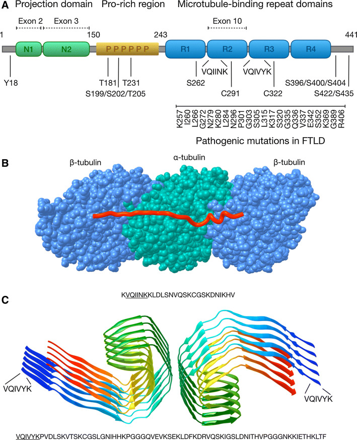
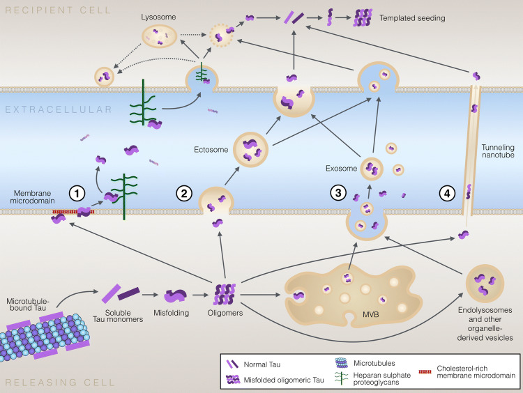
Verification
Claims the paper makes
The claims and statistics of this paper haven’t been checked yet.
Run the check — free, no account needed. It reads the open-access text and checks the sources it cites.
Claims other papers make about this one
Each is checked against this paper’s own text: does it say what the citing paper says it does?
The source backs part of the claim, not all of it — or backs it for a different population, condition or comparison.
In the citing paper
“Thus, it is suggested that CSF p-tau biomarkers likely indicate an active process of tau secretion, and one that is correlated with cerebral Aβ deposition in early disease.20, 21, 22”
From Cerebrospinal fluid p-tau231 as an early indicator of emerging pathology in Alzheimer's disease (2022) · cites this paper as [22]
Experimental — accuracy not yet measured
Self-check
Percentages, sums and reported differences re-computed from the paper’s own numbers.
The self-check needs the paper’s full text.
Citations
Cites
Neuropathological stageing of Alzheimer-related changes.
Braak, H., & Braak, E. (1991) · Acta Neuropathologica · doi:10.1007/bf00308809 · Cited by 16,123 · cited 1×
“It is well established that in AD the development of tau pathology occurs in a hierarchical pattern of accumulation starting from the layer II of entorhinal cortex (EC) and spreading toward cortical regions, as originally proposed by the findings of Heiko and Eva Braak [107].”
ResultAmyloid oligomers exacerbate tau pathology in a mouse model of tauopathy
Selenica, M.-L. B., Brownlow, M., Jimenez, J. P., Lee, D. C., Pena, G., Dickey, C. A., Gordon, M. N., & Morgan, D. (2013) · Neurodegenerative Diseases · doi:10.1159/000337230 · Cited by 10,359
Iliff, J. J., Wang, M., Liao, Y., Plogg, B. A., Peng, W., Gundersen, G. A., Benveniste, H., Vates, G. E., Deane, R., Goldman, S. A., et al. (2012) · Science Translational Medicine · doi:10.1126/scitranslmed.3003748 · Cited by 5,824 · cited 1×
“One important contributor to clearance of tau aggregates from the brain is the brain glymphatic system, a brain-wide interstitial solute clearance system composed of astrocytes, paravascular routes and dural lymphatics [230, 231].”
BackgroundFrontotemporal lobar degeneration: A consensus on clinical diagnostic criteria
Neary, D., Snowden, J. S., Gustafson, L., Passant, U., Stuss, D., Black, S., Freedman, M., Kertesz, A., Robert, P. H., Albert, M., et al. (1998) · Neurology · doi:10.1212/wnl.51.6.1546 · Cited by 5,047
Association of missense and 5′-splice-site mutations in tau with the inherited dementia FTDP-17
Hutton, M., Lendon, C. L., Rizzu, P., Baker, M., Froelich, S., Houlden, H., Pickering-Brown, S., Chakraverty, S., Isaacs, A., Grover, A., et al. (1998) · Nature · doi:10.1038/31508 · Cited by 3,498 · cited 3×
“First mutations in the MAPT gene were found in 1998 [31, 32]”
BackgroundA protein factor essential for microtubule assembly
Weingarten, M. D., Lockwood, A. H., Hwo, S. Y., & Kirschner, M. W. (1975) · Proceedings of the National Academy of Sciences · doi:10.1073/pnas.72.5.1858 · Cited by 3,089
A Liquid-to-Solid Phase Transition of the ALS Protein FUS Accelerated by Disease Mutation
Patel, A., Lee, H. O., Jawerth, L. M., Maharana, S., Jahnel, M., Hein, M. Y., Stoynov, S., Mahamid, J., Saha, S., Franzmann, T. M., et al. (2015) · Cell · doi:10.1016/j.cell.2015.07.047 · Cited by 2,927
Microglia Function in the Central Nervous System During Health and Neurodegeneration
Colonna, M., & Butovsky, O. (2017) · Annual Review of Immunology · doi:10.1146/annurev-immunol-051116-052358 · Cited by 2,894
Molliex, A., Temirov, J. P., Lee, J., Coughlin, M., Kanagaraj, A., Kim, H. J., Mittag, T., & Taylor, J. P. (2015) · Cell · doi:10.1016/j.cell.2015.09.015 · Cited by 2,690
Tau in physiology and pathology
Wang, Y., & Mandelkow, E. (2016) · Nature Reviews Neuroscience · doi:10.1038/nrn.2015.1 · Cited by 2,251
Research works printed but not linked: 3
- Liu F, Gong CX (2008) Tau exon 10 alternative splicing and tauopathies. Mol Neurodegener 3:8 · DOI · Paper page
- Grundke-Iqbal I, Iqbal K, Tung YC, Quinlan M, Wisniewski HM, Binder LI (1986) Abnormal phosphorylation of the microtubule-associated protein tau (tau) in Alzheimer cytoskeletal pathology. Proc Natl Acad Sci USA 83(13):4913–4917 · DOI · Paper page
- Yamada K et al (2014) Neuronal activity regulates extracellular tau in vivo. J Exp Med 211(3):387–393 · DOI
Cited by
Sorted by citations
Phosphorylated Tau in Alzheimer’s Disease and Other Tauopathies
Rawat, P., Sehar, U., Bisht, J., Selman, A., Culberson, J., & Reddy, P. H. (2022) · International Journal of Molecular Sciences · doi:10.3390/ijms232112841 · Cited by 509
“At the protein level, K257T, G272V, P301L, V337M, and R406W mutations alter the binding properties of the microtubule, and N279K, N296H, P301S, S305I mutations at the RNA level shifts the splicing towards overproduction of 4R tau [97].”
BackgroundThe expanding amyloid family: Structure, stability, function, and pathogenesis
Sawaya, M. R., Hughes, M. P., Rodriguez, J. A., Riek, R., & Eisenberg, D. S. (2021) · Cell · doi:10.1016/j.cell.2021.08.013 · Cited by 475
Role of tau protein in Alzheimer’s disease: The prime pathological player
Muralidar, S., Ambi, S. V., Sekaran, S., Thirumalai, D., & Palaniappan, B. (2020) · International Journal of Biological Macromolecules · doi:10.1016/j.ijbiomac.2020.07.327 · Cited by 278
Alzheimer’s disease brain-derived extracellular vesicles spread tau pathology in interneurons
Ruan, Z., Pathak, D., Venkatesan Kalavai, S., Yoshii-Kitahara, A., Muraoka, S., Bhatt, N., Takamatsu-Yukawa, K., Hu, J., Wang, Y., Hersh, S., et al. (2021) · Brain · doi:10.1093/brain/awaa376 · Cited by 283
“secreted primarily in free form, while a minor fraction of tau is found in EVs as observed in the CSF or blood of control subjects and Alzheimer’s disease patients (Arai et al., 1998; Saman et al., 2012; Zetterberg et al., 2013; Fiandaca et al., 2015; Ruan and Ikezu, 2019; Brunello et al., 2020).”
BackgroundAcetylated tau inhibits chaperone-mediated autophagy and promotes tau pathology propagation in mice
Caballero, B., Bourdenx, M., Luengo, E., Diaz, A., Sohn, P. D., Chen, X., Wang, C., Juste, Y. R., Wegmann, S., Patel, B., et al. (2021) · Nature Communications · doi:10.1038/s41467-021-22501-9 · Cited by 223
Novak, P., Kovacech, B., Katina, S., Schmidt, R., Scheltens, P., Kontsekova, E., Ropele, S., Fialova, L., Kramberger, M., Paulenka-Ivanovova, N., et al. (2021) · Nature Aging · doi:10.1038/s43587-021-00070-2 · Cited by 187
Cerebrospinal fluid p-tau231 as an early indicator of emerging pathology in Alzheimer's disease
Ashton, N. J., Benedet, A. L., Pascoal, T. A., Karikari, T. K., Lantero-Rodriguez, J., Brum, W. S., Mathotaarachchi, S., Therriault, J., Savard, M., Chamoun, M., et al. (2022) · eBioMedicine · doi:10.1016/j.ebiom.2022.103836 · Cited by 184
Role of natural products for the treatment of Alzheimer’s disease
Noori, T., Dehpour, A. R., Sureda, A., Sobarzo-Sanchez, E., & Shirooie, S. (2021) · European Journal of Pharmacology · doi:10.1016/j.ejphar.2021.173974 · Cited by 141
Buchholz, S., & Zempel, H. (2024) · Alzheimer s & Dementia · doi:10.1002/alz.13784 · Cited by 154
“309 As TAU spreading has been shown to be mediated (among other pathways) via EVs (reviewed, e.g., in 310 ),theseinitialresultssupportourhypothesisthatratherindividual TAU isoforms mediate TAU toxicity in disease context.”
BackgroundBiomolecular condensates can both accelerate and suppress aggregation of α-synuclein
Lipiński, W. P., Visser, B. S., Robu, I., Fakhree, M. A. A., Lindhoud, S., Claessens, M. M. A. E., & Spruijt, E. (2022) · Science Advances · doi:10.1126/sciadv.abq6495 · Cited by 131
“This has been observed for amyloid-β(1-42) (33) and may occur also for other amyloidogenic proteins.”
Extracellular Monomeric Tau Is Internalized by Astrocytes
Perea, J. R., López, E., Díez-Ballesteros, J. C., Ávila, J., Hernández, F., & Bolós, M. (2019) · Frontiers in Neuroscience · doi:10.3389/fnins.2019.00442 · Cited by 114
Tilavonemab in early Alzheimer’s disease: results from a phase 2, randomized, double-blind study
Florian, H., Wang, D., Arnold, S. E., Boada, M., Guo, Q., Jin, Z., Zheng, H., Fisseha, N., Kalluri, H. V., Rendenbach‐Mueller, B., et al. (2023) · Brain · doi:10.1093/brain/awad024 · Cited by 143
“Further research is needed to understand the cellular mechanisms involved in the secretion and spreading of tau aggregates between cells.(7)”
BackgroundLimorenko, G., & Lashuel, H. A. (2021) · Chemical Society Reviews · doi:10.1039/d1cs00127b · Cited by 168
Tau deposition patterns are associated with functional connectivity in primary tauopathies
Franzmeier, N., Brendel, M., Beyer, L., Slemann, L., Kovács, G. G., Arzberger, T., Kurz, C., Respondek, G., Ječmenica‐Lukić, M., Biel, D., et al. (2022) · Nature Communications · doi:10.1038/s41467-022-28896-3 · Cited by 120
Microglia in Alzheimer’s Disease in the Context of Tau Pathology
Perea, J. R., Bolós, M., & Ávila, J. (2020) · Biomolecules · doi:10.3390/biom10101439 · Cited by 134
“Propagation commonly involves the monomer and oligomer forms of tau, which are susceptible to aberrant folding and post-translational modifications such as truncation and phosphorylation [118].”
BackgroundTau Isoforms: Gaining Insight into MAPT Alternative Splicing
Corsi, A., Bombieri, C., Valenti, M. T., & Romanelli, M. G. (2022) · International Journal of Molecular Sciences · doi:10.3390/ijms232315383 · Cited by 118
“Conversely, under pathological conditions, this mechanism contributes to the spread of altered forms of Tau across different brain areas and thus to the spatio-temporal disease progression in tauopathies [65,66].”
BackgroundThe effect of insomnia on development of Alzheimer’s disease
Sadeghmousavi, S., Eskian, M., Rahmani, F., & Rezaei, N. (2020) · Journal of Neuroinflammation · doi:10.1186/s12974-020-01960-9 · Cited by 122
Role of Chemokines in the Development and Progression of Alzheimer’s Disease
Wojcieszak, J., Kuczyńska, K., & Zawilska, J. B. (2022) · Journal of Molecular Neuroscience · doi:10.1007/s12031-022-02047-1 · Cited by 97
“Additionally, pathological tau can be secreted to extracellular space and enter surrounding neurons or glial cells further impairing axonal transport and causing neuronal synaptic loss thus contributing to progression of the disease (Brunello et al. 2019).”
BackgroundSynaptic tau: A pathological or physiological phenomenon?
Robbins, M., Clayton, E. L., & Schierle, G. S. K. (2021) · Acta Neuropathologica Communications · doi:10.1186/s40478-021-01246-y · Cited by 128
“It is thus interesting to note that several endocytosisrelated pathways are involved in Tau trafficking and thus may explain why Tau pathology is linked to these different pathways (for a review see [50].”
BackgroundPossible Mechanisms of Tau Spread and Toxicity in Alzheimer’s Disease
Zhang, H., Cao, Y., Ma, L., Wei, Y., & Li, H. (2021) · Frontiers in Cell and Developmental Biology · doi:10.3389/fcell.2021.707268 · Cited by 109
Tau as a Biomarker of Neurodegeneration
Holper, S., Watson, R., & Yassi, N. (2022) · International Journal of Molecular Sciences · doi:10.3390/ijms23137307 · Cited by 121
“Specifically, Aβmay actively drive the secretion of tau from the neuron, accounting for the pathological CSF tau concentrations seen early in AD pathogenesis, before frank neurodegeneration [95].”
BackgroundHamano, T., Enomoto, S., Shirafuji, N., Ikawa, M., Yamamura, O., Yen, S.-H. C., & Nakamoto, Y. (2021) · International Journal of Molecular Sciences · doi:10.3390/ijms22147475 · Cited by 122
“At present, it is unclear which of the above mechanisms are involved in tau synaptic release, and whether the same mechanism is responsible for physiological tau synaptic release and pathological tau synaptic release [60].”
BackgroundShulman, M. B., Kong, J., O'Gorman, J. T., Ratti, E., Rajagovindan, R., Viollet, L., Huang, E., Sharma, S., Racine, A. M., Czerkowicz, J., et al. (2023) · Nature Aging · doi:10.1038/s43587-023-00523-w · Cited by 100
Nand, S., Tiwari, N. K., Singh, P., Mishra, D., Singh, A. K., Hooshmandi, E., Vamanu, E., & Singh, M. P. (2021) · Frontiers in Neuroscience · doi:10.3389/fnins.2021.777347 · Cited by 96
“and displaying the prion-like characteristics of Tau pathology (Brunello et al., 2020).”
BackgroundKim, O. Y., & Song, J. (2023) · Life Sciences · doi:10.1016/j.lfs.2023.122356 · Cited by 96
Cerebrovascular disease promotes tau pathology in Alzheimer’s disease
Laing, K. K., Simoes, S., Baena-Caldas, G. P., Lao, P. J., Kothiya, M., Igwe, K. C., Chesebro, A. G., Houck, A. L., Pedraza, L., Hernandez, A. I., et al. (2020) · Brain Communications · doi:10.1093/braincomms/fcaa132 · Cited by 94
Promiscuous Roles of Autophagy and Proteasome in Neurodegenerative Proteinopathies
Limanaqi, F., Biagioni, F., Gambardella, S., Familiari, P., Frati, A., & Fornai, F. (2020) · International Journal of Molecular Sciences · doi:10.3390/ijms21083028 · Cited by 93
“Sci. 2020, 21, 3028; doi:10.3390/ijms21083028 www.mdpi.com/journal/ijms of specific disease-associated proteins include alpha-synuclein (α-syn) in synucleinopathies such as PD and multiple system atrophy (MSA), amyloid-beta (Aβ) and tau in AD, polyglutamine-expanded huntingtin in HD, TAR-DNA-binding protein of 43 kDa…”
BackgroundInfluentialVesga-Jiménez, D. J., Martin, C., Barreto, G. E., Aristizábal-Pachón, A. F., Pinzón, A. M., & González, J. (2022) · International Journal of Molecular Sciences · doi:10.3390/ijms23052577 · Cited by 105
“Abnormal tangles in the brain are formed with tau protein in the hyperphosphorylated form they are unable to bind to tubulin and microtubules, disrupting the neuronal cytoskeleton, as well as reducing microtubules and neurofilaments [187,188].”
BackgroundLiu, S., Hossinger, A., Heumüller, S.-E., Hornberger, A., Buravlova, O., Konstantoulea, K., Müller, S. A., Paulsen, L., Rousseau, F., Schymkowitz, J., et al. (2021) · Nature Communications · doi:10.1038/s41467-021-25855-2 · Cited by 72
Tauopathies: Deciphering Disease Mechanisms to Develop Effective Therapies
Silva, M. C., & Haggarty, S. J. (2020) · International Journal of Molecular Sciences · doi:10.3390/ijms21238948 · Cited by 89
Rai, S. K., Khanna, R., Avni, A., & Mukhopadhyay, S. (2023) · Proceedings of the National Academy of Sciences · doi:10.1073/pnas.2216338120 · Cited by 76
“The truncations of tau protein and human PrP (23-231) were created using full-length variants of the respective constructs.”
BackgroundMethodProteostasis Disturbances and Inflammation in Neurodegenerative Diseases
Sonninen, T.-M., Goldsteins, G., Laham-Karam, N., Koıstınaho, J., & Lehtonen, Š. (2020) · Cells · doi:10.3390/cells9102183 · Cited by 89
“Another hallmark of AD pathology, tauopathy, has been demonstrated to spread between the brain cells by direct secretion, ectosomal, and exosomal mechanisms [87].”
BackgroundThe extracellular chaperone Clusterin enhances Tau aggregate seeding in a cellular model
Yuste‐Checa, P., Trinkaus, V. A., Riera-Tur, I., Imamoglu, R., Schaller, T., Wang, H., Dudanova, I., Hipp, M. S., Bracher, A., & Hartl, F. U. (2021) · Nature Communications · doi:10.1038/s41467-021-25060-1 · Cited by 78
Zhang, D., Zhang, W., Chen, M., Gao, X., Yuan, H., Lin, X., Mao, X., Wang, C., Guo, X., Du, Y., et al. (2024) · Neuron · doi:10.1016/j.neuron.2024.02.017 · Cited by 75
A critical appraisal of tau‐targeting therapies for primary and secondary tauopathies
Imbimbo, B. P., Ippati, S., Watling, M., & Balducci, C. (2021) · Alzheimer s & Dementia · doi:10.1002/alz.12453 · Cited by 76
Microglia: Friend and foe in tauopathy
Odfalk, K. F., Bieniek, K. F., & Hopp, S. C. (2022) · Progress in Neurobiology · doi:10.1016/j.pneurobio.2022.102306 · Cited by 60
Kazim, S. F., Sharma, A., Saroja, S. R., Seo, J. H., Larson, C. S., Ramakrishnan, A., Wang, M., Blitzer, R. D., Shen, L., Peña, C. J., et al. (2021) · Biological Psychiatry · doi:10.1016/j.biopsych.2021.02.973 · Cited by 75
Human Tau Isoforms and Proteolysis for Production of Toxic Tau Fragments in Neurodegeneration
Boyarko, B., & HOOK, V. Y. H. (2021) · Frontiers in Neuroscience · doi:10.3389/fnins.2021.702788 · Cited by 81
Role of the Lipid Membrane and Membrane Proteins in Tau Pathology
Bok, E., Leem, E., Lee, B.-R., Lee, J. M., Yoo, C. J., Lee, E. M., & Kim, J. (2021) · Frontiers in Cell and Developmental Biology · doi:10.3389/fcell.2021.653815 · Cited by 61
Targeting tau in Alzheimer's disease: from mechanisms to clinical therapy
Ye, J., Wan, H., Chen, S., & Liu, G. (2023) · Neural Regeneration Research · doi:10.4103/1673-5374.385847 · Cited by 74
“…and AD, thereby supporting a critical involvement of glial cells and neuroinflammation in tauopathies (Zhong et al., 2017; Pimenova et al., 2018; Brunello et al., 2020; Wang et al., 2020b, 2022; Griciuc and Tanzi, 2021; Haass, 2021; Jin et al., 2021; Lee et al., 2021; Wang and Ye, 2021; Zhao et…”
Neuropathological assessment of the Alzheimer spectrum
Jellinger, K. A. (2020) · Journal of Neural Transmission · doi:10.1007/s00702-020-02232-9 · Cited by 85
Li, Z., Yin, B., Zhang, S., Lan, Z., & Zhang, L. (2023) · European Journal of Medicinal Chemistry · doi:10.1016/j.ejmech.2023.115817 · Cited by 54
Lim, D. W., Lee, J., Lee, C.-H., & Kim, Y. T. (2024) · International Journal of Molecular Sciences · doi:10.3390/ijms252011223 · Cited by 70
“Tau proteins contribute to maintaining the morphology of neurons, particularly by stabilizing microtubules within axons and dendrites to support cellular structures [31].”
BackgroundFranzmeier, N., Roemer, S. N., Bernhardt, A. M., Dehsarvi, A., Dewenter, A., Steward, A., Biel, D., Frontzkowski, L. K., Zhu, Z., Gnörich, J. S., et al. (2025) · Molecular Neurodegeneration · doi:10.1186/s13024-025-00822-3 · Cited by 70
“CSF p-tau increases have previously been shown to be more closely linked to PET-assessed fibrillar Aβ than to fibrillar tau pathology [58] and to reflect Aβ-associated active secretion of newly synthesized p-tau molecules, rather than passive spill out from dead neurons [43, 59].”
Franzmeier, N., Dehsarvi, A., Steward, A., Biel, D., Dewenter, A., Roemer, S. N., Wagner, F., Groß, M., Brendel, M., Moscoso, A., et al. (2024) · Nature Communications · doi:10.1038/s41467-023-44374-w · Cited by 63
Super-resolution microscopy: a closer look at synaptic dysfunction in Alzheimer disease
Padmanabhan, P., Kneynsberg, A., & Götz, J. (2021) · Nature reviews. Neuroscience · doi:10.1038/s41583-021-00531-y · Cited by 59
Tau and Membranes: Interactions That Promote Folding and Condensation
Sallaberry, C. A., Voss, B. J., Majewski, J. P., Biernat, J., Mandelkow�, E., Chi, E. Y., & Zanden, C. M. V. (2021) · Frontiers in Cell and Developmental Biology · doi:10.3389/fcell.2021.725241 · Cited by 64
“Microtubule binding is commonly ascribed to the RD, but it should be noted that strong binding requires components of the flanking PXXP and C-terminal domains (Butner and Kirschner, 1991; Gustke et al., 1994; Preuss et al., 1997).”
BackgroundHsp40s play complementary roles in the prevention of tau amyloid formation
Irwin, R., Faust, O., Petrovic, I. M., Wolf, S. G., Hofmann, H., & Rosenzweig, R. (2021) · eLife · doi:10.7554/elife.69601 · Cited by 66
Venkataraman, L., Fair, S. R., McElroy, C. A., Hester, M. E., & Fu, H. (2020) · Stem Cell Reviews and Reports · doi:10.1007/s12015-020-10068-9 · Cited by 55
Reactivated endogenous retroviruses promote protein aggregate spreading
Liu, S., Heumüller, S.-E., Hossinger, A., Müller, S. A., Buravlova, O., Lichtenthaler, S. F., Denner, P., & Vorberg, I. M. (2023) · Nature Communications · doi:10.1038/s41467-023-40632-z · Cited by 48
The evolving landscape of Alzheimer’s disease therapy: From Aβ to tau
Courade, J., Zetterberg, H., Höglinger, G. U., & Dewachter, I. (2025) · Cell · doi:10.1016/j.cell.2025.11.033 · Cited by 57
Tau seeding and spreading in vivo is supported by both AD-derived fibrillar and oligomeric tau
de Gérando, A. M., Welikovitch, L. A., Khasnavis, A., Commins, C., Glynn, C. A., Chun, J. E., Perbet, R., & Hyman, B. T. (2023) · Acta Neuropathologica · doi:10.1007/s00401-023-02600-1 · Cited by 54
“Considering the several different pathways of pathological tau secretion and spreading [10] as well as the involvement of neuronal activity in tau pathology progression [12, 46, 63], the rate of progression of SARK and HMW tau could be influenced by multiple, yet unexplored, biological factors.”
BackgroundRecent Advances in Targeting Transition Metals (Copper, Iron, and Zinc) in Alzheimer’s Disease
Singh, R., Panghal, A., Jadhav, K., Thakur, A., Verma, R. K., Singh, C., Goyal, M., Kumar, J., & Namdeo, A. G. (2024) · Molecular Neurobiology · doi:10.1007/s12035-024-04256-8 · Cited by 50
“These fundamental mechanisms have been used as therapeutic targets in the treatment of tau related pathologies and other neurodegenerative diseases [132, 133].”
Huang, Y., Driedonks, T. A. P., Cheng, L., Turchinovich, A., Rajapaksha, H., Arab, T., Powell, B. H., Pletniková, O., Redding‐Ochoa, J., Troncoso, J. C., et al. (2022) · Journal of Alzheimer s Disease · doi:10.3233/jad-220322 · Cited by 58
“EVs have been widely reported to propagate misfolded tau in neurodegenerative disease [15, 44].”
BackgroundTong, B. C.-K., Huang, A. S., Wu, A. J., Iyaswamy, A., Ho, O. K.-Y., Kong, A. H.-Y., Sreenivasmurthy, S. G., Zhu, Z., Su, C., Liu, J., et al. (2022) · Journal of Biomedical Science · doi:10.1186/s12929-022-00871-6 · Cited by 47
“It is believed that pathological tau is liberated from cells via direct secretion of tau clusters, ectosomes, or exosomes or via junctions with adjacent cells [66].”
The pleiotropic roles of autophagy in Alzheimer’s disease: From pathophysiology to therapy
Festa, B. P., Barbosa, A. D., Rob, M., & Rubinsztein, D. C. (2021) · Current Opinion in Pharmacology · doi:10.1016/j.coph.2021.07.011 · Cited by 41
Modulating disease-relevant tau oligomeric strains by small molecules
Cascio, F. L., García, S., Montalbano, M., Puangmalai, N., McAllen, S., Pace, A., Piccionello, A. P., & Kayed, R. (2020) · Journal of Biological Chemistry · doi:10.1074/jbc.ra120.014630 · Cited by 53
“Therefore, more investigations are needed to better understand how tau is released in the extracellular space to be then internalized into neighboring or anatomically connected cells and sequentially template for further aggregation within those cells (74, 75).”
BackgroundDas, R., Balmik, A., & Chinnathambi, S. (2020) · ASN Neuro · doi:10.1177/1759091420981204 · Cited by 41
“The Tau NFTs containing neurons flag phosphatidylserine on its surface and activates the signaling cascade for apoptosis (Brunello et al., 2020).”
BackgroundTau protein as a diagnostic and prognostic biomarker in amyotrophic lateral sclerosis
Agnello, L., Colletti, T., Sasso, B. L., Vidali, M., Spataro, R., Gambino, C. M., Giglio, R. V., Piccoli, T., Bivona, G., Bella, V. L., et al. (2021) · European Journal of Neurology · doi:10.1111/ene.14789 · Cited by 42
Wang, Z., Patel, V. N., Song, X., Xu, Y., Kaminski, A. M., Doan, V. U., Su, G., Liao, Y.-E., Mah, D., Zhang, F., et al. (2023) · Science Advances · doi:10.1126/sciadv.adf6232 · Cited by 41
Marx, G. A., Koenigsberg, D., McKenzie, A. T., Kauffman, J., Hanson, R. W., Whitney, K. R., Signaevsky, M., Prastawa, M. W., Iida, M. A., White, C. L., et al. (2022) · Acta Neuropathologica Communications · doi:10.1186/s40478-022-01457-x · Cited by 45
“This approach to measuring disease burden has the theoretical potential to capture mechanisms of p-tau spread through a given region, which is currently under investigation by several other groups [47–54].”
BackgroundXiao, D., Li, J., Ren, Z., Dai, M., Jiang, Y., Qiu, T., Zhang, H., Chen, Y., Zhang, Y., Zhang, Y., et al. (2024) · Alzheimer s & Dementia · doi:10.1002/alz.14158 · Cited by 38
“57 Such detrimental effects could be amplified by the inefficient glymphatic waste clearance, 58 Some limitations should be mentioned for this study.”
BackgroundUnconventional Protein Secretion Dependent on Two Extracellular Vesicles: Exosomes and Ectosomes
Meldolesi, J. (2022) · Frontiers in Cell and Developmental Biology · doi:10.3389/fcell.2022.877344 · Cited by 43
Metabolomics in degenerative brain diseases
Escobar, M. Q., Pontes, J. G. D. M., & Tasić, L. (2021) · Brain Research · doi:10.1016/j.brainres.2021.147704 · Cited by 41
The chaperone Clusterin in neurodegeneration−friend or foe?
Yuste‐Checa, P., Bracher, A., & Hartl, F. U. (2022) · BioEssays · doi:10.1002/bies.202100287 · Cited by 37
“reach the extracellular environment via release from dying cells or upon active secretion by neurons, which may occur through a transsynaptic mechanism[8] or in a manner facilitated by chaperones, such as the HSP40 protein DnaJC5.”
BackgroundMelatonin: Regulation of Biomolecular Condensates in Neurodegenerative Disorders
Loh, D., & Reiter, R. J. (2021) · Antioxidants · doi:10.3390/antiox10091483 · Cited by 45
Tau internalization: A complex step in tau propagation
Zhao, J., Wu, H., & Tang, X. (2021) · Ageing Research Reviews · doi:10.1016/j.arr.2021.101272 · Cited by 47
Advanced nano delivery system for stem cell therapy for Alzheimer's disease
Pan, Y., Li, L., Cao, N., Liao, J., Chen, H., & Zhang, M. (2024) · Biomaterials · doi:10.1016/j.biomaterials.2024.122852 · Cited by 33
Gong, B., Zhang, W., Cong, W., Gu, Y., Ji, W., Yin, T., Zhou, H., Hu, H., Zhuang, J., Luo, Y., et al. (2024) · Advanced Healthcare Materials · doi:10.1002/adhm.202400149 · Cited by 31
Brain Fog: a Narrative Review of the Most Common Mysterious Cognitive Disorder in COVID-19
Mir, M. A. (2023) · Molecular Neurobiology · doi:10.1007/s12035-023-03715-y · Cited by 38
“Tau aggregates have the ability to spread from cell to cell, causing targeted misfolding and aggregation of healthy tau molecules in previously healthy cells, causing this in a manner similar to prion infection aims to spread tau pathology in different regions of the brain [109, 110].”
Astrocytes in depression and Alzheimer’s disease
Liao, Y., Xing, Q., Li, Q., Zhang, J., Pan, R., & Yuan, Z. (2021) · Frontiers of Medicine · doi:10.1007/s11684-021-0875-0 · Cited by 44
The propagation mechanisms of extracellular tau in Alzheimer’s disease
Wei, Y., Liu, M., & Wang, D. (2021) · Journal of Neurology · doi:10.1007/s00415-021-10573-y · Cited by 40
“Extracellular tau may mediate tau spreading and serve as a biomarker for AD [2].”
BackgroundA dynamical systems approach for multiscale synthesis of Alzheimer’s pathogenesis
Rollo, J. L., Crawford, J. W., & Hardy, J. (2023) · Neuron · doi:10.1016/j.neuron.2023.04.018 · Cited by 31
Dysfunctional microglia and tau pathology in Alzheimer’s disease
Ayyubova, G. (2022) · Reviews in the Neurosciences · doi:10.1515/revneuro-2022-0087 · Cited by 36
Overlaps and divergences between tauopathies and synucleinopathies: a duet of neurodegeneration
Li, W., & Li, J. (2024) · Translational Neurodegeneration · doi:10.1186/s40035-024-00407-y · Cited by 37
“Tau has physiological functions of stabilizing microtubules and maintaining axonal transport in highly polarized neurons [73].”
BackgroundRoles of the cytoskeleton in human diseases
Li, M., Li, P., Wang, Z., Liu, L., Cao, M., Cui, J., Wu, F., & Yang, J. (2023) · Molecular Biology Reports · doi:10.1007/s11033-022-08025-5 · Cited by 35
Single-Cell Spatial Proteomic Imaging for Human Neuropathology
Vijayaragavan, K., Cannon, B. J., Tebaykin, D., Bosse, M. M., Baranski, A., Oliveria, J. P., Bukhari, S. A., Mrdjen, D., Corces, M. R., McCaffrey, E., et al. (2022) · Acta Neuropathologica Communications · doi:10.1186/s40478-022-01465-x · Cited by 40
“For instance, immunogenicity towards Aβ or Tau oligomers in CN and CIND could vary from ADD samples [52, 58, 59], or ADD may have a more reactive microglial-dependent immune response relative to those present in the CN and CIND hippocampus [60].”
Vieweg, S., Mahul-Mellier, A.-L., Ruggeri, F., Riguet, N., Deguire, S. M., Chiki, A., Cendrowska, U., Dietler, G., & Lashuel, H. (2021) · bioRxiv · doi:10.1101/2021.02.15.431207 · Cited by 31
“Several studies have shown that the secretion and cell-to-cell propagation of protein aggregates is mediated by their interactions with cell membranes and other membranous structures [116, 117].”
BackgroundTau in Alzheimer’s Disease: Pathological Alterations and an Attractive Therapeutic Target
Gu, J., & Liu, F. (2020) · Current Medical Science · doi:10.1007/s11596-020-2282-1 · Cited by 41
Altered amyloid-β and tau proteins in neural-derived plasma exosomes in obstructive sleep apnea
Sun, H., Gao, Y., Li, M., Zhang, S., Shen, T., Yuan, X., Shang, X., Li, Z., & Zhang, J. (2022) · Sleep Medicine · doi:10.1016/j.sleep.2022.03.021 · Cited by 30
Sastre, A. A., Montoro, M. L., Lacerda, H. M., Llavero, F., & Zugaza, J. L. (2021) · International Journal of Molecular Sciences · doi:10.3390/ijms22094425 · Cited by 36
Amro, Z., Yool, A. J., & Collins‐Praino, L. E. (2021) · Brain Behavior & Immunity - Health · doi:10.1016/j.bbih.2021.100242 · Cited by 39
Atabi, F., Moassesfar, M., Nakhaie, T., Bagherian, M., Hosseinpour, N., & Hashemi, M. (2025) · Diabetology & Metabolic Syndrome · doi:10.1186/s13098-025-01930-2 · Cited by 37
“These aggregates disrupt neuronal function by sequestering normal tau and other microtubule-associated proteins [65] (Fig.”
Galpern, W. R., Triana‐Baltzer, G., Li, L., Kolen, K. V., Timmers, M., Haeverans, K., Janssens, L., Kolb, H. C., Nandy, P., Aida, K., et al. (2024) · The Journal of Prevention of Alzheimer s Disease · doi:10.14283/jpad.2024.163 · Cited by 38
“However, the mechanism of tau spread is not known; it is possible that alternative mechanisms such as tunneling nanotubes or exosomes may be relevant (12), in which case tau seeds would evade the ISF where the antibody is presumed to act.”
An Overview of Astrocyte Responses in Genetically Induced Alzheimer’s Disease Mouse Models
Spanos, F., & Liddelow, S. A. (2020) · Cells · doi:10.3390/cells9112415 · Cited by 32
Congdon, E. E., Jiang, Y., & Sigurdsson, E. M. (2021) · Seminars in Cell and Developmental Biology · doi:10.1016/j.semcdb.2021.12.002 · Cited by 38
Role of the endolysosomal pathway and exosome release in tau propagation
Yan, M., & Zheng, T. (2021) · Neurochemistry International · doi:10.1016/j.neuint.2021.104988 · Cited by 36
Carbamate as a potential anti‐Alzheimer's pharmacophore: A review
Singh, Y. P., Kumar, N., Chauhan, B. S., & Garg, P. (2023) · Drug Development Research · doi:10.1002/ddr.22113 · Cited by 36
“But, under extreme conditions, hyperphosphorylated tau loses its microtubule ‐ stabilizing and axonal transport purpose (Brunello et al., 2020).”
BackgroundCerebrospinal fluid: A specific biofluid for the biosensing of Alzheimer's diseases biomarkers
Mirzaie, A., Nasrollahpour, H., Khalilzadeh, B., Jamali, A. A., Spiteri, R. J., Yousefi, H., Işıldak, İ., & Rahbarghazi, R. (2023) · TrAC Trends in Analytical Chemistry · doi:10.1016/j.trac.2023.117174 · Cited by 28
α– Linolenic acid modulates phagocytosis and endosomal pathways of extracellular Tau in microglia
Desale, S. E., & Chinnathambi, S. (2021) · Cell Adhesion & Migration · doi:10.1080/19336918.2021.1898727 · Cited by 63
“mechanisms have the tendency to propagate the disease [27,58,59].”
BackgroundEimer, W. A., Rodriguez, A. S., DeFao, M. T., Ehricke, S., Park, J., Kumar, D. K. V., Shanmugam, N. K. N., Singh, S., Sawhney, T., Moir, R. D., et al. (2025) · Nature Neuroscience · doi:10.1038/s41593-025-02157-0 · Cited by 32
Amyloid-β oligomers increase the binding and internalization of tau oligomers in human synapses
Kadamangudi, S., Marcatti, M., Zhang, W.-R., Fracassi, A., Kayed, R., Limon, A., & Taglialatela, G. (2024) · Acta Neuropathologica · doi:10.1007/s00401-024-02839-2 · Cited by 25
Oatman, S. R., Reddy, J. S., Quicksall, Z., Carrasquillo, M. M., Wang, X., Liu, C., Yamazaki, Y., Nguyen, T., Malphrus, K. G., Heckman, M. G., et al. (2023) · Molecular Neurodegeneration · doi:10.1186/s13024-022-00592-2 · Cited by 27
Global brain activity and its coupling with cerebrospinal fluid flow is related to tau pathology
Han, F., Lee, J., Chen, X., Ziontz, J., Ward, T. J., Landau, S. M., Baker, S. L., Harrison, T. M., Jagust, W. J., & Initiative, A. D. N. (2024) · Alzheimer s & Dementia · doi:10.1002/alz.14296 · Cited by 42
“67 In short, global brain activity and specific neural and physiological factors play a critical role insupportingCSFflowandthuscouldaffectADprogressionbymoder-ating the “accumulation-removal balance” of toxic proteins, such as A β and tau.”
Moreira, L., Costa, C., Pires, J., Teixeira, J. P., & Fraga, S. (2021) · Mutation Research/Reviews in Mutation Research · doi:10.1016/j.mrrev.2021.108385 · Cited by 35
Cathepsin D: A Candidate Link between Amyloid ?-protein and Tauopathy in Alzheimer Disease
Suire, C. N., & Leissring, M. A. (2021) · Journal of Experimental Neurology · doi:10.33696/neurol.2.029 · Cited by 15
“Among several identified trafficking mechanisms, endogenous tau has been shown to be secreted via unconventional protein secretion pathways from primary neurons [26-31].”
BackgroundKrawczuk, D., Kulczyńska‐Przybik, A., & Mroczko, B. (2024) · International Journal of Molecular Sciences · doi:10.3390/ijms25158132 · Cited by 26
“Tau is a soluble protein that occurs in six main isoforms in the human central nervous system (CNS) [48].”
BackgroundAutophagy-Related Pathways in Vesicular Unconventional Protein Secretion
Noh, S. H., Kim, Y. J., & Lee, M. G. (2022) · Frontiers in Cell and Developmental Biology · doi:10.3389/fcell.2022.892450 · Cited by 29
“The tau secretion through the UPS pathway involves both non-vesicular and vesicular mechanisms (Brunello et al., 2020); and several recent studies suggest that autophagy machinery participates in the vesicular routemediated tau secretion (Mohamed et al., 2014; Kang et al., 2019; Chen et al., 2020).”
BackgroundTashima, T. (2022) · Pharmaceutics · doi:10.3390/pharmaceutics14020411 · Cited by 27
“In fact: (i) some tau proteins are present in exosomes secreted from cells (Figure 7) [18].”
BackgroundInfluentialFonseca, C., Ettcheto, M., Bicker, J., Fernandes, M. J., Falcão, A. C., Camins, A., & Fortuna, A. C. (2023) · Ageing Research Reviews · doi:10.1016/j.arr.2023.101998 · Cited by 31
Metadata
- Authors
- Brunello, C. A., Merezhko, M., Uronen, R.-L., & Huttunen, H. J.
- Affiliations
- University of Helsinki (FI)
- Corresponding author
- Huttunen, Henri J.
- Venue
- Cellular and Molecular Life Sciences
- Published
- 2019-10-30
- Volume
- 77
- Issue
- 9
- Pages
- 1721-1744
- ISSN
- 1420-682X (ISSN-L)
- 1420-9071
- PMC
- PMC7190606
- OpenAlex
- W2982627322
- Cited by
- 304
- References
- 267
- Citations by year
- 2026: 36 · 2025: 45 · 2024: 56 · 2023: 38 · 2022: 46 · 2021: 57 · 2020: 25 · 2019: 1
- FWCI
- 3.80
- Type
- journal article
- Language
- en
- Open access
- hybrid
- doi.org
- Locations
- Cellular and Molecular Life Sciences (publishedVersion, cc-by)
- PubMed Central (publishedVersion, cc-by)
- PubMed (publishedVersion)
- Työväentutkimus Vuosikirja (submittedVersion, cc-by)
- Funders
- Academy of Finland (296409)
- Alfred Kordelinin Säätiö
- Topics
- Life Sciences › Neuroscience › Cellular and Molecular Neuroscience › Alzheimer's disease research and treatments
- Life Sciences › Biochemistry, Genetics and Molecular Biology › Molecular Biology › Prion Diseases and Protein Misfolding
- Life Sciences › Neuroscience › Neurology › Neuroinflammation and Neurodegeneration Mechanisms
- Keywords
- tau protein, tauopathy, tau secretion, cell-to-cell transmission, prion-like propagation, tau aggregation, exosomes, ectosome, vesicular secretion, extracellular tau, endocytosis, phagocytosis, pinocytosis, Alzheimer's disease, frontotemporal lobar degeneration, neurodegeneration
- MeSH terms
- Animals, Humans, tau Proteins, Disease Progression, Tauopathies, Protein Aggregation, Pathological
About these checks
Registry, retraction, Cites and statistics results are computed, not judged, with one exception: a printed reference that no index lists is read once by a model before it counts as a citation error. Claims are extracted by a model and kept only when their quote appears verbatim in the text; each cited source is then checked, with a model’s help, for whether it says what the claim relies on. Those verdicts can be wrong — read the claim’s quote and the cited source before relying on one.
Statistics and arithmetic are recomputed, not judged.
Confidence bands are shown only for fields calibrated on our annotated test set. None is calibrated yet, so no band is shown.
What counts as a claim ›How we measure confidence ›How we check this ›
Are you an author, or do you see a problem with this page? Request a review
