

The DOI matches its Crossref record; 260 of the 267 references it cites were found in a registry or an index.
Mechanisms of secretion and spreading of pathological tau protein
Open accessAbstract
Accumulation of misfolded and aggregated forms of tau protein in the brain is a neuropathological hallmark of tauopathies, such as Alzheimer’s disease and frontotemporal lobar degeneration. Tau aggregates have the ability to transfer from one cell to another and to induce templated misfolding and aggregation of healthy tau molecules in previously healthy cells, thereby propagating tau pathology across different brain areas in a prion-like manner. The molecular mechanisms involved in cell-to-cell transfer of tau aggregates are diverse, not mutually exclusive and only partially understood. Intracellular accumulation of misfolded tau induces several mechanisms that aim to reduce the cellular burden of aggregated proteins and also promote secretion of tau aggregates. However, tau may also be released from cells physiologically unrelated to protein aggregation. Tau secretion involves multiple vesicular and non-vesicle-mediated pathways, including secretion directly through the plasma membrane. Consequently, extracellular tau can be found in various forms, both as a free protein and in vesicles, such as exosomes and ectosomes. Once in the extracellular space, tau aggregates can be internalized by neighboring cells, both neurons and glial cells, via endocytic, pinocytic and phagocytic mechanisms. Importantly, accumulating evidence suggests that prion-like propagation of misfolding protein pathology could provide a general mechanism for disease progression in tauopathies and other related neurodegenerative diseases. Here, we review the recent literature on cellular mechanisms involved in cell-to-cell transfer of tau, with a particular focus in tau secretion.
Overview
The claims and statistics of this paper haven’t been checked yet.
Run the check — free, no account needed. It reads the open-access text and checks the sources it cites.
Integrity signals
Registry
Cites
Statistics
Figures
Verification
Claims the paper makes
The claims and statistics of this paper haven’t been checked yet.
Run the check — free, no account needed. It reads the open-access text and checks the sources it cites.
Claims other papers make about this one
Each is checked against this paper’s own text: does it say what the citing paper says it does?
The source backs part of the claim, not all of it — or backs it for a different population, condition or comparison.
In the citing paper
“Thus, it is suggested that CSF p-tau biomarkers likely indicate an active process of tau secretion, and one that is correlated with cerebral Aβ deposition in early disease.20, 21, 22”
From Cerebrospinal fluid p-tau231 as an early indicator of emerging pathology in Alzheimer's disease (2022) · cites this paper as [22]
Experimental — accuracy not yet measured
Self-check
Percentages, sums and reported differences re-computed from the paper’s own numbers.
The self-check needs the paper’s full text.
Citations
Cites
Neuropathological stageing of Alzheimer-related changes.
Braak, H., & Braak, E. (1991) · Acta Neuropathologica · doi:10.1007/bf00308809 · Cited by 16,123 · cited 1×
“It is well established that in AD the development of tau pathology occurs in a hierarchical pattern of accumulation starting from the layer II of entorhinal cortex (EC) and spreading toward cortical regions, as originally proposed by the findings of Heiko and Eva Braak [107].”
ResultAmyloid oligomers exacerbate tau pathology in a mouse model of tauopathy
Selenica, M.-L. B., Brownlow, M., Jimenez, J. P., Lee, D. C., Pena, G., Dickey, C. A., Gordon, M. N., & Morgan, D. (2013) · Neurodegenerative Diseases · doi:10.1159/000337230 · Cited by 10,359
Iliff, J. J., Wang, M., Liao, Y., Plogg, B. A., Peng, W., Gundersen, G. A., Benveniste, H., Vates, G. E., Deane, R., Goldman, S. A., et al. (2012) · Science Translational Medicine · doi:10.1126/scitranslmed.3003748 · Cited by 5,824 · cited 1×
“One important contributor to clearance of tau aggregates from the brain is the brain glymphatic system, a brain-wide interstitial solute clearance system composed of astrocytes, paravascular routes and dural lymphatics [230, 231].”
BackgroundFrontotemporal lobar degeneration: A consensus on clinical diagnostic criteria
Neary, D., Snowden, J. S., Gustafson, L., Passant, U., Stuss, D., Black, S., Freedman, M., Kertesz, A., Robert, P. H., Albert, M., et al. (1998) · Neurology · doi:10.1212/wnl.51.6.1546 · Cited by 5,047
Association of missense and 5′-splice-site mutations in tau with the inherited dementia FTDP-17
Hutton, M., Lendon, C. L., Rizzu, P., Baker, M., Froelich, S., Houlden, H., Pickering-Brown, S., Chakraverty, S., Isaacs, A., Grover, A., et al. (1998) · Nature · doi:10.1038/31508 · Cited by 3,498 · cited 3×
“First mutations in the MAPT gene were found in 1998 [31, 32]”
BackgroundA protein factor essential for microtubule assembly
Weingarten, M. D., Lockwood, A. H., Hwo, S. Y., & Kirschner, M. W. (1975) · Proceedings of the National Academy of Sciences · doi:10.1073/pnas.72.5.1858 · Cited by 3,089
A Liquid-to-Solid Phase Transition of the ALS Protein FUS Accelerated by Disease Mutation
Patel, A., Lee, H. O., Jawerth, L. M., Maharana, S., Jahnel, M., Hein, M. Y., Stoynov, S., Mahamid, J., Saha, S., Franzmann, T. M., et al. (2015) · Cell · doi:10.1016/j.cell.2015.07.047 · Cited by 2,927
Microglia Function in the Central Nervous System During Health and Neurodegeneration
Colonna, M., & Butovsky, O. (2017) · Annual Review of Immunology · doi:10.1146/annurev-immunol-051116-052358 · Cited by 2,894
Molliex, A., Temirov, J. P., Lee, J., Coughlin, M., Kanagaraj, A., Kim, H. J., Mittag, T., & Taylor, J. P. (2015) · Cell · doi:10.1016/j.cell.2015.09.015 · Cited by 2,690
Tau in physiology and pathology
Wang, Y., & Mandelkow, E. (2016) · Nature Reviews Neuroscience · doi:10.1038/nrn.2015.1 · Cited by 2,251
Cryo-EM structures of tau filaments from Alzheimer’s disease
Fitzpatrick, A. W. P., Falcon, B., He, S., Murzin, A. G., Murshudov, G., Garringer, H. J., Crowther, R. A., Ghetti, B., Goedert, M., & Scheres, S. H. W. (2017) · Nature · doi:10.1038/nature23002 · Cited by 2,061
Nanotubular Highways for Intercellular Organelle Transport
Rustom, A., Saffrich, R., Markovic, I., Walther, P., & Gerdes, H.-H. (2004) · Science · doi:10.1126/science.1093133 · Cited by 2,038
International Parkinson's Disease Genomics Consortium (IPDGC), Nalls, M. A., Parkinson's Study Group (PSG) Parkinson's Research: The Organized GENetics Initiative (PROGENI), 23andMe, GenePD, NeuroGenetics Research Consortium (NGRC), Hussman Institute of Human Genomics (HIHG), Investigator, T. A. J. D., (CHARGE), C. F. H. A. A. R. I. G. E., North American Brain Expression Consortium (NABEC), et al. (2014) · Nature Genetics · doi:10.1038/ng.3043 · Cited by 1,895 · cited 1×
“Moreover, genetic studies have linked variants of the tau gene, MAPT, to susceptibility of developing sporadic PD [80, 81], and mutations in MAPT are associated with dementia with parkinsonism [31].”
BackgroundMasters, C. L., Bateman, R., Blennow, K., Rowe, C. C., Sperling, R. A., & Cummings, J. L. (2015) · Nature Reviews Disease Primers · doi:10.1038/nrdp.2015.56 · Cited by 1,853
Dendritic Function of Tau Mediates Amyloid-β Toxicity in Alzheimer's Disease Mouse Models
Ittner, L. M., Ke, Y. D., Delerue, F., Bi, M., Gladbach, A., van Eersel, J., Wölfing, H., Chieng, B. C., Christie, M. J., Napier, I. A., et al. (2010) · Cell · doi:10.1016/j.cell.2010.06.036 · Cited by 1,821
Transmission and spreading of tauopathy in transgenic mouse brain
Clavaguera, F., Bolmont, T., Crowther, R. A., Abramowski, D., Frank, S., Probst, A., Fraser, G., Stalder, A. K., Beibel, M., Staufenbiel, M., et al. (2009) · Nature Cell Biology · doi:10.1038/ncb1901 · Cited by 1,803
Self-propagation of pathogenic protein aggregates in neurodegenerative diseases
Jucker, M., & Walker, L. C. (2013) · Nature · doi:10.1038/nature12481 · Cited by 1,663
Depletion of microglia and inhibition of exosome synthesis halt tau propagation
Asai, H., Ikezu, S., Tsunoda, S., Medalla, M., Luebke, J., Haydar, T., Wolozin, B., Butovsky, O., Kügler, S., & Ikezu, T. (2015) · Nature Neuroscience · doi:10.1038/nn.4132 · Cited by 1,591
Endosomal escape pathways for delivery of biologicals
Varkouhi, A. K., Scholte, M., Storm, G., & Haisma, H. J. (2011) · Journal of Controlled Release · doi:10.1016/j.jconrel.2010.11.004 · Cited by 1,561
The glymphatic pathway in neurological disorders.
Rasmussen, M. K., Mestre, H., & Nedergaard, M. (2018) · The Lancet Neurology · doi:10.1016/s1474-4422(18)30318-1 · Cited by 1,560
Formation of Neurofibrillary Tangles in P301L Tau Transgenic Mice Induced by Aβ42 Fibrils
Götz, J., Chen, F., van Dorpe, J., & Nitsch, R. M. (2001) · Science · doi:10.1126/science.1062097 · Cited by 1,526 · cited 1×
“For example, in mouse model of AD, soluble Aβ promotes development of tau pathology [75, 76].”
BackgroundMutation in the tau gene in familial multiple system tauopathy with presenile dementia
Spillantini, M. G., Murrell, J. R., Goedert, M., Farlow, M. R., Klug, A., & Ghetti, B. (1998) · Proceedings of the National Academy of Sciences · doi:10.1073/pnas.95.13.7737 · Cited by 1,474
Propagation of Tau Misfolding from the Outside to the Inside of a Cell
Frost, B., Jacks, R. L., & Diamond, M. I. (2009) · Journal of Biological Chemistry · doi:10.1074/jbc.m808759200 · Cited by 1,252
Verret, L., Mann, E. O., Hang, G. B., Barth, A. M. I., Cobos, I., Ho, K., Devidze, N., Masliah, E., Kreitzer, A. C., Mody, I., et al. (2012) · Cell · doi:10.1016/j.cell.2012.02.046 · Cited by 1,216
Tau in Alzheimer Disease and Related Tauopathies
Iqbal, K., Liu, F., Gong, C., & Grundke, I. (2010) · Current Alzheimer Research · doi:10.2174/156720510793611592 · Cited by 1,163
Stress granules: the Tao of RNA triage
Anderson, P., & Kedersha, N. (2008) · Trends in Biochemical Sciences · doi:10.1016/j.tibs.2007.12.003 · Cited by 1,161
Impairment of Glymphatic Pathway Function Promotes Tau Pathology after Traumatic Brain Injury
Iliff, J. J., Chen, M. J., Plog, B. A., Zeppenfeld, D. M., Soltero, M., Yang, L., Singh, I., Deane, R., & Nedergaard, M. (2014) · The Journal of Neuroscience · doi:10.1523/jneurosci.3020-14.2014 · Cited by 1,159
Hoover, B. R., Reed, M. N., Su, J., Penrod, R. D., Kotilinek, L. A., Grant, M. K., Pitstick, R., Carlson, G. A., Lanier, L. M., Yuan, L.-L., et al. (2010) · Neuron · doi:10.1016/j.neuron.2010.11.030 · Cited by 1,118
Tau protein liquid–liquid phase separation can initiate tau aggregation
Wegmann, S., Eftekharzadeh, B., Tepper, K., Zoltowska, K. M., Bennett, R. E., Dujardin, S., Laskowski, P. R., MacKenzie, D., Kamath, T., Commins, C., et al. (2018) · The EMBO Journal · doi:10.15252/embj.201798049 · Cited by 1,102
Goedert, M., Jakes, R., Spillantini, M. G., Hasegawa, M., Smith, M. J., & Crowther, R. A. (1996) · Nature · doi:10.1038/383550a0 · Cited by 1,036
Mutation-specific functional impairments in distinct tau isoforms of hereditary FTDP-17.
Hong, M., Zhukareva, V., Vogelsberg-Ragaglia, V., Wszolek, Z., Reed, L., Miller, B., Geschwind, D., Bird, T., McKeel, D., Goate, A., et al. (1998) · Science · doi:10.1126/science.282.5395.1914 · Cited by 1,024
von Bergen, M., Friedhoff, P., Biernat, J., Heberle, J., Mandelkow, E.-M., & Mandelkow, E. (2000) · Proceedings of the National Academy of Sciences · doi:10.1073/pnas.97.10.5129 · Cited by 1,021
Roles of tau protein in health and disease
Guo, T., Noble, W., & Hanger, D. P. (2017) · Acta Neuropathologica · doi:10.1007/s00401-017-1707-9 · Cited by 1,003
Trans-Synaptic Spread of Tau Pathology In Vivo
Liu, L., Drouet, V., Wu, J., Witter, M. P., Small, S. A., Clelland, C. L. T., & Duff, K. (2012) · PLoS ONE · doi:10.1371/journal.pone.0031302 · Cited by 994
Serio, T., Cashikar, A., Kowal, A., Sawicki, G., Moslehi, J., Serpell, L., Arnsdorf, M., & Lindquist, S. (2000) · Science · doi:10.1126/science.289.5483.1317 · Cited by 988
Distinct Tau Prion Strains Propagate in Cells and Mice and Define Different Tauopathies
Sanders, D. W., Kaufman, S. K., DeVos, S. L., Sharma, A. M., Mirbaha, H., Li, A., Barker, S. J., Foley, A. C., Thorpe, J. R., Serpell, L. C., et al. (2014) · Neuron · doi:10.1016/j.neuron.2014.04.047 · Cited by 986
Saman, S., Kim, W., Raya, M., Visnick, Y., Miro, S., Saman, S., Jackson, B., McKee, A. C., Alvarez, V. E., Lee, N. C. Y., et al. (2012) · Journal of Biological Chemistry · doi:10.1074/jbc.m111.277061 · Cited by 985
Amyloid deposition precedes tangle formation in a triple transgenic model of Alzheimer's disease
Oddo, S. (2003) · Neurobiology of Aging · doi:10.1016/j.neurobiolaging.2003.08.012 · Cited by 980
Tau phosphorylation: the therapeutic challenge for neurodegenerative disease
Hanger, D. P., Anderton, B. H., & Noble, W. (2009) · Trends in Molecular Medicine · doi:10.1016/j.molmed.2009.01.003 · Cited by 965
Fiandaca, M. S., Kapogiannis, D., Mapstone, M., Boxer, A., Eitan, E., Schwartz, J. B., Abner, E. L., Petersen, R. C., Federoff, H. J., Miller, B. L., et al. (2015) · Alzheimer's & Dementia · doi:10.1016/j.jalz.2014.06.008 · Cited by 959
Exosomes and Ectosomes in Intercellular Communication
Meldolesi, J. (2018) · Current Biology · doi:10.1016/j.cub.2018.01.059 · Cited by 944
Goedert, M., & Jakes, R. (1990) · The EMBO Journal · doi:10.1002/j.1460-2075.1990.tb07870.x · Cited by 929
Christianson, H. C., Svensson, K. J., van Kuppevelt, T. H., Li, J.-P., & Belting, M. (2013) · Proceedings of the National Academy of Sciences · doi:10.1073/pnas.1304266110 · Cited by 903
Morris, M., Maeda, S., Vossel, K., & Mucke, L. (2011) · Neuron · doi:10.1016/j.neuron.2011.04.009 · Cited by 899
Exosomes are released by cultured cortical neurones
Fauré, J., Lachenal, G., Court, M., Hirrlinger, J., Chatellard-Causse, C., Blot, B., Grange, J., Schoehn, G., Goldberg, Y., Boyer, V., et al. (2006) · Molecular and Cellular Neuroscience · doi:10.1016/j.mcn.2005.12.003 · Cited by 896
Physical and chemical properties of purified tau factor and the role of tau in microtubule assembly
Cleveland, D. W., Hwo, S. Y., & Kirschner, M. W. (1977) · Journal of Molecular Biology · doi:10.1016/0022-2836(77)90214-5 · Cited by 881
Initiation and Synergistic Fibrillization of Tau and Alpha-Synuclein
Giasson, B. I., Forman, M. S., Higuchi, M., Golbe, L. I., Graves, C. L., Kotzbauer, P. T., Trojanowski, J. Q., & Lee, V. M.-Y. (2003) · Science · doi:10.1126/science.1082324 · Cited by 879
Heparan sulfate proteoglycans mediate internalization and propagation of specific proteopathic seeds
Holmes, B. B., DeVos, S. L., Kfoury, N., Li, M., Jacks, R., Yanamandra, K., Ouidja, M. O., Brodsky, F. M., Marasa, J., Bagchi, D. P., et al. (2013) · Proceedings of the National Academy of Sciences · doi:10.1073/pnas.1301440110 · Cited by 859
Ebneth, A., Godemann, R., Stamer, K., Illenberger, S., Trinczek, B., Mandelkow, E.-M., & Mandelkow, E. (1998) · The Journal of Cell Biology · doi:10.1083/jcb.143.3.777 · Cited by 820
Neuronal activity enhances tau propagation and tau pathology in vivo
Wu, J. W., Hussaini, S. A., Bastille, I. M., Rodriguez, G. A., Mrejeru, A., Rilett, K., Sanders, D. W., Cook, C., Fu, H., Boonen, R. A. C. M., et al. (2016) · Nature Neuroscience · doi:10.1038/nn.4328 · Cited by 820
The sleep-wake cycle regulates brain interstitial fluid tau in mice and CSF tau in humans
Holth, J. K., Fritschi, S. K., Wang, C., Pedersen, N. P., Cirrito, J. R., Mahan, T. E., Finn, M. B., Manis, M., Geerling, J. C., Fuller, P. M., et al. (2019) · Science · doi:10.1126/science.aav2546 · Cited by 813
Brain homogenates from human tauopathies induce tau inclusions in mouse brain
Clavaguera, F., Akatsu, H., Fraser, G. M., Crowther, R. A., Frank, S., Hench, J., Probst, A., Winkler, D. T., Reichwald, J., Staufenbiel, M., et al. (2013) · Proceedings of the National Academy of Sciences · doi:10.1073/pnas.1301175110 · Cited by 792
Ambadipudi, S., Biernat, J., Riedel, D., Mandelkow, E., & Zweckstetter, M. (2017) · Nature Communications · doi:10.1038/s41467-017-00480-0 · Cited by 782 · cited 1×
“It is also important to note that, similarly to other disease-associated low complexity proteins [208–210], tau can undergo liquid–liquid phase separation, where tau separates into liquid-like droplets similarly as in a water–oil demixing event [58, 211].”
ResultLiu, F., Grundke‐Iqbal, I., Iqbal, K., & Gong, C. (2005) · European Journal of Neuroscience · doi:10.1111/j.1460-9568.2005.04391.x · Cited by 759
Tau and Aβ imaging, CSF measures, and cognition in Alzheimer’s disease
Brier, M. R., Gordon, B., Friedrichsen, K., McCarthy, J., Stern, A., Christensen, J., Owen, C., Aldea, P., Su, Y., Hassenstab, J., et al. (2016) · Science Translational Medicine · doi:10.1126/scitranslmed.aaf2362 · Cited by 734
Post-translational modifications of tau protein: Implications for Alzheimer's disease
Martin, L., Latypova, X., & Terro, F. (2011) · Neurochemistry International · doi:10.1016/j.neuint.2010.12.023 · Cited by 683
The release and trans-synaptic transmission of Tau via exosomes
Wang, Y., Balaji, V., Kaniyappan, S., Krüger, L., Irsen, S., Tepper, K., Chandupatla, R., Maetzler, W., Schneider, A., Mandelkow, E., et al. (2017) · Molecular Neurodegeneration · doi:10.1186/s13024-016-0143-y · Cited by 674
Frontotemporal dementia and parkinsonism linked to chromosome 17: A consensus conference
Foster, N. L., Wilhelmsen, K., Sima, A. A. F., Jones, M. Z., D'Amato, C., & Gilman, S. (1997) · Annals of Neurology · doi:10.1002/ana.410410606 · Cited by 664
Iba, M., Guo, J. L., McBride, J. D., Zhang, B., Trojanowski, J. Q., & Lee, V. M.-Y. (2013) · The Journal of Neuroscience · doi:10.1523/jneurosci.2642-12.2013 · Cited by 664
Brandt, R., Léger, J. G., & Lee, G. S. (1995) · The Journal of Cell Biology · doi:10.1083/jcb.131.5.1327 · Cited by 663
Progressive supranuclear palsy: clinicopathological concepts and diagnostic challenges
Williams, D. R., & Lees, A. J. (2009) · The Lancet Neurology · doi:10.1016/s1474-4422(09)70042-0 · Cited by 650
Dickey, C. A., Kamal, A., Lundgren, K., Klosak, N., Bailey, R. M., Dunmore, J., Ash, P., Shoraka, S., Zlatkovic, J., Eckman, C. B., et al. (2007) · Journal of Clinical Investigation · doi:10.1172/jci29715 · Cited by 642
Physiological release of endogenous tau is stimulated by neuronal activity
Pooler, A. M., Phillips, E. C., Lau, D. H. W., Noble, W., & Hanger, D. P. (2013) · The EMBO Reports · doi:10.1038/embor.2013.15 · Cited by 642
Seeding of Normal Tau by Pathological Tau Conformers Drives Pathogenesis of Alzheimer-like Tangles
Guo, J. L., & Lee, V. M.-Y. (2011) · Journal of Biological Chemistry · doi:10.1074/jbc.m110.209296 · Cited by 636
Lachenal, G. M., Pernet‐Gallay, K., Chivet, M., Hemming, F. J., Belly, A., Bodon, G., Blot, B., Haase, G., Goldberg, Y., & Sadoul, R. (2010) · Molecular and Cellular Neuroscience · doi:10.1016/j.mcn.2010.11.004 · Cited by 617
Maphis, N., Xu, G., Kokiko-Cochran, O. N., Jiang, S., Cardona, A., Ransohoff, R. M., Lamb, B. T., & Bhaskar, K. (2015) · Brain · doi:10.1093/brain/awv081 · Cited by 595
Epileptic activity in Alzheimer’s disease: Causes and clinical relevance
Vossel, K. A., Tartaglia, M. C., Nygaard, H. B., Zeman, A. Z., & Miller, B. L. (2017) · The Lancet Neurology · doi:10.1016/s1474-4422(17)30044-3 · Cited by 590
Pathways of Unconventional Protein Secretion
Rabouille, C. (2016) · Trends in Cell Biology · doi:10.1016/j.tcb.2016.11.007 · Cited by 573
Kirschner, D. A., Abraham, C. R., & Selkoe, D. J. (1986) · Proceedings of the National Academy of Sciences · doi:10.1073/pnas.83.2.503 · Cited by 570
Schneider, A., Biernat, J., von Bergen, M., Mandelkow, E., & Mandelkow, E.-M. (1999) · Biochemistry · doi:10.1021/bi981874p · Cited by 564
Kampers, T., Friedhoff, P., Biernat, J., Mandelkow, E.-M., & Mandelkow, E. (1996) · FEBS Letters · doi:10.1016/s0014-5793(96)01386-5 · Cited by 548
Wu, J. W., Herman, M., Liu, L., Simoes, S., Acker, C. M., Figueroa, H., Steinberg, J. I., Margittai, M., Kayed, R., Zurzolo, C., et al. (2013) · Journal of Biological Chemistry · doi:10.1074/jbc.m112.394528 · Cited by 537
Tau stabilizes microtubules by binding at the interface between tubulin heterodimers
Kadavath, H., Hofele, R. V., Biernat, J., Kumar, S., Tepper, K., Urlaub, H., Mandelkow, E., & Zweckstetter, M. (2015) · Proceedings of the National Academy of Sciences · doi:10.1073/pnas.1504081112 · Cited by 524
Goedert, J. J., Kessler, C. M., Aledort, L. M., Biggar, R. J., Andes, W. A., White, G., Drummond, J. E., Vaidya, K., Mann, D. L., Eyster, M. E., et al. (1989) · New England Journal of Medicine · doi:10.1056/nejm198910263211701 · Cited by 522
Alzheimer brain-derived tau oligomers propagate pathology from endogenous tau
Lasagna-Reeves, C. A., Castillo-Carranza, D. L., Sengupta, U., Guerrero-Munoz, M. J., Kiritoshi, T., Neugebauer, V., Jackson, G. R., & Kayed, R. (2012) · Scientific Reports · doi:10.1038/srep00700 · Cited by 502
Invited review: Neuropathology of tauopathies: principles and practice
Kovacs, G. G. (2015) · Neuropathology and Applied Neurobiology · doi:10.1111/nan.12208 · Cited by 492
Ahmed, Z., Cooper, J., Murray, T. K., Garn, K., McNaughton, E., Clarke, H., Parhizkar, S., Ward, M. A., Cavallini, A., Jackson, S., et al. (2014) · Acta Neuropathologica · doi:10.1007/s00401-014-1254-6 · Cited by 484
Chaperones increase association of tau protein with microtubules
Dou, F., Netzer, W. J., Tanemura, K., Li, F., Hartl, F. U., Takashima, A., Gouras, G. K., Greengard, P., & Xu, H. (2003) · Proceedings of the National Academy of Sciences · doi:10.1073/pnas.242720499 · Cited by 462
Ghetti, B. F., Oblak, A. L., Boeve, B. F., Johnson, K. A., Dickerson, B. C., & Goedert, M. (2014) · Neuropathology and Applied Neurobiology · doi:10.1111/nan.12213 · Cited by 458
Guo, J. L., Narasimhan, S., Changolkar, L., He, Z., Stieber, A., Zhang, B., Gathagan, R. J., Iba, M., McBride, J. D., Trojanowski, J. Q., et al. (2016) · Journal of Experimental Medicine · doi:10.1084/jem.20160833 · Cited by 440
Alonso, A. D. C., Grundke-Iqbal, I., Barra, H. S., & Iqbal, K. (1997) · Proceedings of the National Academy of Sciences · doi:10.1073/pnas.94.1.298 · Cited by 437
Tau association with synaptic vesicles causes presynaptic dysfunction
Zhou, L., McInnes, J., Wierda, K., Holt, M., Herrmann, A. G., Jackson, R. J., Wang, Y.-C., Swerts, J., Beyens, J., Miskiewicz, K., et al. (2017) · Nature Communications · doi:10.1038/ncomms15295 · Cited by 434
Schwarz, A. J., Yu, P., Miller, B. B., Shcherbinin, S., Dickson, J., Navitsky, M., Joshi, A. D., Devous, M. D., & Mintun, M. S. (2016) · Brain · doi:10.1093/brain/aww023 · Cited by 425
Pérez, M., Valpuesta, J., Medina, M., de Garcini, E. M., & Ávila, J. (1996) · Journal of Neurochemistry · doi:10.1046/j.1471-4159.1996.67031183.x · Cited by 424
Neuropathologic differentiation of progressive supranuclear palsy and corticobasal degeneration
Dickson, D. W. (1999) · Journal of Neurology · doi:10.1007/bf03161076 · Cited by 424
Nuclear Tau, a Key Player in Neuronal DNA Protection
Sultan, A., Nesslany, F., Violet, M., Bégard, S., Loyens, A., Talahari, S., Mansuroglu, Z., Marzin, D., Sergeant, N., Humez, S., et al. (2011) · Journal of Biological Chemistry · doi:10.1074/jbc.m110.199976 · Cited by 421
Near-atomic model of microtubule-tau interactions
Kellogg, E. H., Hejab, N. M. A., Poepsel, S., Downing, K. H., DiMaio, F., & Nogales, E. (2018) · Science · doi:10.1126/science.aat1780 · Cited by 416
Colocalization of Tau and Alpha-Synuclein Epitopes in Lewy Bodies
Ishizawa, T., Mattila, P. M., Davies, P., Wang, D., & Dickson, D. W. (2003) · Journal of Neuropathology & Experimental Neurology · doi:10.1093/jnen/62.4.389 · Cited by 402
Decreased synaptic proteins in neuronal exosomes of frontotemporal dementia and Alzheimer's disease
Goetzl, E. J., Kapogiannis, D., Schwartz, J. B., Lobach, I. V., Goetzl, L., Abner, E. L., Jicha, G. A., Karydas, A. M., Boxer, A., & Miller, B. L. (2016) · The FASEB Journal · doi:10.1096/fj.201600816r · Cited by 398
Total and phosphorylated tau protein as biological markers of Alzheimer’s disease
Hampel, H., Blennow, K., Shaw, L. M., Hoessler, Y. C., Zetterberg, H., & Trojanowski, J. Q. (2010) · Experimental Gerontology · doi:10.1016/j.exger.2009.10.010 · Cited by 389
Neuropathology of variants of progressive supranuclear palsy
Dickson, D. W., Ahmed, Z., Algom, A. A., Tsuboi, Y., & Josephs, K. A. (2010) · Current Opinion in Neurology · doi:10.1097/wco.0b013e32833be924 · Cited by 386
Alzheimer's DiseasePreprint
Selkoe, D. J. (2011) · Cold Spring Harbor Perspectives in Biology · doi:10.1101/cshperspect.a004457 · Cited by 384
Schweers, O., Mandelkow, E., Biernat, J., & Mandelkow, E. (1995) · Proceedings of the National Academy of Sciences · doi:10.1073/pnas.92.18.8463 · Cited by 384 · cited 1×
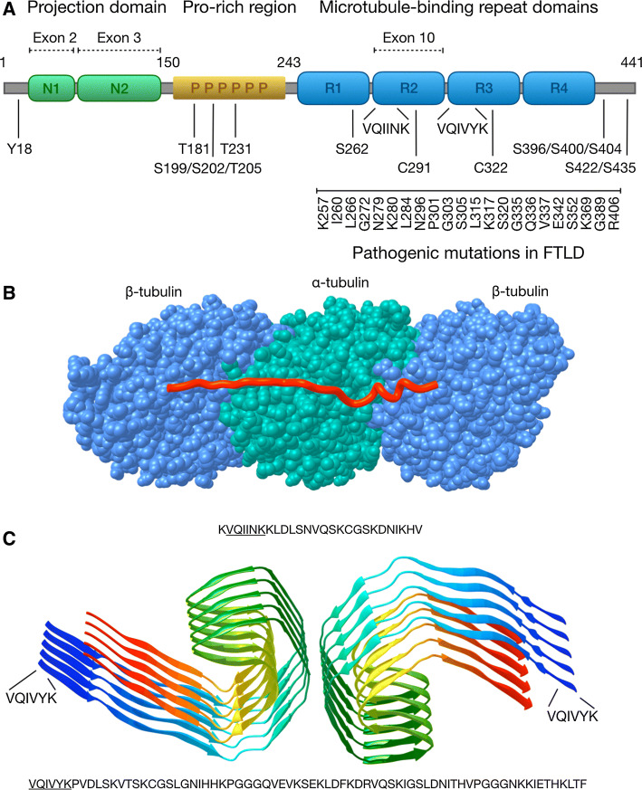
“Of particular importance are also two cysteine residues located in R2 and R3, which form disulfide bridges during tau dimerization and which have been found to constitute a critical step in the formation of tau fibrils [44].”
BackgroundGWAS of Cerebrospinal Fluid Tau Levels Identifies Risk Variants for Alzheimer’s Disease
Cruchaga, C., Kauwe, J. S. K., Harari, O., Jin, S. C., Cai, Y., Karch, C. M., Benitez, B. A., Jeng, A. T., Skorupa, T., Carrell, D., et al. (2013) · Neuron · doi:10.1016/j.neuron.2013.02.026 · Cited by 383
Winston, C. N., Goetzl, E. J., Akers, J. C., Carter, B. S., Rockenstein, E. M., Galasko, D. R., Masliah, E., & Rissman, R. A. (2016) · Alzheimer s & Dementia Diagnosis Assessment & Disease Monitoring · doi:10.1016/j.dadm.2016.04.001 · Cited by 379
Preparation and Characterization of Neurotoxic Tau Oligomers
Lasagna-Reeves, C. A., Castillo-Carranza, D. L., Guerrero-Muñoz, M. J., Jackson, G. R., & Kayed, R. (2010) · Biochemistry · doi:10.1021/bi1016233 · Cited by 359
Arendt, T., Stieler, J., Strijkstra, A. M., Hut, R. A., Rüdiger, J., Van der Zee, E. A., Harkany, T., Holzer, M., & Härtig, W. (2003) · The Journal of Neuroscience · doi:10.1523/jneurosci.23-18-06972.2003 · Cited by 357 · cited 1×
“In line with these results, Arendt and colleagues suggested that hyperphosphorylation of tau may have a physiological role, possibly related to synaptic plasticity [73].”
ResultDing, T. T., Lee, S., Rochet, J., & Lansbury, P. T. (2002) · Biochemistry · doi:10.1021/bi020139h · Cited by 356 · cited 1×
“Interestingly, other disease-associated proteins, such as α-synuclein and Aβ, also have the ability to form bacteriotoxin-like annular protofibrils and pore-like structures in membranes [183, 184], suggesting that this mechanism could play a central role in unconventional secretion of several misfolded aggregating pro…”
BackgroundDynamics of exosome internalization and trafficking
Tian, T., Zhu, Y., Hu, F., Wang, Y., Huang, N., & Xiao, Z. (2012) · Journal of Cellular Physiology · doi:10.1002/jcp.24304 · Cited by 351
Interaction of tau with the RNA-Binding Protein TIA1 Regulates tau Pathophysiology and Toxicity
Vanderweyde, T. E., Apicco, D. J., Youmans-Kidder, K., Ash, P. E. A., Cook, C. N., da Rocha, E. L., Jansen‐West, K. R., Frame, A. A., Citro, A., Leszyk, J., et al. (2016) · Cell Reports · doi:10.1016/j.celrep.2016.04.045 · Cited by 346 · cited 1×
“Interestingly, TIA-1, an RNA-binding protein centrally involved in the formation of stress granules [214], has been shown to directly modulate tau pathology in vitro and in vivo [215, 216].”
BackgroundHuman Astrocytes Transfer Aggregated Alpha-Synuclein via Tunneling Nanotubes
Rostami, J., Holmqvist, S., Lindström, V., Sigvardson, J., Westermark, G. T., Ingelsson, M., Bergström, J., Roybon, L., & Erlandsson, A. (2017) · The Journal of Neuroscience · doi:10.1523/jneurosci.0983-17.2017 · Cited by 339
Kawarabayashi, T., Shoji, M., Younkin, L. H., Wen-Lang, L., Dickson, D. W., Murakami, T., Matsubara, E., Abe, K., Ashe, K. H., & Younkin, S. G. (2004) · The Journal of Neuroscience · doi:10.1523/jneurosci.5543-03.2004 · Cited by 320
Tau in cerebrospinal fluid: A potential diagnostic marker in Alzheimer's disease
Arai, H., Terajima, M., Miura, M., Higuchi, S., Muramatsu, T., Machida, N., Seiki, H., Takase, S., Clark, C., Lee, V. M., et al. (1995) · Annals of Neurology · doi:10.1002/ana.410380414 · Cited by 314
Widespread brain tau and its association with ageing, Braak stage and Alzheimer’s dementia
Lowe, V. J., Wiste, H. J., Senjem, M. L., Weigand, S. D., Therneau, T. M., Boeve, B. F., Josephs, K. A., Fang, P., Pandey, M. K., Murray, M. E., et al. (2017) · Brain · doi:10.1093/brain/awx320 · Cited by 311
Henderson, M. X., Cornblath, E. J., Darwich, A., Zhang, B., Brown, H., Gathagan, R. J., Sandler, R. M., Bassett, D. S., Trojanowski, J. Q., & Lee, V. M. Y. (2019) · Nature Neuroscience · doi:10.1038/s41593-019-0457-5 · Cited by 310
Impairment of the glymphatic system after diabetes
Jiang, Q., Zhang, L., Ding, G., Davoodi-Bojd, E., Li, Q., Li, L., Sadry, N., Nedergaard, M., Chopp, M., & Zhang, Z. (2017) · Journal of Cerebral Blood Flow & Metabolism · doi:10.1177/0271678x16654702 · Cited by 308
Microtubule-associated protein tau is essential for long-term depression in the hippocampus
Kimura, T., Whitcomb, D. J., Jo, J., Regan, P., Piers, T., Heo, S., Brown, C., Hashikawa, T., Murayama, M., Seok, H., et al. (2014) · Philosophical Transactions of the Royal Society B: Biological Sciences · doi:10.1098/rstb.2013.0144 · Cited by 307
Tunneling nanotube (TNT)-mediated neuron-to neuron transfer of pathological Tau protein assemblies
Tardivel, M., Bégard, S., Bousset, L., Dujardin, S., Coens, A., Melki, R., Buée, L., & Colin, M. (2016) · Acta Neuropathologica Communications · doi:10.1186/s40478-016-0386-4 · Cited by 305
A critical appraisal of the pathogenic protein spread hypothesis of neurodegeneration
Walsh, D. M., & Selkoe, D. J. (2016) · Nature reviews. Neuroscience · doi:10.1038/nrn.2016.13 · Cited by 301
Tau Pathology in Two Dutch Families with Mutations in the Microtubule-Binding Region of Tau
Spillantini, M. G., Crowther, R. A., Kamphorst, W., Heutink, P., & Swieten, J. C. V. (1998) · American Journal Of Pathology · doi:10.1016/s0002-9440(10)65721-5 · Cited by 293
The role of microglia in processing and spreading of bioactive tau seeds in Alzheimer’s disease
Hopp, S. C., Lin, Y., Oakley, D., Roe, A. D., DeVos, S. L., Hanlon, D., & Hyman, B. T. (2018) · Journal of Neuroinflammation · doi:10.1186/s12974-018-1309-z · Cited by 286
Reducing the RNA binding protein TIA1 protects against tau-mediated neurodegeneration in vivo
Apicco, D. J., Ash, P. E. A., Maziuk, B., LeBlang, C., Medalla, M., Al Abdullatif, A., Ferragud, A., Botelho, E., Ballance, H. I., Dhawan, U., et al. (2018) · Nature Neuroscience · doi:10.1038/s41593-017-0022-z · Cited by 281
Polanco, J. C., Scicluna, B. J., Hill, A. F., & Götz, J. (2016) · Journal of Biological Chemistry · doi:10.1074/jbc.m115.709485 · Cited by 280
Synaptic Contacts Enhance Cell-to-Cell Tau Pathology Propagation
Calafate, S., Buist, A., Miśkiewicz, K., Vijayan, V. A., Daneels, G., Strooper, B. D., de Wit, J., Verstreken, P., & Moechars, D. (2015) · Cell Reports · doi:10.1016/j.celrep.2015.04.043 · Cited by 277 · cited 4×
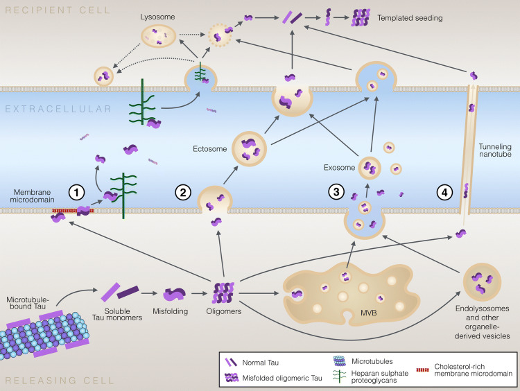
“However, the formation of tunneling nanotubes—or cellular contacts in other forms—is not required for cell-to-cell transfer of tau oligomers and aggregates, as several studies have shown that tau pathology can be transferred from one cell to another via the extracellular medium without cell-to-cell contacts or presenc…”
BackgroundInfluentialBraak, H., & Braak, E. (1989) · Neuropathology and Applied Neurobiology · doi:10.1111/j.1365-2990.1989.tb01146.x · Cited by 276
Template-assisted filament growth by parallel stacking of tau
Margittai, M., & Langen, R. (2004) · Proceedings of the National Academy of Sciences · doi:10.1073/pnas.0401911101 · Cited by 276
Unconventional Secretion Mediates the Trans-cellular Spreading of Tau
Katsinelos, T., Zeitler, M., Dimou, E., Karakatsani, A., Müller, H., Nachman, E., Steringer, J. P., de Almodóvar, C. R., Nickel, W., & Jahn, T. R. (2018) · Cell Reports · doi:10.1016/j.celrep.2018.04.056 · Cited by 274
Boluda, S., Iba, M., Zhang, B., Raible, K. M., Lee, V. M. ‐., & Trojanowski, J. Q. (2014) · Acta Neuropathologica · doi:10.1007/s00401-014-1373-0 · Cited by 268
Loss of Bin1 Promotes the Propagation of Tau Pathology
Calafate, S., Flavin, W., Verstreken, P., & Moechars, D. (2016) · Cell Reports · doi:10.1016/j.celrep.2016.09.063 · Cited by 265 · cited 1×
“Tau is indeed capable of rupturing the endosome membrane [200, 201], although the molecular mechanisms of this process, or which forms of tau mediate it, have not been fully elucidated.”
BackgroundEndocytic vesicle rupture is a conserved mechanism of cellular invasion by amyloid proteins
Flavin, W. P., Bousset, L., Green, Z. C., Chu, Y., Skarpathiotis, S., Chaney, M. J., Kordower, J. H., Melki, R., & Campbell, E. M. (2017) · Acta Neuropathologica · doi:10.1007/s00401-017-1722-x · Cited by 260
Liquid–liquid phase separation of tau protein: The crucial role of electrostatic interactions
Boyko, S., Qi, X., Chen, T., Surewicz, K. A., & Surewicz, W. K. (2019) · Journal of Biological Chemistry · doi:10.1074/jbc.ac119.009198 · Cited by 259
Single-molecule investigation of the interference between kinesin, tau and MAP2c
Seitz, A., Kojima, H., Oiwa, K., Mandelkow, E.-M., Song, Y.-H., & Mandelkow, E. (2002) · The EMBO Journal · doi:10.1093/emboj/cdf503 · Cited by 257
TFEB enhances astroglial uptake of extracellular tau species and reduces tau spreading
Martini-Stoica, H., Cole, A. L., Swartzlander, D. B., Chen, F., Wan, Y.-W., Bajaj, L., Bader, D. A., Lee, V. M. Y., Trojanowski, J. Q., Liu, Z., et al. (2018) · Journal of Experimental Medicine · doi:10.1084/jem.20172158 · Cited by 253
Klein, C., Krämer, E.-M., Cardine, A.-M., Schraven, B., Brandt, R., & Trotter, J. (2002) · The Journal of Neuroscience · doi:10.1523/jneurosci.22-03-00698.2002 · Cited by 250
Phosphate analysis and dephosphorylation of modified tau associated with paired helical filaments
Ksiȩżak-Reding, H., Liu, W.-K., & Yen, S.-H. C. (1992) · Brain Research · doi:10.1016/0006-8993(92)91476-u · Cited by 249
Anionic Micelles and Vesicles Induce Tau Fibrillization in Vitro
Chirita, C. N., Necula, M., & Kuret, J. (2003) · Journal of Biological Chemistry · doi:10.1074/jbc.m301663200 · Cited by 249
Gómez‐Ramos, A., Dı́az-Hernández, M., Rubio, A., Miras‐Portugal, M. T., & Ávila, J. (2008) · Molecular and Cellular Neuroscience · doi:10.1016/j.mcn.2007.12.010 · Cited by 245
Ectosomes: A New Mechanism for Non-Exosomal Secretion of Tau Protein
Dujardin, S., Bégard, S., Caillierez, R., Lachaud, C., Delattre, L., Carrier, S., Loyens, A., Galas, M., Bousset, L., Melki, R., et al. (2014) · PLoS ONE · doi:10.1371/journal.pone.0100760 · Cited by 242 · cited 7×
“In this study, microglia internalized neuronally secreted tau from the extracellular space and packed it in exosomes—which appear to contain the most toxic forms of tau [127, 155].”
BackgroundInfluentialLuo, W., Liu, W., Hu, X., Hanna, M., Caravaca, A., & Paul, S. M. (2015) · Scientific Reports · doi:10.1038/srep11161 · Cited by 241
Constitutive secretion of tau protein by an unconventional mechanism
Chai, X., Dage, J. L., & Citron, M. (2012) · Neurobiology of Disease · doi:10.1016/j.nbd.2012.05.021 · Cited by 238
Potential role of orexin and sleep modulation in the pathogenesis of Alzheimer’s disease
Roh, J. H., Jiang, H., Finn, M. B., Stewart, F. R., Mahan, T. E., Cirrito, J. R., Heda, A., Snider, B. J., Li, M., Yanagisawa, M., et al. (2014) · Journal of Experimental Medicine · doi:10.1084/jem.20141788 · Cited by 237
Delacourte, A., Sergeant, N., Wattez, A., Gauvreau, D., & Robitaille, Y. (1998) · Annals of Neurology · doi:10.1002/ana.410430209 · Cited by 235 · cited 1×
“1 3 known tauopathy characterized by inclusions of 3R-tau is Pick’s disease [89].”
BackgroundDirect Evidence of Internalization of Tau by Microglia In Vitro and In Vivo
Bolós, M., Llorens-Martín, M., Jurado-Arjona, J., Hernández, F., Rábano, A., & Avila, J. (2016) · Journal of Alzheimer's Disease · doi:10.3233/jad-150704 · Cited by 234
Parkinsonism and impaired axonal transport in a mouse model of frontotemporal dementia
Ittner, L. M., Fath, T., Ke, Y. D., Bi, M., van Eersel, J., Li, K. M., Gunning, P. W., & Götz, J. (2008) · Proceedings of the National Academy of Sciences · doi:10.1073/pnas.0808084105 · Cited by 230
Maas, T., Eidenmüller, J., & Brandt, R. (2000) · Journal of Biological Chemistry · doi:10.1074/jbc.m000389200 · Cited by 226
CSF biomarkers predict a more malignant outcome in Alzheimer disease
Wallin, A., Blennow, K., Zetterberg, H., Londos, E., Minthon, L., & Hansson, O. (2010) · Neurology · doi:10.1212/wnl.0b013e3181dd4dd8 · Cited by 223
Dural lymphatics regulate clearance of extracellular tau from the CNS
Patel, T. K., Habimana-Griffin, L., Gao, X., Xu, B. J., Achilefu, S., Alitalo, K. K., McKee, C. A., Sheehan, P. W., Musiek, E. S., Xiong, C., et al. (2019) · Molecular Neurodegeneration · doi:10.1186/s13024-019-0312-x · Cited by 220
Wills, J., Jones, J. M., Haggerty, T., Duka, V., Joyce, J. N., & Sidhu, A. (2010) · Experimental Neurology · doi:10.1016/j.expneurol.2010.06.017 · Cited by 217
Ferrer, I., López-González, I., Carmona, M., Arregui, L., Dalfó, E., Torrejón-Escribano, B., Diehl, R., & Kovacs, G. G. (2014) · Journal of Neuropathology & Experimental Neurology · doi:10.1097/nen.0000000000000030 · Cited by 217
Mirbaha, H., Holmes, B. B., Sanders, D. W., Bieschke, J., & Diamond, M. I. (2015) · Journal of Biological Chemistry · doi:10.1074/jbc.m115.652693 · Cited by 215
Fontaine, S. N., Zheng, D., Sabbagh, J. J., Martin, M. D., Chaput, D., Darling, A. L., Trotter, J. H., Stothert, A. R., Nordhues, B. A., Lussier, A. L., et al. (2016) · The EMBO Journal · doi:10.15252/embj.201593489 · Cited by 214 · cited 2×
“Several studies suggest that while a fraction of the total extracellular tau is indeed found in exosomes or ectosomes, a large fraction of the extracellular tau pool exists in vesicle-free form [149, 155, 157, 158] (Table 2).”
BackgroundGoedert, M., & Spillantini, M. G. (2017) · Molecular Brain · doi:10.1186/s13041-017-0298-7 · Cited by 213
Tau Internalization is Regulated by 6-O Sulfation on Heparan Sulfate Proteoglycans (HSPGs)
Rauch, J. N., Chen, J. J., Sorum, A. W., Miller, G. M., Sharf, T., See, S. K., Hsieh-Wilson, L. C., Kampmann, M., & Kosik, K. S. (2018) · Scientific Reports · doi:10.1038/s41598-018-24904-z · Cited by 211
Alzheimer-like Changes in Microtubule-associated Protein Tau Induced by Sulfated Glycosaminoglycans
Hasegawa, M., Crowther, R. A., Jakes, R., & Goedert, M. (1997) · Journal of Biological Chemistry · doi:10.1074/jbc.272.52.33118 · Cited by 207
Abounit, S., Wu, J. W., Duff, K., Victoria, G. S., & Zurzolo, C. (2016) · Prion · doi:10.1080/19336896.2016.1223003 · Cited by 206
Muñoz-García, D., & Ludwin, S. (1984) · Annals of Neurology · doi:10.1002/ana.410160408 · Cited by 205
Tau protein and tau aggregation inhibitors
Bulic, B., Pickhardt, M., Mandelkow, E., & Mandelkow�, E. (2010) · Neuropharmacology · doi:10.1016/j.neuropharm.2010.01.016 · Cited by 190
Retarded Axonal Transport of R406W Mutant Tau in Transgenic Mice with a Neurodegenerative Tauopathy
Zhang, B., Higuchi, M., Yoshiyama, Y., Ishihara, T., Forman, M. S., Martinez, D., Joyce, S. C., Trojanowski, J. Q., & Lee, V. M. ‐. (2004) · Journal of Neuroscience · doi:10.1523/jneurosci.0797-04.2004 · Cited by 187
Evans, L., Wassmer, T., Fraser, G. M., Smith, J. C., Perkinton, M. S., Billinton, A., & Livesey, F. J. (2018) · Cell Reports · doi:10.1016/j.celrep.2018.03.021 · Cited by 187 · cited 1×
“In line with this, a recent study demonstrated that different tau species can be internalized by different cellular mechanisms [195].”
ResultDetection of Aggregation-Competent Tau in Neuron-Derived Extracellular Vesicles
Guix, F. X., Corbett, G. T., Cha, D., Mustapić, M., Liu, W., Mengel, D., Chen, Z., Aïkawa, E., Young‐Pearse, T. L., Kapogiannis, D., et al. (2018) · International Journal of Molecular Sciences · doi:10.3390/ijms19030663 · Cited by 186
Extracellular Tau Levels Are Influenced by Variability in Tau That Is Associated with Tauopathies
Karch, C. M. M., Jeng, A. T., & Goate, A. (2012) · Journal of Biological Chemistry · doi:10.1074/jbc.m112.380642 · Cited by 185
Exosomes taken up by neurons hijack the endosomal pathway to spread to interconnected neurons
Polanco, J. C., Li, C., Durisic, N., Sullivan, R., & Götz, J. (2018) · Acta Neuropathologica Communications · doi:10.1186/s40478-018-0514-4 · Cited by 185
Differential Assembly of Human Tau Isoforms in the Presence of Arachidonic Acid
King, M. E., Gamblin, T. C., Kuret, J., & Binder, L. I. (2000) · Journal of Neurochemistry · doi:10.1046/j.1471-4159.2000.0741749.x · Cited by 179
Li, X., Hu, Y., Wang, Z.-H., Luo, Y., Zhang, Y., Liu, X., Feng, Q., Wang, Q., Ye, K., Liu, G., et al. (2016) · Scientific Reports · doi:10.1038/srep24756 · Cited by 173
Delisle, M., Murrell, J. R., Richardson, R., Trofatter, J. A., Rascol, O., Soulages, X., Mohr, M., Calvas, P., & Ghetti, B. F. (1999) · Acta Neuropathologica · doi:10.1007/s004010051052 · Cited by 171
Caspase-cleaved Tau Expression Induces Mitochondrial Dysfunction in Immortalized Cortical Neurons
Quintanilla, R. A., Matthews-Roberson, T. A., Dolan, P. J., & Johnson, G. V. W. (2009) · Journal of Biological Chemistry · doi:10.1074/jbc.m808908200 · Cited by 170
Proteostasis of tau. Tau overexpression results in its secretion via membrane vesicles
Simón, D., García‐García, E., Royo, F., Falcón‐Pérez, J. M., & Ávila, J. (2011) · FEBS Letters · doi:10.1016/j.febslet.2011.11.022 · Cited by 166
Komori, T., Arai, N., Oda, M., Nakayama, H., Mori, H., Yagishita, S., Takahashi, T., Amano, N., Murayama, S., Murakami, S., et al. (1998) · Acta Neuropathologica · doi:10.1007/s004010050911 · Cited by 166
Secretion of tau via an unconventional non-vesicular mechanism
Merezhko, M., Brunello, C. A., Yan, X., Vihinen, H., Jokitalo, E., Uronen, R.-L., & Huttunen, H. J. (2018) · Cell Reports · doi:10.1016/j.celrep.2018.10.078 · Cited by 166
Self-citationALZHEIMER'S DISEASE: PAIRED HELICAL FILAMENTS AND CYTOMEMBRANES
Gray, E. G., PAULA‐BARBOSA, M., & Roher, A. E. (1987) · Neuropathology and Applied Neurobiology · doi:10.1111/j.1365-2990.1987.tb00174.x · Cited by 160
Hyperphosphorylation and Cleavage at D421 Enhance Tau Secretion
Plouffe, V., Mohamed, N., Rivest‐McGraw, J., Bertrand, J., Lauzon, M., & Leclerc, N. M. (2012) · PLoS ONE · doi:10.1371/journal.pone.0036873 · Cited by 159
CSF biomarkers predict rate of cognitive decline in Alzheimer disease
Kester, M. I., van der Vlies, A. E., Blankenstein, M. A., Pijnenburg, Y. A. L., van Elk, E. J., Scheltens, P., & van der Flier, W. M. (2009) · Neurology · doi:10.1212/wnl.0b013e3181bd8271 · Cited by 159
Retiring the term FTDP-17 as MAPT mutations are genetic forms of sporadic frontotemporal tauopathies
Forrest, S. L., Kril, J. J., Stevens, C. H., Kwok, J. B., Hallupp, M., Kim, W. S., Huang, Y. M., McGinley, C. V., Werka, H., Kiernan, M. C., et al. (2017) · Brain · doi:10.1093/brain/awx328 · Cited by 156
Argyrophilic grain disease: A late‐onset dementia with distinctive features among tauopathies
Tolnay, M., & Clavaguera, F. (2004) · Neuropathology · doi:10.1111/j.1440-1789.2004.00591.x · Cited by 154
Victoria, G. S., & Zurzolo, C. (2017) · The Journal of Cell Biology · doi:10.1083/jcb.201701047 · Cited by 152
Tau Enhances α-Synuclein Aggregation and Toxicity in Cellular Models of Synucleinopathy
Badiola, N., de Oliveira, R. M., Herrera, F., Guardia‐Laguarta, C., Gonçalves, S. A., Pera, M., Suárez‐Calvet, M., Clarimón, J., Outeiro, T. F., & Lleó, A. (2011) · PLoS ONE · doi:10.1371/journal.pone.0026609 · Cited by 150 · cited 1×
“Despite the fact that tau and α-synuclein are distinct proteins that contribute to different disease-specific pathologies, multiple lines of evidence suggest that tau and α-synuclein interact, modulate the aggregation of each other and coexist in pathological inclusions in human brain [103].”
BackgroundJones, E. M., Dubey, M. K., Camp, P. J., Vernon, B. C., Biernat, J., Mandelkow�, E., Majewski, J. P., & Chi, E. Y. (2012) · Biochemistry · doi:10.1021/bi201857v · Cited by 149
Discovery and characterization of stable and toxic Tau/phospholipid oligomeric complexes
Ait‐Bouziad, N., Lv, G., Mahul‐Mellier, A., Xiao, S., Zorludemir, G., Eliezer, D., Walz, T., & Lashuel, H. A. (2017) · Nature Communications · doi:10.1038/s41467-017-01575-4 · Cited by 149
Dynamic association of tau with neuronal membranes is regulated by phosphorylation
Pooler, A. M., Usardi, A., Evans, C. J., Philpott, K. L., Noble, W., & Hanger, D. P. (2012) · Neurobiology of Aging · doi:10.1016/j.neurobiolaging.2011.01.005 · Cited by 148
Sposito, T., Preza, E., Mahoney, C. J., Setó-Salvia, N., Ryan, N. S., Morris, H. R., Arber, C., Devine, M. J., Houlden, H., Warner, T. T., et al. (2015) · Human Molecular Genetics · doi:10.1093/hmg/ddv246 · Cited by 148 · cited 1×
“, N279K, N296H, P301S, S305I mutations) [33, 34].”
Tai, H., Wang, B. Y., Serrano‐Pozo, A., Frosch, M. P., Spires‐Jones, T. L., & Hyman, B. T. (2014) · Acta Neuropathologica Communications · doi:10.1186/s40478-014-0146-2 · Cited by 147 · cited 2×
“Furthermore, in animal models pathological tau appears to be more localized to synapses compared to non-pathological tau [63], and synaptosomes isolated from human AD brains were shown to contain more phosphorylated and aggregated tau than in healthy controls [64].”
BackgroundDysregulation of RNA Binding Protein Aggregation in Neurodegenerative Disorders
Maziuk, B. F., Ballance, H. I., & Wolozin, B. L. (2017) · Frontiers in Molecular Neuroscience · doi:10.3389/fnmol.2017.00089 · Cited by 147
Santa‐María, I., Varghese, M., Ksiȩżak-Reding, H., Dzhun, A., Wang, J., & Pasinetti, G. M. (2012) · Journal of Biological Chemistry · doi:10.1074/jbc.m111.323279 · Cited by 145
Ramachandran, G., & Udgaonkar, J. B. (2011) · Journal of Biological Chemistry · doi:10.1074/jbc.m111.271874 · Cited by 143
Neuronal apolipoprotein E4 increases cell death and phosphorylated tau release in alzheimer disease
Wadhwani, A. R., Affaneh, A., Van Gulden, S., & Kessler, J. A. (2019) · Annals of Neurology · doi:10.1002/ana.25455 · Cited by 142
Avila, J., Jiménez, J. S., Sayas, C. L., Bolós, M., Zabala, J. C., Rivas, G., & Hernández, F. (2016) · Frontiers in Aging Neuroscience · doi:10.3389/fnagi.2016.00262 · Cited by 140
Amyloid-β Annular Protofibrils Evade Fibrillar Fate in Alzheimer Disease Brain
Lasagna‐Reeves, C. A., Glabe, C. G., & Kayed, R. (2011) · Journal of Biological Chemistry · doi:10.1074/jbc.m111.236257 · Cited by 139 · cited 1×
“Interestingly, other disease-associated proteins, such as α-synuclein and Aβ, also have the ability to form bacteriotoxin-like annular protofibrils and pore-like structures in membranes [183, 184], suggesting that this mechanism could play a central role in unconventional secretion of several misfolded aggregating pro…”
BackgroundGauthier‐Kemper, A., Weissmann, C., Golovyashkina, N., Sebö-Lemke, Z., Drewes, G., Gerke, V., Heinisch, J. J., & Brandt, R. (2011) · The Journal of Cell Biology · doi:10.1083/jcb.201007161 · Cited by 137
Kouri, N., Murray, M. E., Hassan, A., Rademakers, R., Uitti, R. J., Boeve, B. F., Graff‐Radford, N. R., Wszołek, Z. K., Litvan, I., Josephs, K. A., et al. (2011) · Brain · doi:10.1093/brain/awr234 · Cited by 137
Induction of a transmissible tau pathology by traumatic brain injury
Zanier, E. R., Bertani, I., Sammali, E., Pischiutta, F., Chiaravalloti, M. A., Vegliante, G., Masone, A., Corbelli, A., Smith, D. H., Menon, D. K., et al. (2018) · Brain · doi:10.1093/brain/awy193 · Cited by 136 · cited 3×
“AD Alzheimer’s disease AGD Argyrophylic grain disease CBD Corticobasal degeneration CSF Cerebrospinal fluid EM Electron microscopy ER Endoplasmic reticulum FGF2 Fibroblast growth factor 2 FTD Frontotemporal dementia FTDP-17 Frontotemporal dementia with Parkinsonism in chromosome 17 GAG Glycosaminoglycan HSPG Heparin s…”
BackgroundPre‐synaptic C‐terminal truncated tau is released from cortical synapses in Alzheimer's disease
Sokolow, S., Henkins, K. M., Bilousova, T., Gonzalez, B., Vinters, H. V., Miller, C. A., Cornwell, L., Poon, W. W., & Gylys, K. H. (2015) · Journal of Neurochemistry · doi:10.1111/jnc.12991 · Cited by 133
Neuronal exosomes reveal Alzheimer's disease biomarkers in Down syndrome
Hamlett, E. D., Goetzl, E. J., Ledreux, A., Vasilevko, V., Boger, H. A., LaRosa, A., Clark, D., Carroll, S. L., Carmona‐Iragui, M., Fortea, J., et al. (2017) · Alzheimer's & Dementia · doi:10.1016/j.jalz.2016.08.012 · Cited by 129
Cruchaga, C., Kauwe, J. S. K., Mayo, K., Spiegel, N., Bertelsen, S., Nowotny, P., Shah, A. R., Abraham, R., Hollingworth, P., Harold, D., et al. (2010) · PLoS Genetics · doi:10.1371/journal.pgen.1001101 · Cited by 128
Li, H.-R., Chen, T.-C., Hsiao, C.-L., Shi, L., Chou, C., & Huang, J. (2017) · Biochimica et Biophysica Acta (BBA) - Proteins and Proteomics · doi:10.1016/j.bbapap.2017.10.001 · Cited by 128
Expression of microtubule-associated proteins MAP2 and tau in cultured rat brain oligodendrocytes
Müller, R., Heinrich, M., Heck, S., Blohm, D., & Richter-Landsberg, C. (1997) · Cell and Tissue Research · doi:10.1007/s004410050809 · Cited by 127
The Role of the Lipid Bilayer in Tau Aggregation
Elbaum‐Garfinkle, S., Ramlall, T. F., & Rhoades, E. R. (2010) · Biophysical Journal · doi:10.1016/j.bpj.2010.03.013 · Cited by 127
Nucleation-dependent Tau Filament Formation
Congdon, E. E., Kim, S., Bonchak, J., Songrug, T., Anastasios Matzavinos, A., & Kuret, J. (2008) · Journal of Biological Chemistry · doi:10.1074/jbc.m800247200 · Cited by 126
Georgieva, E. R., Xiao, S., Borbat, P. P., Freed, J. H., & Eliezer, D. (2014) · Biophysical Journal · doi:10.1016/j.bpj.2014.07.046 · Cited by 124
Soluble Aβ Promotes Wild-Type Tau Pathology In Vivo
Chabrier, M. A., Blurton-Jones, M., Agazaryan, A. A., Nerhus, J. L., Martinez-Coria, H., & LaFerla, F. M. (2012) · The Journal of Neuroscience · doi:10.1523/jneurosci.0172-12.2012 · Cited by 123 · cited 1×
“For example, in mouse model of AD, soluble Aβ promotes development of tau pathology [75, 76].”
Tau isoform composition influences rate and extent of filament formation
Zhong, Q., Congdon, E. E., Nagaraja, H. N., & Kuret, J. (2012) · Journal of Biological Chemistry · doi:10.1074/jbc.m112.364067 · Cited by 122
Di, J., Cohen, L. S., Corbo, C. P., Phillips, G. R., Idrissi, A. E., & Alonso, A. D. C. (2016) · Scientific Reports · doi:10.1038/srep20833 · Cited by 120
Neuropathology of Pick’s disease
Dickson, D. W. (2001) · Neurology · doi:10.1212/wnl.56.suppl_4.s16 · Cited by 119
DNAJC5 facilitates USP19-dependent unconventional secretion of misfolded cytosolic proteins
Xu, Y., Cui, L., DiBello, A., Wang, L., Lee, J., Saidi, L. F., Lee, J., & Ye, Y. (2018) · Cell Discovery · doi:10.1038/s41421-018-0012-7 · Cited by 117
Extracellular Monomeric Tau Is Internalized by Astrocytes
Perea, J. R., López, E., Díez-Ballesteros, J. C., Ávila, J., Hernández, F., & Bolós, M. (2019) · Frontiers in Neuroscience · doi:10.3389/fnins.2019.00442 · Cited by 114
mTor mediates tau localization and secretion: implication for Alzheimer’s disease
Tang, Z., Ioja, E., Bereczki, E., Hultenby, K., Li, C., Guan, Z., Winblad, B., & Pei, J.-J. (2015) · Biochimica et Biophysica Acta (BBA) - Molecular Cell Research · doi:10.1016/j.bbamcr.2015.03.003 · Cited by 112
Chronic Sleep Disruption Advances the Temporal Progression of Tauopathy in P301S Mutant Mice
Zhu, Y., Zhan, G., Fenik, P., Brandes, M., Bell, P., François, N., Shulman, K., & Veasey, S. C. (2018) · Journal of Neuroscience · doi:10.1523/jneurosci.0275-18.2018 · Cited by 110
Sepulveda-Diaz, J. E., Naini, S. M. A., Huynh, M. B., Ouidja, M. O., Yanicostas, C., Chantepie, S., Villares, J., Lamari, F., Jospin, E., Kuppevelt, T. H. V., et al. (2015) · Brain · doi:10.1093/brain/awv056 · Cited by 108 · cited 1×
“Moreover, expression and activities of several enzymes participating in generation and modification of sulfated glycosaminoglycans, particularly heparan sulfate sulfotransferases, have been reported to be altered in AD brain and to affect accumulation of phosphorylated tau species [228, 229].”
BackgroundYamada, T., & McGeer, P. (1990) · Neuroscience Letters · doi:10.1016/0304-3940(90)90028-8 · Cited by 101
Pallas‐Bazarra, N., Jurado‐Arjona, J., Navarrete, M., Esteban, J. A., Hernández, F., Ávila, J., & Llorens‐Martín, M. (2016) · The EMBO Journal · doi:10.15252/embj.201593518 · Cited by 101
Baker, S., Polanco, J. C., & Götz, J. (2016) · Journal of Alzheimer's Disease · doi:10.3233/jad-160371 · Cited by 98
Research works printed but not linked: 3
- Liu F, Gong CX (2008) Tau exon 10 alternative splicing and tauopathies. Mol Neurodegener 3:8 · DOI · Paper page
- Grundke-Iqbal I, Iqbal K, Tung YC, Quinlan M, Wisniewski HM, Binder LI (1986) Abnormal phosphorylation of the microtubule-associated protein tau (tau) in Alzheimer cytoskeletal pathology. Proc Natl Acad Sci USA 83(13):4913–4917 · DOI · Paper page
- Yamada K et al (2014) Neuronal activity regulates extracellular tau in vivo. J Exp Med 211(3):387–393 · DOI
Cited by
Sorted by citations
Phosphorylated Tau in Alzheimer’s Disease and Other Tauopathies
Rawat, P., Sehar, U., Bisht, J., Selman, A., Culberson, J., & Reddy, P. H. (2022) · International Journal of Molecular Sciences · doi:10.3390/ijms232112841 · Cited by 509
“At the protein level, K257T, G272V, P301L, V337M, and R406W mutations alter the binding properties of the microtubule, and N279K, N296H, P301S, S305I mutations at the RNA level shifts the splicing towards overproduction of 4R tau [97].”
BackgroundThe expanding amyloid family: Structure, stability, function, and pathogenesis
Sawaya, M. R., Hughes, M. P., Rodriguez, J. A., Riek, R., & Eisenberg, D. S. (2021) · Cell · doi:10.1016/j.cell.2021.08.013 · Cited by 475
Role of tau protein in Alzheimer’s disease: The prime pathological player
Muralidar, S., Ambi, S. V., Sekaran, S., Thirumalai, D., & Palaniappan, B. (2020) · International Journal of Biological Macromolecules · doi:10.1016/j.ijbiomac.2020.07.327 · Cited by 278
Alzheimer’s disease brain-derived extracellular vesicles spread tau pathology in interneurons
Ruan, Z., Pathak, D., Venkatesan Kalavai, S., Yoshii-Kitahara, A., Muraoka, S., Bhatt, N., Takamatsu-Yukawa, K., Hu, J., Wang, Y., Hersh, S., et al. (2021) · Brain · doi:10.1093/brain/awaa376 · Cited by 283
“secreted primarily in free form, while a minor fraction of tau is found in EVs as observed in the CSF or blood of control subjects and Alzheimer’s disease patients (Arai et al., 1998; Saman et al., 2012; Zetterberg et al., 2013; Fiandaca et al., 2015; Ruan and Ikezu, 2019; Brunello et al., 2020).”
BackgroundAcetylated tau inhibits chaperone-mediated autophagy and promotes tau pathology propagation in mice
Caballero, B., Bourdenx, M., Luengo, E., Diaz, A., Sohn, P. D., Chen, X., Wang, C., Juste, Y. R., Wegmann, S., Patel, B., et al. (2021) · Nature Communications · doi:10.1038/s41467-021-22501-9 · Cited by 223
Metadata
- Authors
- Brunello, C. A., Merezhko, M., Uronen, R.-L., & Huttunen, H. J.
- Affiliations
- University of Helsinki (FI)
- Corresponding author
- Huttunen, Henri J.
- Venue
- Cellular and Molecular Life Sciences
- Published
- 2019-10-30
- Volume
- 77
- Issue
- 9
- Pages
- 1721-1744
- ISSN
- 1420-682X (ISSN-L)
- 1420-9071
- PMC
- PMC7190606
- OpenAlex
- W2982627322
- Cited by
- 304
- References
- 267
- Citations by year
- 2026: 36 · 2025: 45 · 2024: 56 · 2023: 38 · 2022: 46 · 2021: 57 · 2020: 25 · 2019: 1
- FWCI
- 3.80
- Type
- journal article
- Language
- en
- Open access
- hybrid
- doi.org
- Locations
- Cellular and Molecular Life Sciences (publishedVersion, cc-by)
- PubMed Central (publishedVersion, cc-by)
- PubMed (publishedVersion)
- Työväentutkimus Vuosikirja (submittedVersion, cc-by)
- Funders
- Academy of Finland (296409)
- Alfred Kordelinin Säätiö
- Topics
- Life Sciences › Neuroscience › Cellular and Molecular Neuroscience › Alzheimer's disease research and treatments
- Life Sciences › Biochemistry, Genetics and Molecular Biology › Molecular Biology › Prion Diseases and Protein Misfolding
- Life Sciences › Neuroscience › Neurology › Neuroinflammation and Neurodegeneration Mechanisms
- Keywords
- tau protein, tauopathy, tau secretion, cell-to-cell transmission, prion-like propagation, tau aggregation, exosomes, ectosome, vesicular secretion, extracellular tau, endocytosis, phagocytosis, pinocytosis, Alzheimer's disease, frontotemporal lobar degeneration, neurodegeneration
- MeSH terms
- Animals, Humans, tau Proteins, Disease Progression, Tauopathies, Protein Aggregation, Pathological
About these checks
Registry, retraction, Cites and statistics results are computed, not judged, with one exception: a printed reference that no index lists is read once by a model before it counts as a citation error. Claims are extracted by a model and kept only when their quote appears verbatim in the text; each cited source is then checked, with a model’s help, for whether it says what the claim relies on. Those verdicts can be wrong — read the claim’s quote and the cited source before relying on one.
Statistics and arithmetic are recomputed, not judged.
Confidence bands are shown only for fields calibrated on our annotated test set. None is calibrated yet, so no band is shown.
What counts as a claim ›How we measure confidence ›How we check this ›
Are you an author, or do you see a problem with this page? Request a review
