

The DOI matches its Crossref record; 260 of the 267 references it cites were found in a registry or an index.
Mechanisms of secretion and spreading of pathological tau protein
Open accessAbstract
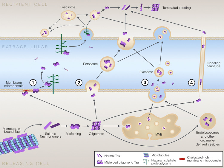
Accumulation of misfolded and aggregated forms of tau protein in the brain is a neuropathological hallmark of tauopathies, such as Alzheimer’s disease and frontotemporal lobar degeneration. Tau aggregates have the ability to transfer from one cell to another and to induce templated misfolding and aggregation of healthy tau molecules in previously healthy cells, thereby propagating tau pathology across different brain areas in a prion-like manner. The molecular mechanisms involved in cell-to-cell transfer of tau aggregates are diverse, not mutually exclusive and only partially understood. Intracellular accumulation of misfolded tau induces several mechanisms that aim to reduce the cellular burden of aggregated proteins and also promote secretion of tau aggregates. However, tau may also be released from cells physiologically unrelated to protein aggregation. Tau secretion involves multiple vesicular and non-vesicle-mediated pathways, including secretion directly through the plasma membrane. Consequently, extracellular tau can be found in various forms, both as a free protein and in vesicles, such as exosomes and ectosomes. Once in the extracellular space, tau aggregates can be internalized by neighboring cells, both neurons and glial cells, via endocytic, pinocytic and phagocytic mechanisms. Importantly, accumulating evidence suggests that prion-like propagation of misfolding protein pathology could provide a general mechanism for disease progression in tauopathies and other related neurodegenerative diseases. Here, we review the recent literature on cellular mechanisms involved in cell-to-cell transfer of tau, with a particular focus in tau secretion.
Overview
The claims and statistics of this paper haven’t been checked yet.
Run the check — free, no account needed. It reads the open-access text and checks the sources it cites.
Integrity signals
Registry
Cites
Statistics
Figures
Verification
Claims the paper makes
The claims and statistics of this paper haven’t been checked yet.
Run the check — free, no account needed. It reads the open-access text and checks the sources it cites.
Claims other papers make about this one
Each is checked against this paper’s own text: does it say what the citing paper says it does?
The source backs part of the claim, not all of it — or backs it for a different population, condition or comparison.
In the citing paper
“Thus, it is suggested that CSF p-tau biomarkers likely indicate an active process of tau secretion, and one that is correlated with cerebral Aβ deposition in early disease.20, 21, 22”
From Cerebrospinal fluid p-tau231 as an early indicator of emerging pathology in Alzheimer's disease (2022) · cites this paper as [22]
Experimental — accuracy not yet measured
Self-check
Percentages, sums and reported differences re-computed from the paper’s own numbers.
The self-check needs the paper’s full text.
Citations
Cites
Neuropathological stageing of Alzheimer-related changes.
Braak, H., & Braak, E. (1991) · Acta Neuropathologica · doi:10.1007/bf00308809 · Cited by 16,123 · cited 1×
“It is well established that in AD the development of tau pathology occurs in a hierarchical pattern of accumulation starting from the layer II of entorhinal cortex (EC) and spreading toward cortical regions, as originally proposed by the findings of Heiko and Eva Braak [107].”
ResultAmyloid oligomers exacerbate tau pathology in a mouse model of tauopathy
Selenica, M.-L. B., Brownlow, M., Jimenez, J. P., Lee, D. C., Pena, G., Dickey, C. A., Gordon, M. N., & Morgan, D. (2013) · Neurodegenerative Diseases · doi:10.1159/000337230 · Cited by 10,359
Iliff, J. J., Wang, M., Liao, Y., Plogg, B. A., Peng, W., Gundersen, G. A., Benveniste, H., Vates, G. E., Deane, R., Goldman, S. A., et al. (2012) · Science Translational Medicine · doi:10.1126/scitranslmed.3003748 · Cited by 5,824 · cited 1×
“One important contributor to clearance of tau aggregates from the brain is the brain glymphatic system, a brain-wide interstitial solute clearance system composed of astrocytes, paravascular routes and dural lymphatics [230, 231].”
BackgroundFrontotemporal lobar degeneration: A consensus on clinical diagnostic criteria
Neary, D., Snowden, J. S., Gustafson, L., Passant, U., Stuss, D., Black, S., Freedman, M., Kertesz, A., Robert, P. H., Albert, M., et al. (1998) · Neurology · doi:10.1212/wnl.51.6.1546 · Cited by 5,047
Association of missense and 5′-splice-site mutations in tau with the inherited dementia FTDP-17
Hutton, M., Lendon, C. L., Rizzu, P., Baker, M., Froelich, S., Houlden, H., Pickering-Brown, S., Chakraverty, S., Isaacs, A., Grover, A., et al. (1998) · Nature · doi:10.1038/31508 · Cited by 3,498 · cited 3×
“First mutations in the MAPT gene were found in 1998 [31, 32]”
BackgroundA protein factor essential for microtubule assembly
Weingarten, M. D., Lockwood, A. H., Hwo, S. Y., & Kirschner, M. W. (1975) · Proceedings of the National Academy of Sciences · doi:10.1073/pnas.72.5.1858 · Cited by 3,089
A Liquid-to-Solid Phase Transition of the ALS Protein FUS Accelerated by Disease Mutation
Patel, A., Lee, H. O., Jawerth, L. M., Maharana, S., Jahnel, M., Hein, M. Y., Stoynov, S., Mahamid, J., Saha, S., Franzmann, T. M., et al. (2015) · Cell · doi:10.1016/j.cell.2015.07.047 · Cited by 2,927
Microglia Function in the Central Nervous System During Health and Neurodegeneration
Colonna, M., & Butovsky, O. (2017) · Annual Review of Immunology · doi:10.1146/annurev-immunol-051116-052358 · Cited by 2,894
Molliex, A., Temirov, J. P., Lee, J., Coughlin, M., Kanagaraj, A., Kim, H. J., Mittag, T., & Taylor, J. P. (2015) · Cell · doi:10.1016/j.cell.2015.09.015 · Cited by 2,690
Tau in physiology and pathology
Wang, Y., & Mandelkow, E. (2016) · Nature Reviews Neuroscience · doi:10.1038/nrn.2015.1 · Cited by 2,251
Research works printed but not linked: 3
- Liu F, Gong CX (2008) Tau exon 10 alternative splicing and tauopathies. Mol Neurodegener 3:8 · DOI · Paper page
- Grundke-Iqbal I, Iqbal K, Tung YC, Quinlan M, Wisniewski HM, Binder LI (1986) Abnormal phosphorylation of the microtubule-associated protein tau (tau) in Alzheimer cytoskeletal pathology. Proc Natl Acad Sci USA 83(13):4913–4917 · DOI · Paper page
- Yamada K et al (2014) Neuronal activity regulates extracellular tau in vivo. J Exp Med 211(3):387–393 · DOI
Cited by
Sorted by citations
Items 201–272 of 272
Dinamarca, M. C., Colombo, L., Brykczynska, U., Grimm, A., Fruh, I., Hossain, I., Gabriel, D., Eckert, A., Müller, M. M., & Pecho‐Vrieseling, E. (2024) · Frontiers in Molecular Neuroscience · doi:10.3389/fnmol.2023.1287510 · Cited by 7
“…tau (AD), alpha-synuclein ( α -syn) (PD), mutant huntingtin (mHTT) (HD), and TDP-43 (ALS Several cellular mechanisms have been proposed to underlie the inter-neuronal transmission of misfolded proteins (Masnata and Cicchetti, 2017; Brunello et al., 2020; Donnelly et al., 2020; Peng et al., 2020).”
BackgroundSpreading of P301S Aggregated Tau Investigated in Organotypic Mouse Brain Slice Cultures
Korde, D. S., & Humpel, C. (2022) · Biomolecules · doi:10.3390/biom12091164 · Cited by 8
“Since then, evidence from in vivo and in vitro studies has emerged, supporting the proposal, that tau extends throughout the brain via interconnected pathways in a mode similar to the spread of pathological prion proteins [6,12].”
BackgroundFranzmeier, N., Brendel, M., Beyer, L., Kovacs, G., Arzberger, T., Kurz, C., Respondek, G., Lukić, M., Biel, D., Rubinski, A., et al. (2021) · medRxiv · doi:10.1101/2021.08.16.21261523 · Cited by 4
Desale, S. E., & Chinnathambi, S. (2020) · doi:10.21203/rs.3.rs-22830/v1 · Cited by 5
“The aggregated extracellular Tau species secreted by various mechanisms have tendency to propagate the disease [41-43].”
BackgroundHeterotypic phase separation in aggregation: Driver or deterrent?
Jacob, T., & Hoyer, W. (2026) · Biophysical Chemistry · doi:10.1016/j.bpc.2026.107577 · Cited by 3
Characteristics of Scopolamine- and Trimethyltin-Induced Models of Alzheimer’s Disease
Nevmerzhytska, N. M. (2025) · Ukrainian Scientific Medical Youth Journal · doi:10.32345/supplement.2.2025.280-286
Giannelli, S. G., Eroli, F., Loera‐Valencia, R., Leoni, V., Latorre‐Leal, M., Testa, G., Staurenghi, E., Sottero, B., Gamba, P., Maioli, S., et al. (2025) · Neurobiology of Disease · doi:10.1016/j.nbd.2025.107029 · Cited by 3
“…leading to tau-tau interactions, insoluble aggregate formation, and microtubule destabilization (Johnson & Stoothoff, 2004). f Tau deposits determine the dysfunction of axonal transport, the reduction of dendritic spines, and the impairment transmission (Brunello et al., 2020).”
Nemati, R., Sohrabi-Ashlaghi, A., Saberian, P., Sadeghi, M., Mardani, S., Abadi, A., Yaghoobpoor, A., Heydari, A., Khoshroo, N., Rahnama, Y., et al. (2025) · BMC Neurology · doi:10.1186/s12883-025-04140-5 · Cited by 6
“These seeding-competent tau proteins can travel across synapses between neurons, contributing to tau spread in AD [43, 44].”
Data-driven simulation of network-based tau spreading tailored to individual Alzheimer's patients
Kim, S.-W., Cho, H., Lee, Y., Lyoo, C. H., Seong, J., & Initiative, A. D. N. (2024) · Engineering With Computers · doi:10.1007/s00366-024-01988-y · Cited by 2
“The accumulation of tangled tau proteins in the brain is an early indicator of Alzheimer's disease (AD) [1] due to its strong associations with neuronal death [2] and eventual cognitive decline [3].”
New aspects of a small GTPase RAB35 in brain development and function
Maejima, I., & Sato, K. (2024) · Neural Regeneration Research · doi:10.4103/nrr.nrr-d-23-01543 · Cited by 3
“Tau is a member of the microtubule-associated protein family, which is primarily expressed in neurons and promotes the assembly and stabilization of microtubules (Brunello et al., 2020; Yan and Zheng, 2021).”
α-Linolenic acid modulates phagocytosis of extracellular Tau and induces microglial migrationPreprint
Desale, S. E., & Chinnathambi, S. (2020) · bioRxiv (Cold Spring Harbor Laboratory) · doi:10.1101/2020.04.15.042143 · Cited by 12
“The aggregated extracellular Tau species secreted by various mechanisms have tendency to propagate the disease [41-43].”
BackgroundLipid Composition Drastically Alters Tau–Membrane Interaction: Implications for Alzheimer’s Disease
Wu, A., Kandel, N., Li, S., Enoki, T. A., Hewes, K., Palumbo, J., Buttaci, M., Jones, K., Xia, K., Zhao, J., et al. (2026) · JACS Au · doi:10.1021/jacsau.5c01563 · Cited by 2
“11,21,23 This interaction is strongly modulated by membrane lipid composition, particularly negatively charged lipids such as phosphatidylserine (PS), which mediate electrostatic attraction to tau’s positively charged regions.”
Selective vulnerability of human synapses to soluble tau oligomers
Kadamangudi, S., Sánchez-Sánchez, L., Kayed, R., Limón, A., & Taglialatela, G. (2026) · Journal of Alzheimer s Disease · doi:10.1177/13872877261416539 · Cited by 2
“9 One interpretation is that tau, following activity-dependent presynaptic release, 15,78,79 may bind to postsynaptic GABA A R in a sustained agonistic or allosteric fashion, leading to prolonged receptor activation.”
Pan, T., Tan, Y., Dong, X., Tang, Y., Yang, Z., Chen, Y., & Wei, G. (2025) · The Journal of Physical Chemistry B · doi:10.1021/acs.jpcb.5c01590 · Cited by 2
Presenilin deficiency enhances tau phosphorylation and its secretion
Sun, Y., Islam, S., Gao, Y., Nakamura, T., Tomita, T., Michikawa, M., & Zou, K. (2024) · Journal of Neurochemistry · doi:10.1111/jnc.16155 · Cited by 2
“…to localize to the plasma membrane (PM) in a clustered manner, and this localization is enhanced by over-expression or hyperphosphorylation, possibly as a consequence of increased levels of cytosolic tau or enhanced aggregation (Brunello et al., 2020; Mari et al., 2018 ; Merezhko et al., 2018).”
Pilot Study of the Total and Phosphorylated Tau Proteins in Early-Stage Multiple Sclerosis
Navickaitė, I., Pampuščenko, K., Žemgulytė, G., Bilskienė, D., Borutaitė, V., & Balnytė, R. (2024) · Medicina · doi:10.3390/medicina60030416 · Cited by 2
“However, recent research showed that tau can be secreted from neurons independently of cell death [7,15,16].”
BackgroundInfluentialGlucocorticoid stress hormones stimulate vesicle-free Tau secretion and spreading in the brain
Yu, Q., Du, F., Belli, I., Gomes, P. A., Sotiropoulos, I., & Waites, C. (2023) · bioRxiv · doi:10.1101/2023.06.07.544054 · Cited by 2
Feng, H., Liu, Y., Wang, X., Wang, C., & Wang, T. (2023) · BMJ Open · doi:10.1136/bmjopen-2023-072068 · Cited by 2
Transcriptomic signature of Alzheimer’s disease tau seed-induced pathology
Csicsatkova, N., Szalay, P., Matyasova, K., Mate, V., Cente, M., Smolek, T., Brezováková, V., Kawecka, L., Žilka, N., & Jadhav, S. (2021) · General Physiology and Biophysics · doi:10.4149/gpb_2021022 · Cited by 6
“In addition, the mode of transmission such as secretion, mechanism of uptake is progressively unearthed (Saman et al. 2012; Yamada et al. 2014; Tang et al. 2015; Kang et al. 2019; Morozova et al. 2019; Brunello et al. 2020).”
Smuggle tau through a secret(ory) pathway
Xu, H. (2021) · Biochemical Journal · doi:10.1042/bcj20210324 · Cited by 2
Ruan, Z., Pathak, D., Kalavai, S., Yoshii-Kitahara, A., Muraoka, S., Takamatsu-Yukawa, K., Hu, J.-Q., Wang, Y., Hersh, S., Gorantla, S., et al. (2020) · Alzheimer's & Dementia · doi:10.1002/alz.037316 · Cited by 2
Sheng, J., Yan, J., Xu, Y., Xu, X., Zhang, X., Wang, A. G., Liu, B., Ma, J., Duan, X., Zhou, H., et al. (2026) · Cell Death and Disease · doi:10.1038/s41419-026-08983-9 · Cited by 1
“The endo-lysosomal pathway has been implicated as a mechanism for tau secretion [11].”
Setiawan, A. W., Choi, J. T., Park, S., Choi, M., Tjandrawinata, R. R., Hadinata, E., Park, M. N., Ikrar, T., Nurkolis, F., & Kim, B. (2026) · Pharmaceuticals · doi:10.3390/ph19050792 · Cited by 1
“Even when a molecule can enter the brain, it may not distribute into the intracellular compartments where tau aggregation and phosphorylation cascades occur [76,79].”
Jiang, Y., Tetlow, A. M., Lin, Y., Ji, C., Ader, J., Laborc, K. F., Mar, A. C., Pan, R., Kong, X., Congdon, E. E., et al. (2026) · Science Translational Medicine · doi:10.1126/scitranslmed.aea4205 · Cited by 2
An overview of gene and cell therapy approaches for Alzheimer’s disease
Heidari, Z., Zakaee, A., Vafadar, A., Alavimanesh, S., Charami, H., Jamali, Z., Jahromi, A. H., Rakhsha, A., & Savardashtaki, A. (2025) · Metabolic Brain Disease · doi:10.1007/s11011-025-01763-y · Cited by 1
“Tau aggregates have the potential to be taken up by adjacent cells which encompass both neurons and glial cells, through endocytic, pinocytic, and phagocytic mechanisms (Brunello et al. 2020).”
Co‐localization of tau and TDP ‐43 after extracellular vesicle delivery to cells
Aliakbari, F., Volkening, K., Nayeri, Z., Polat, A. Y., Donison, N. S., Palik, J., & Strong, M. J. (2025) · FEBS Journal · doi:10.1111/febs.70336 · Cited by 2
Scalable human neuronal models of tauopathy producing endogenous seed-competent 4R tau
Tsefou, E., Bez, S., Birkle, T., Foiani, M., Watamura, N., Bourdenx, M., Gavriouchkina, D., Turkes, E., Crawford, S., Coneys, R., et al. (2025) · bioRxiv · doi:10.1101/2025.07.11.664346 · Cited by 1
Martínez, P., Jury, N., Patel, H., You, Y., Perkins, A., You, Y., Lee‐Gosselin, A., Vidal, R., & Lasagna‐Reeves, C. A. (2025) · Journal of Alzheimer s Disease · doi:10.1177/13872877251372603 · Cited by 3
“62,63 Our results suggested that pTau-Ser214 is involved in the transsynaptic propagation of pathological tau in neuronal culture with a minor effect to in vivo seed formation.”
Vegas‐Gomez, L., Pizarro, M., Garcia-Martin, J., Arredondo-Alcala, M. A., Bustamante, B., Gonzalez‐Silva, C., Matus, S., Díaz‐Espinoza, R., Gutiérrez, A., Morales, R., et al. (2025) · PLoS ONE · doi:10.1371/journal.pone.0328470 · Cited by 2
“Perivascular tau deposition has been observed in multiple tauopathies, reinforcing the idea that vascular transport plays a crucial role in the systemic-to-CNS propagation of tau aggregates [79,80].”
Corticobasal degeneration: An update
Révész, T., Lees, A. J., & Morris, H. R. (2024) · Ideggyógyászati Szemle · doi:10.18071/isz.77.0379 · Cited by 4
Martínez, P., Jury-Garfe, N., Patel, H., You, Y.-W., Perkins, A., You, Y., Lee‐Gosselin, A., Vidal, R., & Lasagna-Reeves, C. A. (2024) · bioRxiv · doi:10.1101/2024.07.22.604618 · Cited by 1
“Similarly, phosphorylation at specific sites may contribute to a greater propensity of tau to spread/propagate and the pattern in which propagates 58,59 .”
BackgroundAderinto, N., & Olatunji, G. D. (2023) · Neurology Letters · doi:10.61186/nl.2.2.78 · Cited by 1
Murthy, M. N., & Shyamala, B. V. (2023) · Journal of Complementary and Alternative Medical Research · doi:10.9734/jocamr/2023/v22i3461 · Cited by 1
Sodium selenate as a therapeutic for tauopathies: A hypothesis paper
Dilcher, R., Malpas, C. B., Walterfang, M., Velakoulis, D., O'Brien, T. J., & Vivash, L. E. (2022) · Frontiers in Aging Neuroscience · doi:10.3389/fnagi.2022.915460 · Cited by 2
“It was suggested that in FTLD-tau, abnormal hyperphosphorylated tau aggregates detach from microtubules by propagating transcellularly and extracellular in the cerebrospinal fluid (CSF) and thereby spreading tau pathology into different brain areas as the disease progresses (Brunello et al., 2020).”
BackgroundEarly Detection of Tau PathologyPreprint
Parekh, P. A., Badachhape, A., Mu, Q., Bhavane, R. C., Srivastava, M., Stupin, I. V., Bhandari, P., Devkota, L., Eriksen, J. L., Tanifum, E. A., et al. (2021) · bioRxiv (Cold Spring Harbor Laboratory) · doi:10.1101/2021.05.14.444233 · Cited by 1
Endogenous retroviruses promote prion-like spreading of proteopathic seeds
Liu, S., Heumüller, S.-E., Hossinger, A., Müller, S. A., Buravlova, O., Lichtenthaler, S., Denner, P., & Vorberg, I. (2022) · bioRxiv · doi:10.1101/2022.05.06.490866 · Cited by 1
“transmitted to bystander cells via direct cell contact (9).”
BackgroundHuang, Y., Driedonks, T., Cheng, L., Turchinovich, A., Rajapaksha, H., Arab, T., Powell, B. H., Pletnikova, O., Redding, J., Troncoso, J., et al. (2020) · medRxiv · doi:10.1101/2020.12.12.20247890 · Cited by 1
Zhang, J., Chi, H., Wang, T., Zhang, S., Shen, T., Leng, B., Sun, H., Li, Z., & Li, F. (2020) · Research Square · doi:10.21203/rs.3.rs-125106/v1
“AD-associated proteins, such as Aβ and tau protein, are secreted in exosomes during their formation in the brain [16, 17].”
Backgroundα-Linolenic acid induces clearance of Tau seed via Actin-remodeling in Microglia
Desale, S., & Chinnathambi, S. (2020) · doi:10.21203/rs.3.rs-33329/v1 · Cited by 1
“After being secreted in extracellular spaces; Tau monomer, oligomers, and aggregates are found to enter another cell through various cellular uptake mechanisms [6].”
Diabetes Mellitus: Review of the Common, Rare, and Uncommon (Conceptual and Metaphorical) Types
Pliszka, M., & Szablewski, L. (2026) · International Journal of Molecular Sciences · doi:10.3390/ijms27219611
Kozlov, I., Hung, Y., Roy, S., Goud, A. C., Kouřil, R., Wong, Y., & Das, V. (2026) · British Journal of Pharmacology · doi:10.1111/bph.70668
Jacquier‐Sarlin, M. R., Seignobos, M., Rauzier, L., Schaack, B., Laurin, D., & Buisson, A. (2026) · Frontiers in Neuroscience · doi:10.3389/fnins.2026.1963040
Therapeutic targeting of tauopathies: From druggable biology to precision intervention
Zhang, C., Ma, J., Wu, X., Wang, X., Huang, X., Zheng, H., & Zhao, Y. (2026) · Pharmacological Research · doi:10.1016/j.phrs.2026.108432
Zrineh, A., Akwan, R., Darawsheh, R., Nasser, A., & Elsharkawy, M. M. (2026) · Clinical Interventions in Aging · doi:10.2147/cia.s639690
Bhandari, K., Xiong, W., Hou, T., Ke, P. C., & Ding, F. (2026) · The Journal of Physical Chemistry Letters · doi:10.1021/acs.jpclett.6c01930
Clusterin as a biomarker for Alzheimer’s disease
Alpidovskaya, O. V., & Tikhonov, B. V. (2026) · S S Korsakov Journal of Neurology and Psychiatry · doi:10.17116/jnevro202612607122
Kinase-specific phosphorylation of tau outside the amyloid core encodes fibril fold selection
Ramesh, R., Banerjee, S., Balija, M. B. G., Chakraborty, P., Dienemann, C., Landau, M., & Zweckstetter, M. (2026) · bioRxiv · doi:10.64898/2026.08.04.742669
“Tauopathies, including AD, propagate transneuronally via a “prion-like” mechanism, in which misfolded tau assembled in affected neurons can spread to healthy neurons and induce aggrega?on of endogenous tau 22,23 .”
The importance of tau tracers in the diagnosis of neurodegenerative diseases
Sadeghian, S., & Seradj, S. H. (2026) · Results in Chemistry · doi:10.1016/j.rechem.2026.103531
Hernández, D. L. P., Sánchez, Á. A. L., & Juárez, R. G. (2026) · Casos y Revisiones de Salud · doi:10.22201/fesz.26831422e.2026.8.1.6
Tau physiology and pathology: impacts on cellular structures and neurodegenerative diseases
Akbari, V., & Bahramikia, S. (2026) · Inflammopharmacology · doi:10.1007/s10787-026-02328-w
“Several approaches have been explored to modulate tau pathology within neurons, including reducing tau phosphorylation (Liu et al. 2005), inhibiting tau aggregation (Bulic et al. 2010), promoting tau clearance (Wang et al. 2021), and targeting tau propagation (Brunello et al. 2020).”
Bagda, V., Farooz, Z. H., Knuckey, N., South, S., Gribble, S. K., Tomar, M., Bharadwaj, P., Ariyath, A., Taddei, K., Martins, R. N., et al. (2026) · Biomedicines · doi:10.3390/biomedicines14071564
“According to the “prion-like hypothesis”, misfolded tau aggregates propagate between neurons via trans-synaptic transfer or endocytic uptake, where they act as seeds that induce further tau misfolding, thereby driving the progressive spread of pathology in AD [15–18].”
Riou, A., Broeglin, A., Papassotiropoulos, A., Eckert, A., & Grimm, A. (2026) · Cells · doi:10.3390/cells15121101 · Cited by 3
“Bidirectional mitochondrial exchange has been reported in the central nervous system and is also known to participate to the spread of abnormal tau protein to other brain cells [40].”
Tang, Z., Guo, M., Ding, Y., Wen, Y., Li, B., Xiao, Y., Ni, R., Guan, Z.-Z., & Qi, X. (2024) · bioRxiv (Cold Spring Harbor Laboratory) · doi:10.1101/2024.11.24.625068 · Cited by 1
Maslii, V. P., Suprun, T. I., & Beiresh, Y. V. (2026) · Reports of Vinnytsia National Medical University · doi:10.31393/reports-vnmedical-2026-30(1)-29
Melnik, A. P., Торопов, А., Deryabin, P. I., Konyushatova, А. О., Shatrova, A. N., & Бородкина, А. В. (2026) · Cell and Tissue Biology · doi:10.1134/s1990519x26600109
Implications of virus-induced stress granules in tauopathies
Sharma, S., Vandenakker, A., Cortes-Pérez, C. M., Milne, S., & Douville, R. N. (2026) · Translational Neurodegeneration · doi:10.1186/s40035-026-00538-4
“The capacity of tau to stabilize microtubules is compromised by its abnormal phosphorylation, which can remove other microtubule-associated proteins (MAP) and exacerbate microtubule destabilisation [21].”
Tau-mediated Mechanisms in Alzheimerʼs Disease Pathogenesis
Narayanappa, T. S., Ananthanarayana, V., & Chinnathambi, S. (2026) · Molecular Neurobiology · doi:10.1007/s12035-026-06057-7
“While small Tau species propagate between cells and seed endogenous Tau in healthy neurons [25, 108, 113, 116, 117] recent studies using size-exclusion chromatography have isolated high molecular mass Tau species from AD brain tissues and CSF, which exhibit strong seeding competence and efficient…”
InfluentialExtracellular Protein Quality Control in Tau Pathology
Bhunia, P. K., Verma, D., Vimal, P., & Kasturi, P. (2026) · Molecular Neurobiology · doi:10.1007/s12035-026-05834-8
Kumar, V., Kakoty, V., & Wadhwa, P. K. (2026) · Molecular Neurobiology · doi:10.1007/s12035-026-05901-0 · Cited by 1
Nanotechnology Approaches in Neurodegenerative Diseases
Mehanna, S., Habib, O. A., Elsharkawy, H. K., Ghobashy, R. I., Tantawy, M. A., Moustafa, S. T., Abdelazeem, S. A., Ghorab, A. G., Zidan, M. E., Abdel-Gawad, S. A., et al. (2025) · Nanotechnology and Applied Sciences Journal · doi:10.21608/nasj.2025.408057.1010
Selective imaging probes for differential detection of pathological tau polymorphs in tauopathies
Bisi, N., Pinzi, L., & Rastelli, G. (2025) · Drug Discovery Today · doi:10.1016/j.drudis.2025.104352
Paralisi sopranucleare progressiva
Grimaldi, S., Koric, L., & Eusébio, A. (2024) · EMC - Neurologia · doi:10.1016/s1634-7072(24)49601-3
A non-autonomous protein quality control mechanism targeting tau aggregate propagationPreprint
Bluemke, A., Hagemeier, B., Ripkens, K., Schulze, N., Strzala, M., Koci, M., Kaschani, F., Kaiser, M., Erkelenz, M., Schluecker, S., et al. (2024) · bioRxiv (Cold Spring Harbor Laboratory) · doi:10.1101/2024.07.26.605305
“…of seeds caused an approximately 2-fold increase of the FRET signal in acceptor cells, indicating that tau overproduction causes a low level of aggregation (23), while treatment with seeds in the absence of tau overexpression increased the FRET signal by approximately 5-fold in acceptor cells.”
BackgroundParalysie supranucléaire progressive
Grimaldi, S., Koric, L., & Eusebio, A. (2024) · EMC - Neurologia · doi:10.1016/s0246-0378(23)65735-5
Self-assembly of tau fragments as a key pathologic event in tauopathies
Le, L., Lee, J. H., & Lee, M. J. (2024) · Neural Regeneration Research · doi:10.4103/nrr.nrr-d-23-01720
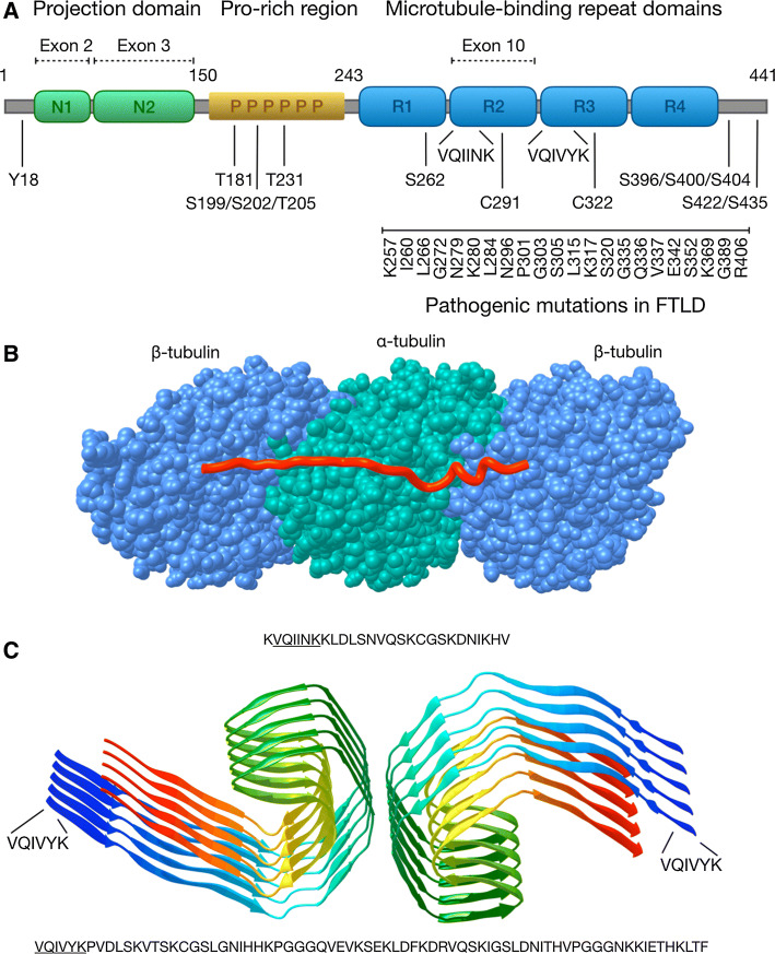
“Initially, four repeats in the microtubule-binding domains, each consisting of 18 amino acids with a Lys-Ile/Cys-Gly-Ser (KxGS) motif, were thought to be sufficient and necessary for the fibrillization of full-length tau (tau-FL) (Brunello et al., 2020).”
BackgroundGlucocorticoid stress hormones stimulate vesicle-free Tau secretion and spreading in the brain
Waites, C., Yu, Q., Du, F., Belli, I., Gomes, P. A., & Sotiropoulos, I. (2023) · Research Square · doi:10.21203/rs.3.rs-3097174/v1
Manthapuri, S. S., & Davis, E. (2021) · Journal of Student Research · doi:10.47611/jsrhs.v10i4.2468
“It is generally agreed that tau inclusions propagate along neuroanatomically connected brain regions, similar to prion diseases [10], [11], [12].”
BackgroundInfluentialIrwin, R., Faust, O., Petrovic, I., Wolf, S., Hofmann, H., & Rosenzweig, R. (2021) · bioRxiv · doi:10.1101/2021.04.11.439324
“In such cases, tau forms large intracellular aggregates, termed neurofibrillary tangles, whose abundance and localization in the brain correlates with cognitive decline (8, 9).”
BackgroundLiu, S., Hossinger, A., Hornberger, A., Buravlova, O., Müller, S. A., Lichtenthaler, S., Neumann, M., Denner, P., & Vorberg, I. (2020) · bioRxiv · doi:10.1101/2020.06.26.173070
Ruan, Z., Pathak, D., Kalavai, S., Yoshii-Kitahara, A., Muraoka, S., Takamatsu-Yukawa, K., Bhatt, N., Hu, J.-Q., Wang, Y., Hersh, S., et al. (2020) · doi:10.1101/2020.03.15.992719
Alzheimer's Disease Pathophysiology and Novel Treatment: An Update
Mo, A. (2024) · International Journal of Pharma Medicine and Biological Sciences · doi:10.18178/ijpmbs.13.1.30-38
“In humans, tau is encoded by the MAPT gene, which is situated on chromosome 17 [38].”
BackgroundLiu, Q.-Q., Ding, S.-K., & Shang, Y. Z. (2020) · International Journal of Medicinal Plants and Natural Products · doi:10.20431/2454-7999.0604001 · Cited by 1
Metadata
- Authors
- Brunello, C. A., Merezhko, M., Uronen, R.-L., & Huttunen, H. J.
- Affiliations
- University of Helsinki (FI)
- Corresponding author
- Huttunen, Henri J.
- Venue
- Cellular and Molecular Life Sciences
- Published
- 2019-10-30
- Volume
- 77
- Issue
- 9
- Pages
- 1721-1744
- ISSN
- 1420-682X (ISSN-L)
- 1420-9071
- PMC
- PMC7190606
- OpenAlex
- W2982627322
- Cited by
- 304
- References
- 267
- Citations by year
- 2026: 36 · 2025: 45 · 2024: 56 · 2023: 38 · 2022: 46 · 2021: 57 · 2020: 25 · 2019: 1
- FWCI
- 3.80
- Type
- journal article
- Language
- en
- Open access
- hybrid
- doi.org
- Locations
- Cellular and Molecular Life Sciences (publishedVersion, cc-by)
- PubMed Central (publishedVersion, cc-by)
- PubMed (publishedVersion)
- Työväentutkimus Vuosikirja (submittedVersion, cc-by)
- Funders
- Academy of Finland (296409)
- Alfred Kordelinin Säätiö
- Topics
- Life Sciences › Neuroscience › Cellular and Molecular Neuroscience › Alzheimer's disease research and treatments
- Life Sciences › Biochemistry, Genetics and Molecular Biology › Molecular Biology › Prion Diseases and Protein Misfolding
- Life Sciences › Neuroscience › Neurology › Neuroinflammation and Neurodegeneration Mechanisms
- Keywords
- tau protein, tauopathy, tau secretion, cell-to-cell transmission, prion-like propagation, tau aggregation, exosomes, ectosome, vesicular secretion, extracellular tau, endocytosis, phagocytosis, pinocytosis, Alzheimer's disease, frontotemporal lobar degeneration, neurodegeneration
- MeSH terms
- Animals, Humans, tau Proteins, Disease Progression, Tauopathies, Protein Aggregation, Pathological
About these checks
Registry, retraction, Cites and statistics results are computed, not judged, with one exception: a printed reference that no index lists is read once by a model before it counts as a citation error. Claims are extracted by a model and kept only when their quote appears verbatim in the text; each cited source is then checked, with a model’s help, for whether it says what the claim relies on. Those verdicts can be wrong — read the claim’s quote and the cited source before relying on one.
Statistics and arithmetic are recomputed, not judged.
Confidence bands are shown only for fields calibrated on our annotated test set. None is calibrated yet, so no band is shown.
What counts as a claim ›How we measure confidence ›How we check this ›
Are you an author, or do you see a problem with this page? Request a review
