

The DOI matches its Crossref record; 260 of the 267 references it cites were found in a registry or an index.
Mechanisms of secretion and spreading of pathological tau protein
Open accessAbstract
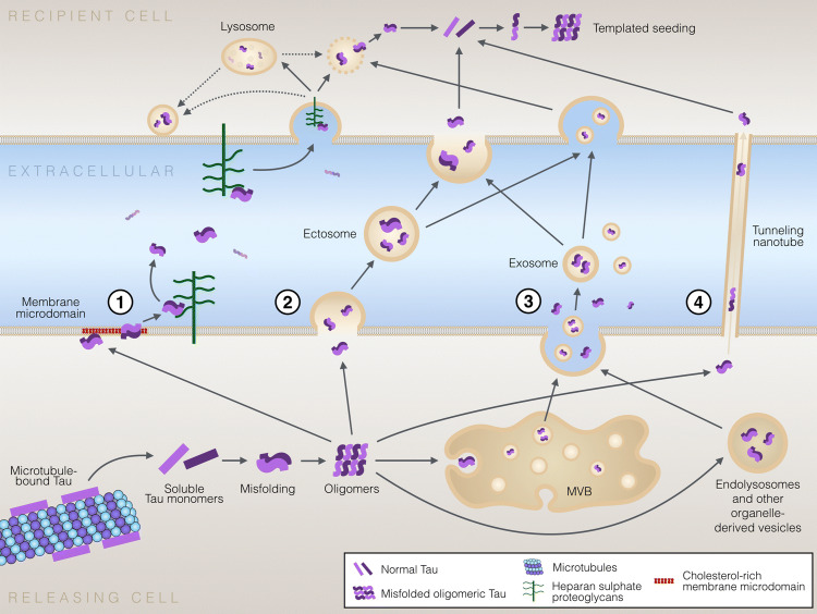
Accumulation of misfolded and aggregated forms of tau protein in the brain is a neuropathological hallmark of tauopathies, such as Alzheimer’s disease and frontotemporal lobar degeneration. Tau aggregates have the ability to transfer from one cell to another and to induce templated misfolding and aggregation of healthy tau molecules in previously healthy cells, thereby propagating tau pathology across different brain areas in a prion-like manner. The molecular mechanisms involved in cell-to-cell transfer of tau aggregates are diverse, not mutually exclusive and only partially understood. Intracellular accumulation of misfolded tau induces several mechanisms that aim to reduce the cellular burden of aggregated proteins and also promote secretion of tau aggregates. However, tau may also be released from cells physiologically unrelated to protein aggregation. Tau secretion involves multiple vesicular and non-vesicle-mediated pathways, including secretion directly through the plasma membrane. Consequently, extracellular tau can be found in various forms, both as a free protein and in vesicles, such as exosomes and ectosomes. Once in the extracellular space, tau aggregates can be internalized by neighboring cells, both neurons and glial cells, via endocytic, pinocytic and phagocytic mechanisms. Importantly, accumulating evidence suggests that prion-like propagation of misfolding protein pathology could provide a general mechanism for disease progression in tauopathies and other related neurodegenerative diseases. Here, we review the recent literature on cellular mechanisms involved in cell-to-cell transfer of tau, with a particular focus in tau secretion.
Overview
The claims and statistics of this paper haven’t been checked yet.
Run the check — free, no account needed. It reads the open-access text and checks the sources it cites.
Integrity signals
Registry
Cites
Statistics
Figures
Verification
Claims the paper makes
The claims and statistics of this paper haven’t been checked yet.
Run the check — free, no account needed. It reads the open-access text and checks the sources it cites.
Claims other papers make about this one
Each is checked against this paper’s own text: does it say what the citing paper says it does?
The source backs part of the claim, not all of it — or backs it for a different population, condition or comparison.
In the citing paper
“Thus, it is suggested that CSF p-tau biomarkers likely indicate an active process of tau secretion, and one that is correlated with cerebral Aβ deposition in early disease.20, 21, 22”
From Cerebrospinal fluid p-tau231 as an early indicator of emerging pathology in Alzheimer's disease (2022) · cites this paper as [22]
Experimental — accuracy not yet measured
Self-check
Percentages, sums and reported differences re-computed from the paper’s own numbers.
The self-check needs the paper’s full text.
Citations
Cites
Items 201–260 of 260
Characterization of tau prion seeding activity and strains from formaldehyde-fixed tissue
Kaufman, S. K., Thomas, T. L., Tredici, K. D., Braak, H., & Diamond, M. I. (2017) · Acta Neuropathologica Communications · doi:10.1186/s40478-017-0442-8 · Cited by 97
Characteristics and consequences of muscarinic receptor activation by tau protein
Gómez‐Ramos, A., Dı́az-Hernández, M., Rubio, A., Díaz‐Hernandéz, J. I., Miras‐Portugal, M. T., & Ávila, J. (2009) · European Neuropsychopharmacology · doi:10.1016/j.euroneuro.2009.04.006 · Cited by 95
TDP-43 expression influences amyloidβ plaque deposition and tau aggregation
Davis, S. A., Gan, K. A., Dowell, J. A., Cairns, N. J., & Gitcho, M. A. (2017) · Neurobiology of Disease · doi:10.1016/j.nbd.2017.04.012 · Cited by 95
Microtubule-associated proteins connect microtubules and neurofilaments in vitro
Aamodt, E. J., & Williams, R. C. (1984) · Biochemistry · doi:10.1021/bi00320a019 · Cited by 91
Internalized Tau sensitizes cells to stress by promoting formation and stability of stress granules
Brunello, C. A., Yan, X., & Huttunen, H. J. (2016) · Scientific Reports · doi:10.1038/srep30498 · Cited by 89
Self-citationEbert, A., Laußmann, M., Wegehingel, S., Kaderali, L., Erfle, H., Reichert, J., Lechner, J., Beer, H., Pepperkok, R., & Nickel, W. (2010) · Traffic · doi:10.1111/j.1600-0854.2010.01059.x · Cited by 89
Gauthier‐Kemper, A., Alonso, M. S., Sündermann, F., Niewidok, B., Fernández, M. P., Bakota, L., Heinisch, J. J., & Brandt, R. (2018) · Journal of Biological Chemistry · doi:10.1074/jbc.ra117.000490 · Cited by 87
Mechanism of Nucleated Conformational Conversion of Aβ42
Fu, Z., Aucoin, D., Davis, J., Nostrand, W. E. V., & Smith, S. O. (2015) · Biochemistry · doi:10.1021/acs.biochem.5b00467 · Cited by 83 · cited 1×
“In the second model, shared with PrP and Aβ [205] and called oligomer-nucleated conformational induction, monomers are not directly assembled into bigger aggregates, but they oligomerize before being incorporated into fibrils [206].”
BackgroundKünze, G., Barré, P., Scheidt, H. A., Thomas, L., Eliezer, D., & Huster, D. (2012) · Biochimica et Biophysica Acta (BBA) - Biomembranes · doi:10.1016/j.bbamem.2012.03.019 · Cited by 81
APOE , MAPT , and COMT and Parkinson’s Disease Susceptibility and Cognitive Symptom Progression
Paul, K. C., Rausch, R., Creek, M. M., Sinsheimer, J. S., Bronstein, J. M., Bordelon, Y. M., & Ritz, B. R. (2016) · Journal of Parkinson s Disease · doi:10.3233/jpd-150762 · Cited by 79
Lasagna‐Reeves, C. A., Sengupta, U., Castillo‐Carranza, D. L., Gerson, J. E., Guerrero‐Munoz, M. J., Troncoso, J. C., Jackson, G. R., & Kayed, R. (2014) · Acta Neuropathologica Communications · doi:10.1186/2051-5960-2-56 · Cited by 78
Neuronal Exosome-Derived Human Tau is Toxic to Recipient Mouse Neurons in vivo
Winston, C. N., Aulston, B. D., Rockenstein, E. M., Adame, A., Prikhodko, O., Dave, K. N., Mishra, P., Rissman, R. A., & Yuan, S. H. (2018) · Journal of Alzheimer s Disease · doi:10.3233/jad-180776 · Cited by 77
SOD1 protein aggregates stimulate macropinocytosis in neurons to facilitate their propagation
Zeineddine, R., Pundavela, J., Corcoran, L., Stewart, E. M., Do‐Ha, D., Bax, M., Guillemin, G. J., Vine, K. L., Hatters, D. M., Ecroyd, H. W., et al. (2015) · Molecular Neurodegeneration · doi:10.1186/s13024-015-0053-4 · Cited by 75
Mercken, M. H., Grynspan, F., & Nixon, R. A. (1995) · FEBS Letters · doi:10.1016/0014-5793(95)00590-6 · Cited by 74
Identification of disulfide cross-linked tau dimer responsible for tau propagation
Kim, D., Lim, S., Haque, M. M., Ryoo, N., Hong, H., Rhim, H., Lee, D., Chang, Y., Lee, J., Cheong, E., et al. (2015) · Scientific Reports · doi:10.1038/srep15231 · Cited by 74
Plasma neuronal exosomal levels of Alzheimer's disease biomarkers in normal aging
Abner, E. L., Jicha, G. A., Shaw, L. M. J., Trojanowski, J. Q., & Goetzl, E. J. (2016) · Annals of Clinical and Translational Neurology · doi:10.1002/acn3.309 · Cited by 73
Tau interacts with Golgi membranes and mediates their association with microtubules
Farah, C. A., Perreault, S., Liazoghli, D., Desjardins, M., Antón, A., Lauzon, M., Paiement, J. M., & Leclerc, N. M. (2006) · Cell Motility and the Cytoskeleton · doi:10.1002/cm.20157 · Cited by 73
Barré, P., & Eliezer, D. (2013) · Protein Science · doi:10.1002/pro.2290 · Cited by 72
Arima, K., Mizutani, T., Alim, A., Tonozuka-Uehara, H., Izumiyama, Y., Hirai, S., & Uéda, K. (2000) · Acta Neuropathologica · doi:10.1007/s004010050002 · Cited by 71
Folding of the Repeat Domain of Tau Upon Binding to Lipid Surfaces
Barré, P., & Eliezer, D. (2006) · Journal of Molecular Biology · doi:10.1016/j.jmb.2006.07.018 · Cited by 69
Huynh, M. B., Ouidja, M. O., Chantepie, S., Carpentier, G., Maïza, A., Zhang, G., Vilares, J., Raisman-Vozari, R., & Papy-Garcia, D. (2019) · PLOS ONE · doi:10.1371/journal.pone.0209573 · Cited by 69
Lee, S., Kim, W., Li, Z., & Hall, G. F. (2012) · International Journal of Alzheimer s Disease · doi:10.1155/2012/172837 · Cited by 67
Matsumoto, S.-E., Motoi, Y., Ishiguro, K., Tabira, T., Kametani, F., Hasegawa, M., & Hattori, N. (2015) · Human Molecular Genetics · doi:10.1093/hmg/ddv351 · Cited by 66
Rodríguez, L. C. D. L. C., Mohamed, N., Desjardins, A., Lippé, R., Fon, E. A., & Leclerc, N. M. (2017) · Journal of Neurochemistry · doi:10.1111/jnc.13994 · Cited by 66
Formation, release, and internalization of stable tau oligomers in cells
Wegmann, S., Nicholls, S. B., Takeda, S., Fan, Z., & Hyman, B. T. (2016) · Journal of Neurochemistry · doi:10.1111/jnc.13866 · Cited by 66
Sharma, A. M., Thomas, T. L., Woodard, D. R., Kashmer, O. M., & Diamond, M. I. (2018) · bioRxiv · doi:10.1101/325043 · Cited by 64
The interaction of α-synuclein and Tau: A molecular conspiracy in neurodegeneration?
Xu, Y., Uronen, R., & Huttunen, H. J. (2018) · Seminars in Cell and Developmental Biology · doi:10.1016/j.semcdb.2018.05.005 · Cited by 64 · cited 3×
“Apart from the potential gain-of-toxic-function relationship that feeds forward the aggregation of each other, tau and α-synuclein may also overlap in their loss-of-function effects via a partially shared protein interactome [68].”
BackgroundInfluentialSelf-citationCroft, C. L., Wade, M. A., Kurbatskaya, K., Mastrandreas, P., Hughes, M. M., Phillips, E. C., Pooler, A. M., Perkinton, M. S., Hanger, D. P., & Noble, W. J. (2017) · Cell Death and Disease · doi:10.1038/cddis.2017.97 · Cited by 62
Novel human neuronal tau model exhibiting neurofibrillary tangles and transcellular propagation
Reilly, P., Winston, C. N., Baron, K. R., Trejo, M., Rockenstein, E. M., Akers, J. C., Kfoury, N., Diamond, M. I., Masliah, E., Rissman, R. A., et al. (2017) · Neurobiology of Disease · doi:10.1016/j.nbd.2017.06.005 · Cited by 61
MAPT S305I mutation: implications for argyrophilic grain disease
Kovács, G., Pittman, A., Révész, T., Luk, C., Lees, A., Kiss, É., Tariska, P., Laszlo, L., Molnár, K., Molnár, M., et al. (2008) · Acta Neuropathologica · doi:10.1007/s00401-007-0322-6 · Cited by 59
Wang, X.-S., Wang, D., Zhao, J., Qu, M.-H., Zhou, X.-H., He, H., & He, R.-Q. (2006) · Protein Peptide Letters · doi:10.2174/092986606777790566 · Cited by 57
Müller, H., Steringer, J. P., Wegehingel, S., Bleicken, S., Münster, M., Dimou, E., Unger, S., Weidmann, G., Andreas, H., García‐Sáez, A. J., et al. (2015) · Journal of Biological Chemistry · doi:10.1074/jbc.m114.622456 · Cited by 56
N-terminal Truncation of Microtubule Associated Protein Tau Dysregulates its Cellular Localization
Paholikova, K., Šalingová, B., Opattová, A., Škrabana, R., Majerová, P., Žilka, N., Kováčech, B., Žilková, M., Baráth, P., & Novák, M. (2014) · Journal of Alzheimer s Disease · doi:10.3233/jad-140996 · Cited by 55
Ion Channel Formation by Tau Protein: Implications for Alzheimer’s Disease and Tauopathies
Patel, N., Ramachandran, S., Azimov, R., Kagan, B. L., & Lal, R. (2015) · Biochemistry · doi:10.1021/acs.biochem.5b00988 · Cited by 53
Steringer, J. P., & Nickel, W. (2018) · Seminars in Cell and Developmental Biology · doi:10.1016/j.semcdb.2018.02.010 · Cited by 53 · cited 1×
“Interestingly, many of these mechanistic aspects of tau secretion have significant similarity to the unconventional secretion of fibroblast growth factor 2 (FGF2), one of the most widely studied unconventionally secreted proteins [160].”
BackgroundAutophagy-Mediated Secretory Pathway is Responsible for Both Normal and Pathological Tau in Neurons
Kang, S., Son, S. M., Baik, S. H., Yang, J., & Mook‐Jung, I. (2019) · Journal of Alzheimer s Disease · doi:10.3233/jad-190180 · Cited by 52
Pedrini, S., Thomas, C., Brautigam, H., Schmeidler, J., Ho, L., Fraser, P., Westaway, D. A., George‐Hyslop, P. S., Martins, R. N., Buxbaum, J. D., et al. (2009) · Molecular Neurodegeneration · doi:10.1186/1750-1326-4-40 · Cited by 50
Spitzer, P., Mulzer, L., Oberstein, T. J., Munoz, L. E., Lewczuk, P., Kornhuber, J., i, M. H., & Maler, J. M. (2019) · Scientific Reports · doi:10.1038/s41598-019-43607-7 · Cited by 49
Parkinsonism–dementia complex of Guam
Steele, J. C. (2005) · Movement Disorders · doi:10.1002/mds.20547 · Cited by 46
FRMD4A–cytohesin signaling modulates the cellular release of tau
Yan, X., Nykänen, N.-P., Brunello, C. A., Haapasalo, A., Hiltunen, M. O., Uronen, R., & Huttunen, H. J. (2016) · Journal of Cell Science · doi:10.1242/jcs.180745 · Cited by 43 · cited 2×
“Several studies suggest that while a fraction of the total extracellular tau is indeed found in exosomes or ectosomes, a large fraction of the extracellular tau pool exists in vesicle-free form [149, 155, 157, 158] (Table 2).”
BackgroundSelf-citationCamero, S., Benítez, M. J., Barrantes, A., Ayuso, J. M., Cuadros, R., Ávila, J., & Jiménez, J. S. (2014) · Journal of Alzheimer s Disease · doi:10.3233/jad-131415 · Cited by 42
Thermodynamics of the Interaction between Alzheimer's Disease Related Tau Protein and DNA
Camero, S., Benítez, M. J., Cuadros, R., Hernández, F., Ávila, J., & Jiménez, J. S. (2014) · PLoS ONE · doi:10.1371/journal.pone.0104690 · Cited by 40
Gibb, W., Luthert, P., & Marsden, C. (1989) · Brain : a journal of neurology · doi:10.1093/brain/112.5.1171 · Cited by 39
Mari, S. A., Wegmann, S., Tepper, K., Hyman, B. T., Mandelkow, E., Mandelkow�, E., & Müller, D. J. (2018) · Nano Letters · doi:10.1021/acs.nanolett.8b01085 · Cited by 39
Sahara, N., Murayama, M., Higuchi, M., Suhara, T., & Takashima, A. (2014) · Frontiers in Neurology · doi:10.3389/fneur.2014.00026 · Cited by 37 · cited 2×
“Furthermore, in animal models pathological tau appears to be more localized to synapses compared to non-pathological tau [63], and synaptosomes isolated from human AD brains were shown to contain more phosphorylated and aggregated tau than in healthy controls [64].”
BackgroundVenuta, G. L., Wegehingel, S., Sehr, P. A., Müller, H., Dimou, E., Steringer, J. P., Grotwinkel, M., Hentze, N., Mayer, M. P., Will, D. W., et al. (2016) · Journal of Biological Chemistry · doi:10.1074/jbc.m116.729384 · Cited by 37
Suppression of tau propagation using an inhibitor that targets the DK-switch of nSMase2
Bilousova, T. V., Elias, C. J., Miyoshi, E., Alam, M. P., Zhu, C., Campagna, J. J., Vadivel, K., Jagodzinska, B. M., Gylys, K. H., & John, V. (2018) · Biochemical and Biophysical Research Communications · doi:10.1016/j.bbrc.2018.03.209 · Cited by 35
Takeda, T. (2017) · Neuropathology · doi:10.1111/neup.12428 · Cited by 34
Bertrand, A., Khan, U., Hoang, D. M., Novikov, D. S., Krishnamurthy, P., Sait, H. B. R., Little, B. W., Sigurdsson, E. M., & Wadghiri, Y. Z. (2012) · NeuroImage · doi:10.1016/j.neuroimage.2012.08.065 · Cited by 33
The Roles of Endo-Lysosomes in Unconventional Protein Secretion
Lee, J., & Ye, Y. (2018) · Cells · doi:10.3390/cells7110198 · Cited by 32
Glypican‐1 facilitates prion conversion in lipid rafts
Hooper, N. M. (2010) · Journal of Neurochemistry · doi:10.1111/j.1471-4159.2010.06936.x · Cited by 29
Distinct Microglial Responses in Two Transgenic Murine Models of TAU Pathology
Romero‐Molina, C., Navarro, V., Sanchez-Varo, R. M., Jiménez, S., Fernandez-Valenzuela, J. J., Sanchez-Mico, M. V., Muñoz‐Castro, C., Gutiérrez, A., Vitórica, J., & Vizuete, M. (2018) · Frontiers in Cellular Neuroscience · doi:10.3389/fncel.2018.00421 · Cited by 25
Cardona‐Gómez, G. P., Arango-Dávila, C. A., Gallego-Gómez, J. C., Barrera-Ocampo, A. A., Pimienta, H. J., & García‐Segura, L. M. (2006) · Neuroreport · doi:10.1097/01.wnr.0000230508.78467.96 · Cited by 25 · cited 1×
“It is also possible that tau localized at the PM occupies a role as a signaling regulator, via its interaction with membrane receptors [19, 20].”
BackgroundWang, B., & Han, S. (2018) · Molecular and Cellular Neuroscience · doi:10.1016/j.mcn.2018.02.002 · Cited by 23
Phosphorylation Pattern of tau Associated with Distinct Changes of the Growth Cone Cytoskeleton
Šimić, G., Diana, A., & Hof, P. R. (2003) · Progress in molecular and subcellular biology · doi:10.1007/978-3-642-55557-2_2 · Cited by 17
Dicke, S. S., Tatge, L., Engen, P., Culp, M., & Masterson, L. R. (2017) · Biochemical and Biophysical Research Communications · doi:10.1016/j.bbrc.2017.10.007 · Cited by 13
Lonati, E., Sala, G., Tresoldi, V., Coco, S., Salerno, D., Milani, C., Losurdo, M., Farina, F., Botto, L., Ferrarese, C., et al. (2018) · Journal of Molecular Neuroscience · doi:10.1007/s12031-018-1199-7 · Cited by 12
Inert and seed-competent tau monomers suggest structural origins of aggregationPreprint
Mirbaha, H., Chen, D., Morozova, O. A., Ruff, K. M., Sharma, A., Liu, X., Pappu, R. V., Colby, D. W., Mirzaei, H., Joachimiak, Ł. A., et al. (2017) · bioRxiv (Cold Spring Harbor Laboratory) · doi:10.1101/163394 · Cited by 10
Systems biology of the secretory pathway: What have we learned so far?
Farhan, H. (2015) · Biology of the Cell · doi:10.1111/boc.201400065 · Cited by 10 · cited 1×
“Conventional secretion mechanisms include the classical secretory route, also known as endoplasmic reticulum (ER)–Golgi pathway, for proteins that carry a signal peptide, which directs them to the ER membrane [141].”
BackgroundS3‐03‐05: Transmission and Spreading of Tauopathy in Transgenic Mouse Brain
Tolnay, M. (2011) · Alzheimer s & Dementia · doi:10.1016/j.jalz.2011.05.2444
Research works printed but not linked: 3
- Liu F, Gong CX (2008) Tau exon 10 alternative splicing and tauopathies. Mol Neurodegener 3:8 · DOI · Paper page
- Grundke-Iqbal I, Iqbal K, Tung YC, Quinlan M, Wisniewski HM, Binder LI (1986) Abnormal phosphorylation of the microtubule-associated protein tau (tau) in Alzheimer cytoskeletal pathology. Proc Natl Acad Sci USA 83(13):4913–4917 · DOI · Paper page
- Yamada K et al (2014) Neuronal activity regulates extracellular tau in vivo. J Exp Med 211(3):387–393 · DOI
Cited by
Sorted by citations
Phosphorylated Tau in Alzheimer’s Disease and Other Tauopathies
Rawat, P., Sehar, U., Bisht, J., Selman, A., Culberson, J., & Reddy, P. H. (2022) · International Journal of Molecular Sciences · doi:10.3390/ijms232112841 · Cited by 509
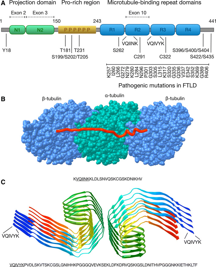
“At the protein level, K257T, G272V, P301L, V337M, and R406W mutations alter the binding properties of the microtubule, and N279K, N296H, P301S, S305I mutations at the RNA level shifts the splicing towards overproduction of 4R tau [97].”
BackgroundThe expanding amyloid family: Structure, stability, function, and pathogenesis
Sawaya, M. R., Hughes, M. P., Rodriguez, J. A., Riek, R., & Eisenberg, D. S. (2021) · Cell · doi:10.1016/j.cell.2021.08.013 · Cited by 475
Role of tau protein in Alzheimer’s disease: The prime pathological player
Muralidar, S., Ambi, S. V., Sekaran, S., Thirumalai, D., & Palaniappan, B. (2020) · International Journal of Biological Macromolecules · doi:10.1016/j.ijbiomac.2020.07.327 · Cited by 278
Alzheimer’s disease brain-derived extracellular vesicles spread tau pathology in interneurons
Ruan, Z., Pathak, D., Venkatesan Kalavai, S., Yoshii-Kitahara, A., Muraoka, S., Bhatt, N., Takamatsu-Yukawa, K., Hu, J., Wang, Y., Hersh, S., et al. (2021) · Brain · doi:10.1093/brain/awaa376 · Cited by 283
“secreted primarily in free form, while a minor fraction of tau is found in EVs as observed in the CSF or blood of control subjects and Alzheimer’s disease patients (Arai et al., 1998; Saman et al., 2012; Zetterberg et al., 2013; Fiandaca et al., 2015; Ruan and Ikezu, 2019; Brunello et al., 2020).”
BackgroundAcetylated tau inhibits chaperone-mediated autophagy and promotes tau pathology propagation in mice
Caballero, B., Bourdenx, M., Luengo, E., Diaz, A., Sohn, P. D., Chen, X., Wang, C., Juste, Y. R., Wegmann, S., Patel, B., et al. (2021) · Nature Communications · doi:10.1038/s41467-021-22501-9 · Cited by 223
Metadata
- Authors
- Brunello, C. A., Merezhko, M., Uronen, R.-L., & Huttunen, H. J.
- Affiliations
- University of Helsinki (FI)
- Corresponding author
- Huttunen, Henri J.
- Venue
- Cellular and Molecular Life Sciences
- Published
- 2019-10-30
- Volume
- 77
- Issue
- 9
- Pages
- 1721-1744
- ISSN
- 1420-682X (ISSN-L)
- 1420-9071
- PMC
- PMC7190606
- OpenAlex
- W2982627322
- Cited by
- 304
- References
- 267
- Citations by year
- 2026: 36 · 2025: 45 · 2024: 56 · 2023: 38 · 2022: 46 · 2021: 57 · 2020: 25 · 2019: 1
- FWCI
- 3.80
- Type
- journal article
- Language
- en
- Open access
- hybrid
- doi.org
- Locations
- Cellular and Molecular Life Sciences (publishedVersion, cc-by)
- PubMed Central (publishedVersion, cc-by)
- PubMed (publishedVersion)
- Työväentutkimus Vuosikirja (submittedVersion, cc-by)
- Funders
- Academy of Finland (296409)
- Alfred Kordelinin Säätiö
- Topics
- Life Sciences › Neuroscience › Cellular and Molecular Neuroscience › Alzheimer's disease research and treatments
- Life Sciences › Biochemistry, Genetics and Molecular Biology › Molecular Biology › Prion Diseases and Protein Misfolding
- Life Sciences › Neuroscience › Neurology › Neuroinflammation and Neurodegeneration Mechanisms
- Keywords
- tau protein, tauopathy, tau secretion, cell-to-cell transmission, prion-like propagation, tau aggregation, exosomes, ectosome, vesicular secretion, extracellular tau, endocytosis, phagocytosis, pinocytosis, Alzheimer's disease, frontotemporal lobar degeneration, neurodegeneration
- MeSH terms
- Animals, Humans, tau Proteins, Disease Progression, Tauopathies, Protein Aggregation, Pathological
About these checks
Registry, retraction, Cites and statistics results are computed, not judged, with one exception: a printed reference that no index lists is read once by a model before it counts as a citation error. Claims are extracted by a model and kept only when their quote appears verbatim in the text; each cited source is then checked, with a model’s help, for whether it says what the claim relies on. Those verdicts can be wrong — read the claim’s quote and the cited source before relying on one.
Statistics and arithmetic are recomputed, not judged.
Confidence bands are shown only for fields calibrated on our annotated test set. None is calibrated yet, so no band is shown.
What counts as a claim ›How we measure confidence ›How we check this ›
Are you an author, or do you see a problem with this page? Request a review
