Cited by 3Genomics and Chromatin DynamicsChromatin Remodeling and CancerFungal and yeast genetics research

The DOI matches its Crossref record; 2 of the 2 references it cites were found in a registry or an index.

Transcriptional activation domains interact with ATPase subunits of yeast chromatin remodelling complexes SWI/SNF, RSC and INO80

Open access

Published version of a preprint

Abstract

Chromatin remodelling complexes (CRC) are ATP-dependent molecular machines important for the dynamic organization of nucleosomes along eukaryotic DNA. CRCs SWI/SNF, RSC and INO80 can move positioned nucleosomes in promoter DNA, leading to nucleosome-depleted regions which facilitate access of general transcription factors. This function is strongly supported by transcriptional activators being able to interact with subunits of various CRCs. In this work we show that SWI/SNF subunits Swi1, Swi2, Snf5 and Snf6 can bind to activation domains of Ino2 required for expression of phospholipid biosynthetic genes in yeast. We identify an activator binding domain (ABD) of ATPase Swi2 and show that this ABD is functionally dispensable, presumably because ABDs of other SWI/SNF subunits can compensate for the loss. In contrast, mutational characterization of the ABD of the Swi2-related ATPase Sth1 revealed that some conserved basic and hydrophobic amino acids within this domain are essential for the function of Sth1. While ABDs of Swi2 and Sth1 define separate functional protein domains, mapping of an ABD within ATPase Ino80 showed co-localization with its HSA domain also required for binding actin-related proteins. Comparative interaction studies finally demonstrated that several unrelated activators each exhibit a specific binding pattern with ABDs of Swi2, Sth1 and Ino80.

Overview

The claims and statistics of this paper haven’t been checked yet.

Run the check — free, no account needed. It reads the open-access text and checks the sources it cites.

A few free checks a day · papers already verified open instantly · results are public

Integrity signals

Registry

Crossref · 10.1007/s00294-024-01300-x

Cites

2 of 2 references verified0 citation errors · 0 not checkable

Statistics

Figures

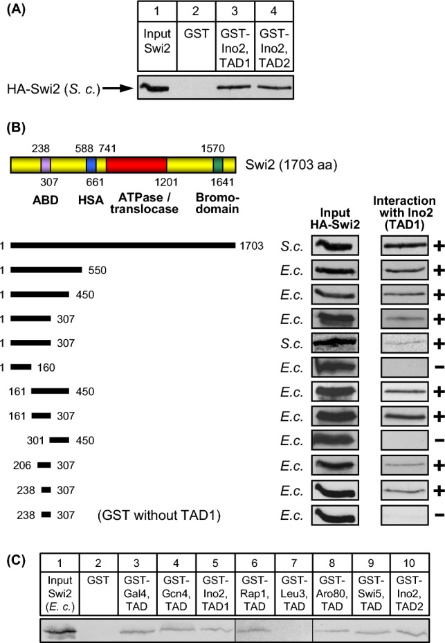
Fig. 1 In vitro interaction assays (GST pull-down) with length variants of Swi2 and various activation domains. A Swi2 interacts with both Ino2 activation domains. Fusion proteins GST-Ino2 TAD1 (aa 1–35; encoded by pSH117) and GST-Ino2 TAD2 (aa 101–135; pSH 118) were synthesized in E. coli, immobilized on GSH sepharose and incubated with yeast protein extracts containing epitope-tagged Swi2 (full-length; synthesized by yeast expression plasmid pES17). B Mapping of Swi2 domains interacting with TAD1 of Ino2. The following expression plasmids were used to synthesize HA-Swi2 variants in E. coli strain BL21 CodonPlus: pMG142 (aa 1–550), pMG140 (aa 1–450), pIB2 (aa 1–307), pIB1 (aa 1–160), pIB3 (aa 161–450), pIB4 (aa 161–307), pIB5 (aa 301–450), pIB9 (aa 206–307) and pIB12 (aa 238–307). Length variant aa 1–307 was also synthesized in S. cerevisiae, using yeast expression plasmid pECW41. ABD, activator binding domain; HSA, helicase/SANT-associated domain. ATPase RecA subdomains are not shown individually. Functional domains SnAC (Snf2 ATP coupling, aa 1302–1369) and AT-hook (aa 1446–1458 and aa 1518–1530) are omitted from the figure. C TADs of various unrelated transcriptional activators interact with Swi2. GST fusions of Ino2 TAD1 (aa 1–35; pSH117), Ino2 TAD2 (aa 101–135; pSH118), Gal4 TAD (aa 768–881; pES20), Gcn4 TAD (aa 9–172; pKH60), Leu3 TAD (aa 841–886; pES5), Rap1 TAD (aa 630–671; pLJ6), Aro80 TAD (aa 846–950; pMG50) and Swi5 TAD (aa 1–85; pDG1) were synthesized in E. coli, immobilized on GSH sepharose and incubated with bacterial protein extract containing epitope-tagged Swi2 length variant aa 161–307 (pIB4). Empty GST plasmid pGEX-2TK was used as a negative control
Fig. 1 In vitro interaction assays (GST pull-down) with length variants of Swi2 and various activation domains. A Swi2 interacts with both Ino2 activation domains. Fusion proteins GST-Ino2 TAD1 (aa 1–35; encoded by pSH117) and GST-Ino2 TAD2 (aa 101–135; pSH 118) were synthesized in E. coli, immobilized on GSH sepharose and incubated with yeast protein extracts containing epitope-tagged Swi2 (full-length; synthesized by yeast expression plasmid pES17). B Mapping of Swi2 domains interacting with TAD1 of Ino2. The following expression plasmids were used to synthesize HA-Swi2 variants in E. coli strain BL21 CodonPlus: pMG142 (aa 1–550), pMG140 (aa 1–450), pIB2 (aa 1–307), pIB1 (aa 1–160), pIB3 (aa 161–450), pIB4 (aa 161–307), pIB5 (aa 301–450), pIB9 (aa 206–307) and pIB12 (aa 238–307). Length variant aa 1–307 was also synthesized in S. cerevisiae, using yeast expression plasmid pECW41. ABD, activator binding domain; HSA, helicase/SANT-associated domain. ATPase RecA subdomains are not shown individually. Functional domains SnAC (Snf2 ATP coupling, aa 1302–1369) and AT-hook (aa 1446–1458 and aa 1518–1530) are omitted from the figure. C TADs of various unrelated transcriptional activators interact with Swi2. GST fusions of Ino2 TAD1 (aa 1–35; pSH117), Ino2 TAD2 (aa 101–135; pSH118), Gal4 TAD (aa 768–881; pES20), Gcn4 TAD (aa 9–172; pKH60), Leu3 TAD (aa 841–886; pES5), Rap1 TAD (aa 630–671; pLJ6), Aro80 TAD (aa 846–950; pMG50) and Swi5 TAD (aa 1–85; pDG1) were synthesized in E. coli, immobilized on GSH sepharose and incubated with bacterial protein extract containing epitope-tagged Swi2 length variant aa 161–307 (pIB4). Empty GST plasmid pGEX-2TK was used as a negative controlCC BY · PubMed Central
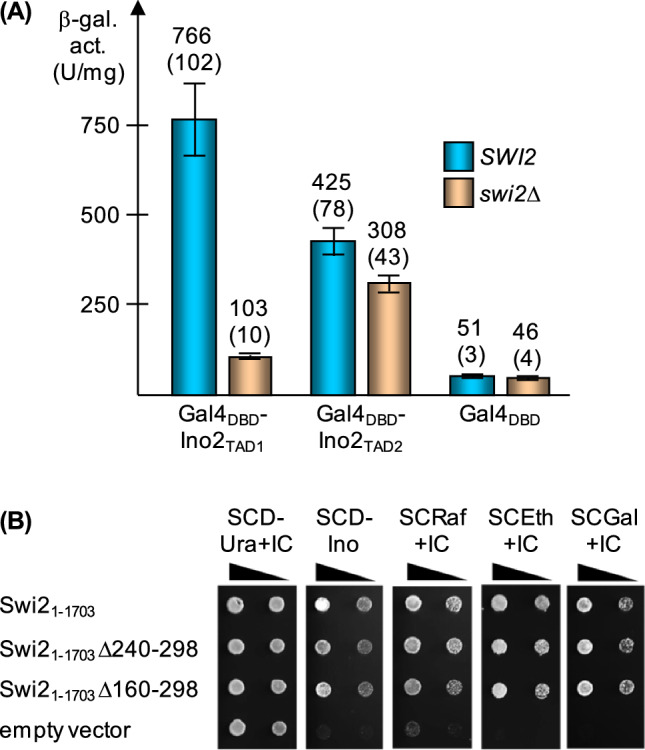
Fig. 2 Influence of SWI2 on gene activation by Ino2. A Expression of a GAL1-lacZ reporter gene in the presence of Gal4DBD-Ino2 hybrid activators. Isogenic strains EWY3 (SWI2 gal4Δ) and EWY4 (swi2Δ gal4Δ) were doubly transformed with reporter plasmid pKH29 (GAL1-lacZ) and effector plasmids pSG14 (GAL4DBD-INO2TAD1), pSG16 (GAL4DBD-INO2TAD2) and pGBD-C1 (GAL4DBD, negative control, basal expression), respectively. Transformants were grown in selective medium under derepressing conditions (0.2% glucose + 1% lactate) until mid-log phase. Specific activities of β-galactosidase are given in U per mg of protein. Activation in vivo was assayed by performing three independent transformations of both strains, using four colonies for each repetition (12 individual enzyme assays). Standard deviations are indicated by error bars and given in parentheses. B Functional complementation of swi2 mutant phenotypes by SWI2 variants devoid of sequences encoding the activator binding domain (ABD). S. cerevisiae strain JS94.8-12 (relevant genotype: ura3 swi2Δ::HIS3) was transformed with plasmids pECW24 (ARS CEN URA3 MET25Pr-SWI2; wild-type), pECW28 (MET25Pr-SWI2Δ240-298), pECW30 (MET25Pr-SWI2Δ160-298) and p416-MET25 (empty vector), respectively. Serial strain dilutions were spotted on the media shown and incubated for one (SCD-Ura + IC), two (SCD-Ino, SCRaf + IC and SCGal + IC) or three days (SCEth + IC). IC, inositol + choline; Ino, inositol; Raf, raffinose; Eth, ethanol; Gal, galactose
Fig. 2 Influence of SWI2 on gene activation by Ino2. A Expression of a GAL1-lacZ reporter gene in the presence of Gal4DBD-Ino2 hybrid activators. Isogenic strains EWY3 (SWI2 gal4Δ) and EWY4 (swi2Δ gal4Δ) were doubly transformed with reporter plasmid pKH29 (GAL1-lacZ) and effector plasmids pSG14 (GAL4DBD-INO2TAD1), pSG16 (GAL4DBD-INO2TAD2) and pGBD-C1 (GAL4DBD, negative control, basal expression), respectively. Transformants were grown in selective medium under derepressing conditions (0.2% glucose + 1% lactate) until mid-log phase. Specific activities of β-galactosidase are given in U per mg of protein. Activation in vivo was assayed by performing three independent transformations of both strains, using four colonies for each repetition (12 individual enzyme assays). Standard deviations are indicated by error bars and given in parentheses. B Functional complementation of swi2 mutant phenotypes by SWI2 variants devoid of sequences encoding the activator binding domain (ABD). S. cerevisiae strain JS94.8-12 (relevant genotype: ura3 swi2Δ::HIS3) was transformed with plasmids pECW24 (ARS CEN URA3 MET25Pr-SWI2; wild-type), pECW28 (MET25Pr-SWI2Δ240-298), pECW30 (MET25Pr-SWI2Δ160-298) and p416-MET25 (empty vector), respectively. Serial strain dilutions were spotted on the media shown and incubated for one (SCD-Ura + IC), two (SCD-Ino, SCRaf + IC and SCGal + IC) or three days (SCEth + IC). IC, inositol + choline; Ino, inositol; Raf, raffinose; Eth, ethanol; Gal, galactoseCC BY · PubMed Central
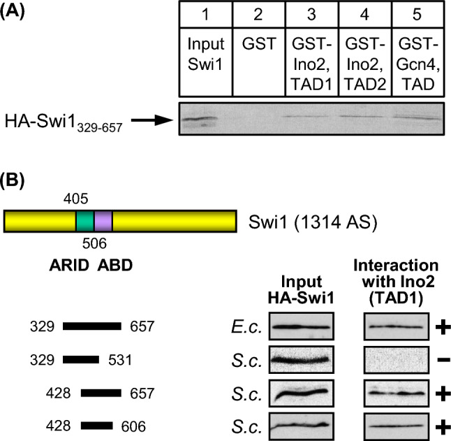
Fig. 3 Swi1 interacts with TAD1 and TAD2 of Ino2. A HA-tagged Swi1 length variant aa 329–657 was synthesized in S. cerevisiae (expression plasmid pECW39) and assayed for binding to GST fusions with Ino2 TAD1 (pSH117), Ino2 TAD2 (pSH118) and Gcn4 TAD (pKH60; positive control). B Swi1 truncations were assayed for binding to GST-Ino2 TAD1 (pSH117). Expression plasmids pJuLu1 (aa 329–657), pMaS5 (aa 329–531), pMaS7 (aa 428–657) and pLM3 (aa 428–606) were used for synthesis of Swi1 variants in E. coli (E.c.) or in S. cerevisiae (S.c.). ABD, activator binding domain; ARID, AT-rich interaction domain
Fig. 3 Swi1 interacts with TAD1 and TAD2 of Ino2. A HA-tagged Swi1 length variant aa 329–657 was synthesized in S. cerevisiae (expression plasmid pECW39) and assayed for binding to GST fusions with Ino2 TAD1 (pSH117), Ino2 TAD2 (pSH118) and Gcn4 TAD (pKH60; positive control). B Swi1 truncations were assayed for binding to GST-Ino2 TAD1 (pSH117). Expression plasmids pJuLu1 (aa 329–657), pMaS5 (aa 329–531), pMaS7 (aa 428–657) and pLM3 (aa 428–606) were used for synthesis of Swi1 variants in E. coli (E.c.) or in S. cerevisiae (S.c.). ABD, activator binding domain; ARID, AT-rich interaction domainCC BY · PubMed Central
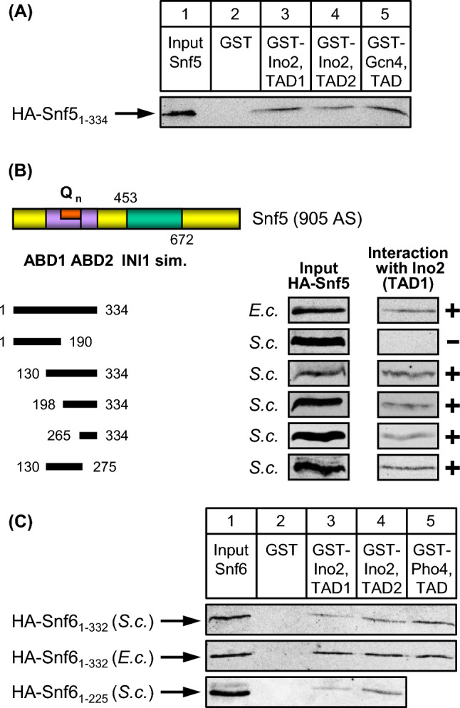
Fig. 4 Snf5 and Snf6 interact with TAD1 and TAD2 of Ino2. A HA-tagged Snf5 length variant aa 1–334 was synthesized in S. cerevisiae (expression plasmid pECW40) and assayed for binding to GST fusions with Ino2 TAD1 (pSH117), Ino2 TAD2 (pSH118) and Gcn4 TAD (pKH60; positive control). B Snf5 truncations were assayed for binding to GST-Ino2 TAD1 (pSH117). Expression plasmids pJuLu2 (aa 1–334), pMaS6 (aa 1–190), pECW49 (aa 130–334), pECW50 (aa 198–334), pLM1 (aa 265–334) and pLM2 (aa 130–275) were used to synthesize Snf5 variants in E. coli (E.c.) or in S. cerevisiae (S.c.). ABD, activator binding domains; Qn, Glutamine-rich region; INI1 sim., region similar to human tumor suppressor INI1. C Snf6 interacts with activators Ino2 and Pho4. Full-length HA-Snf6 was synthesized in S. cerevisiae and in E. coli (expression plasmids pKB1 and pKB5, respectively) and assayed for binding to GST fusions with Ino2 TAD1 (pSH117), Ino2 TAD2 (pSH118) and Pho4 TAD (aa 1–104, pRAR62; positive control). Snf6 truncation variant aa 1–225 (pECW65) was synthesized in S. cerevisiae
Fig. 4 Snf5 and Snf6 interact with TAD1 and TAD2 of Ino2. A HA-tagged Snf5 length variant aa 1–334 was synthesized in S. cerevisiae (expression plasmid pECW40) and assayed for binding to GST fusions with Ino2 TAD1 (pSH117), Ino2 TAD2 (pSH118) and Gcn4 TAD (pKH60; positive control). B Snf5 truncations were assayed for binding to GST-Ino2 TAD1 (pSH117). Expression plasmids pJuLu2 (aa 1–334), pMaS6 (aa 1–190), pECW49 (aa 130–334), pECW50 (aa 198–334), pLM1 (aa 265–334) and pLM2 (aa 130–275) were used to synthesize Snf5 variants in E. coli (E.c.) or in S. cerevisiae (S.c.). ABD, activator binding domains; Qn, Glutamine-rich region; INI1 sim., region similar to human tumor suppressor INI1. C Snf6 interacts with activators Ino2 and Pho4. Full-length HA-Snf6 was synthesized in S. cerevisiae and in E. coli (expression plasmids pKB1 and pKB5, respectively) and assayed for binding to GST fusions with Ino2 TAD1 (pSH117), Ino2 TAD2 (pSH118) and Pho4 TAD (aa 1–104, pRAR62; positive control). Snf6 truncation variant aa 1–225 (pECW65) was synthesized in S. cerevisiaeCC BY · PubMed Central
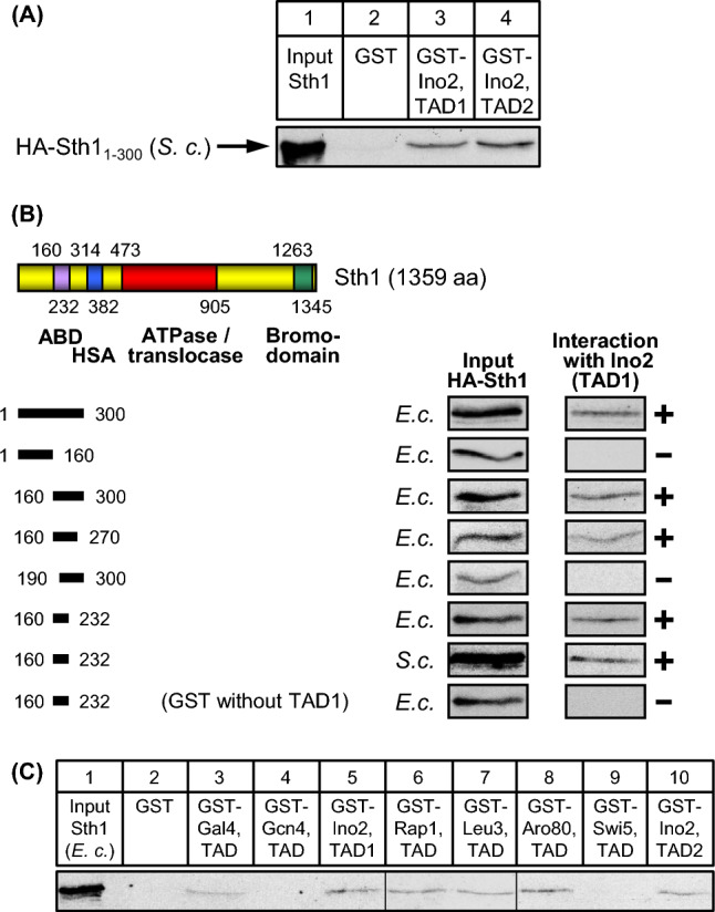
Fig. 5 In vitro interaction assays (GST pull-down) with length variants of Sth1 and various activation domains. A Sth1 interacts with both Ino2 activation domains. GST fusion proteins containing TAD1 or TAD2 of Ino2 were synthesized in E. coli, immobilized on GSH sepharose and incubated with yeast protein extracts containing epitope-tagged Sth1 (aa 1–300; yeast expression plasmid pECW31). B Mapping of Sth1 domains interacting with TAD1 of Ino2. Expression plasmids pECW32 (aa 1–300), pECW34 (aa 1–160), pECW33 (aa 160–300), pMiM4 (aa 160–270), pMiM3 (aa 190–300) and pECW45 (aa 160–232) were used to synthesize HA-Sth1 variants in E. coli strain BL21 CodonPlus. Length variant aa 160–232 was also synthesized in S. cerevisiae (yeast expression plasmid pECW48). ABD, activator binding domain; HSA, helicase/SANT-associated domain. ATPase RecA subdomains are not depicted individually. Functional domain SnAC (aa 1001–1069) is omitted from the figure. C TADs of various unrelated transcriptional activators interact with Sth1. GST fusions of Ino2 TAD1, Ino2 TAD2, Gal4 TAD Gcn4 TAD, Leu3 TAD, Rap1 TAD, Aro80 TAD and Swi5 TAD were synthesized in E. coli, immobilized on GSH sepharose and incubated with bacterial protein extract containing epitope-tagged Sth1 length variant aa 1–300 (pECW32). Empty GST plasmid pGEX-2TK was used as a negative control
Fig. 5 In vitro interaction assays (GST pull-down) with length variants of Sth1 and various activation domains. A Sth1 interacts with both Ino2 activation domains. GST fusion proteins containing TAD1 or TAD2 of Ino2 were synthesized in E. coli, immobilized on GSH sepharose and incubated with yeast protein extracts containing epitope-tagged Sth1 (aa 1–300; yeast expression plasmid pECW31). B Mapping of Sth1 domains interacting with TAD1 of Ino2. Expression plasmids pECW32 (aa 1–300), pECW34 (aa 1–160), pECW33 (aa 160–300), pMiM4 (aa 160–270), pMiM3 (aa 190–300) and pECW45 (aa 160–232) were used to synthesize HA-Sth1 variants in E. coli strain BL21 CodonPlus. Length variant aa 160–232 was also synthesized in S. cerevisiae (yeast expression plasmid pECW48). ABD, activator binding domain; HSA, helicase/SANT-associated domain. ATPase RecA subdomains are not depicted individually. Functional domain SnAC (aa 1001–1069) is omitted from the figure. C TADs of various unrelated transcriptional activators interact with Sth1. GST fusions of Ino2 TAD1, Ino2 TAD2, Gal4 TAD Gcn4 TAD, Leu3 TAD, Rap1 TAD, Aro80 TAD and Swi5 TAD were synthesized in E. coli, immobilized on GSH sepharose and incubated with bacterial protein extract containing epitope-tagged Sth1 length variant aa 1–300 (pECW32). Empty GST plasmid pGEX-2TK was used as a negative controlCC BY · PubMed Central
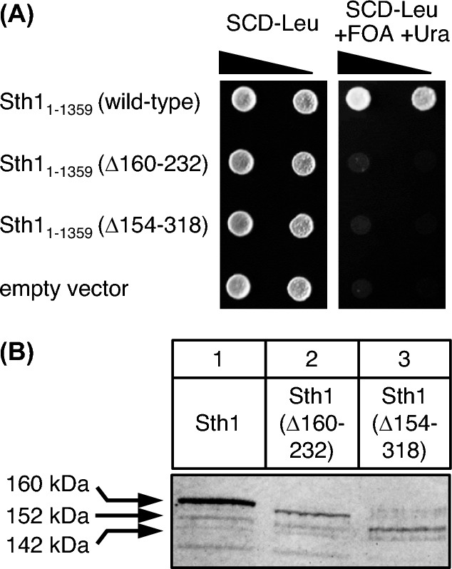
Fig. 6 Assay for functional complementation of sth1 mutation by STH1 variants lacking ABD sequences. A S. cerevisiae strain KSY1 (relevant genotype: ura3 leu2 sth1Δ::HIS3 + [pKS2: ARS CEN URA3 STH1]) was transformed with plasmids pKS4 (ARS CEN LEU2 STH1; wild-type), pKS7 (STH1Δ160-232), pKS8 (STH1Δ154-318) and YCplac111 (empty vector), respectively. Strain dilutions were spotted on SCD-Leu (one day) and SCD-Leu + FOA + Ura (counter-selection against rescue plasmid pKS2, two days). FOA, 5-fluoroorotic acid. B Detection of HA-Sth1 variants in strain C13-ABY.S86, transformed with expression plasmids pKS23 (MET25-HA3-STH1), pKS24 (MET25-HA3-STH1Δ160-232) and pKS25 (MET25-HA3-STH1Δ154-318), respectively. In all lanes, 50 µg of total protein were analyzed
Fig. 6 Assay for functional complementation of sth1 mutation by STH1 variants lacking ABD sequences. A S. cerevisiae strain KSY1 (relevant genotype: ura3 leu2 sth1Δ::HIS3 + [pKS2: ARS CEN URA3 STH1]) was transformed with plasmids pKS4 (ARS CEN LEU2 STH1; wild-type), pKS7 (STH1Δ160-232), pKS8 (STH1Δ154-318) and YCplac111 (empty vector), respectively. Strain dilutions were spotted on SCD-Leu (one day) and SCD-Leu + FOA + Ura (counter-selection against rescue plasmid pKS2, two days). FOA, 5-fluoroorotic acid. B Detection of HA-Sth1 variants in strain C13-ABY.S86, transformed with expression plasmids pKS23 (MET25-HA3-STH1), pKS24 (MET25-HA3-STH1Δ160-232) and pKS25 (MET25-HA3-STH1Δ154-318), respectively. In all lanes, 50 µg of total protein were analyzedCC BY · PubMed Central
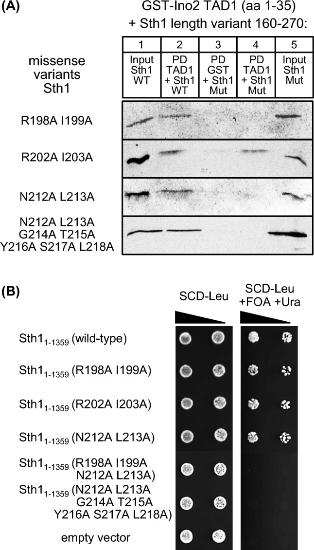
Fig. 7 Functional analysis of missense variants introduced into the ABD of Sth1. A Comparative interaction studies of Ino2 TAD1 and variants of the Sth1 activator binding domain. Fusion protein GST-Ino2 (TAD1) was incubated with bacterial protein extracts containing epitope-tagged Sth1 (plasmid pMiM4, encoding wild-type aa 160–270) as well as variants R198A I199A (pLT3), R202A I203A (pLT2), N212A L213A (pLT4) and N212A L213A G214A T215A Y216A S217A L218A (pLT5). Input samples are shown in lanes 1 (wild-type, wild-type) and lanes 5 (mutant variant, Mut). Pull-down (PD) experiments were analyzed in lanes 2 (wild-type) and lanes 4 (mutant variant). B Plasmid shuffling experiments with STH1 wild-type and missense variants encoding full-length proteins. S. cerevisiae strain KSY1 was transformed with plasmids pKS4 (ARS CEN LEU2 STH1; wild-type), pECW76 (R198A I199A), pECW77 (R202A I203A), pECW78 (N212A L213A), pECW79 (R198A I199A N212A L213A), pECW80 (N212A L213A G214A T215A Y216A S217A L218A) and YCplac111 (empty vector), respectively. Strain dilutions were spotted on SCD-Leu (one day) and SCD-Leu + FOA + Ura (counter-selection against rescue plasmid pKS2, two days). FOA, 5-fluoroorotic acid
Fig. 7 Functional analysis of missense variants introduced into the ABD of Sth1. A Comparative interaction studies of Ino2 TAD1 and variants of the Sth1 activator binding domain. Fusion protein GST-Ino2 (TAD1) was incubated with bacterial protein extracts containing epitope-tagged Sth1 (plasmid pMiM4, encoding wild-type aa 160–270) as well as variants R198A I199A (pLT3), R202A I203A (pLT2), N212A L213A (pLT4) and N212A L213A G214A T215A Y216A S217A L218A (pLT5). Input samples are shown in lanes 1 (wild-type, wild-type) and lanes 5 (mutant variant, Mut). Pull-down (PD) experiments were analyzed in lanes 2 (wild-type) and lanes 4 (mutant variant). B Plasmid shuffling experiments with STH1 wild-type and missense variants encoding full-length proteins. S. cerevisiae strain KSY1 was transformed with plasmids pKS4 (ARS CEN LEU2 STH1; wild-type), pECW76 (R198A I199A), pECW77 (R202A I203A), pECW78 (N212A L213A), pECW79 (R198A I199A N212A L213A), pECW80 (N212A L213A G214A T215A Y216A S217A L218A) and YCplac111 (empty vector), respectively. Strain dilutions were spotted on SCD-Leu (one day) and SCD-Leu + FOA + Ura (counter-selection against rescue plasmid pKS2, two days). FOA, 5-fluoroorotic acidCC BY · PubMed Central
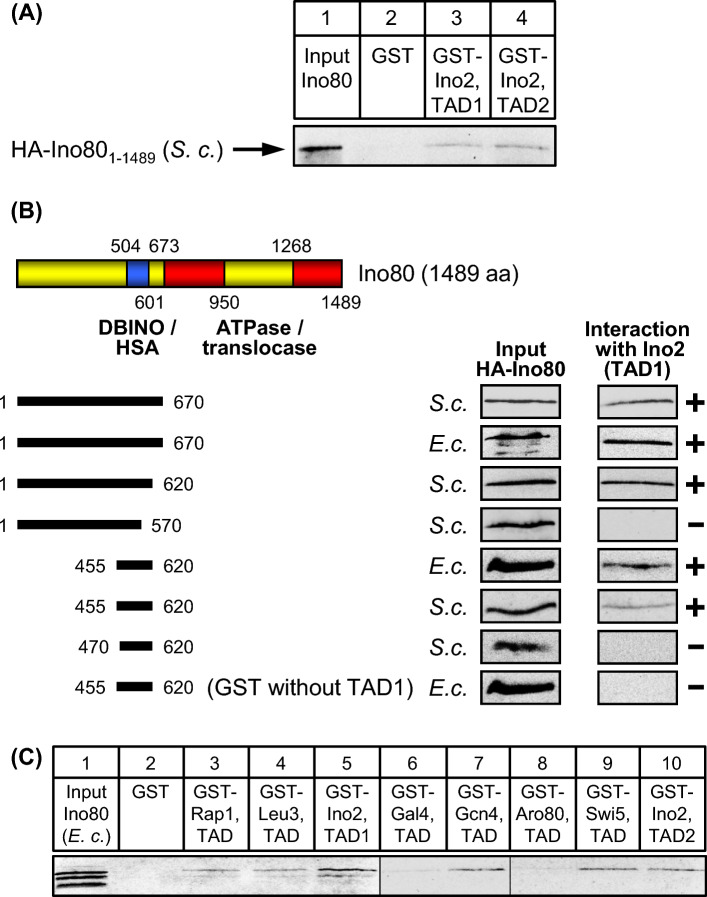
Fig. 8 Ino80 interacts with various activation domains in vitro. A GST pull-down assays with Ino2 activation domains and full-length Ino80 synthesized in yeast (aa 1–1489, expression plasmid pRE82). B Mapping of Ino80 domains interacting with TAD1 of Ino2. Yeast and E. coli expression plasmids pECW38 (aa 1–670, S.c.), pMiM1 (aa 1–670, E.c.), pMM5 (aa 1–620, S.c.), pMM6 (aa 1–570, S.c.), pECW70 (aa 455–620, S.c.), pECW73 (aa 455–620, E.c.) and pECW72 (aa 470–620, S.c.) were used to synthesize HA-Ino80 length variants. DBINO, DNA-binding domain of Ino80. C TADs of various unrelated transcriptional activators interact with Ino80. GST fusions of Ino2 TAD1, Ino2 TAD2, Gal4 TAD Gcn4 TAD, Leu3 TAD, Rap1 TAD, Aro80 TAD and Swi5 TAD were synthesized in E. coli, immobilized on GSH sepharose and incubated with bacterial protein extract containing epitope-tagged Ino80 length variant aa 1–670 (pMiM1). Empty GST plasmid pGEX-2TK was used as a negative control
Fig. 8 Ino80 interacts with various activation domains in vitro. A GST pull-down assays with Ino2 activation domains and full-length Ino80 synthesized in yeast (aa 1–1489, expression plasmid pRE82). B Mapping of Ino80 domains interacting with TAD1 of Ino2. Yeast and E. coli expression plasmids pECW38 (aa 1–670, S.c.), pMiM1 (aa 1–670, E.c.), pMM5 (aa 1–620, S.c.), pMM6 (aa 1–570, S.c.), pECW70 (aa 455–620, S.c.), pECW73 (aa 455–620, E.c.) and pECW72 (aa 470–620, S.c.) were used to synthesize HA-Ino80 length variants. DBINO, DNA-binding domain of Ino80. C TADs of various unrelated transcriptional activators interact with Ino80. GST fusions of Ino2 TAD1, Ino2 TAD2, Gal4 TAD Gcn4 TAD, Leu3 TAD, Rap1 TAD, Aro80 TAD and Swi5 TAD were synthesized in E. coli, immobilized on GSH sepharose and incubated with bacterial protein extract containing epitope-tagged Ino80 length variant aa 1–670 (pMiM1). Empty GST plasmid pGEX-2TK was used as a negative controlCC BY · PubMed Central
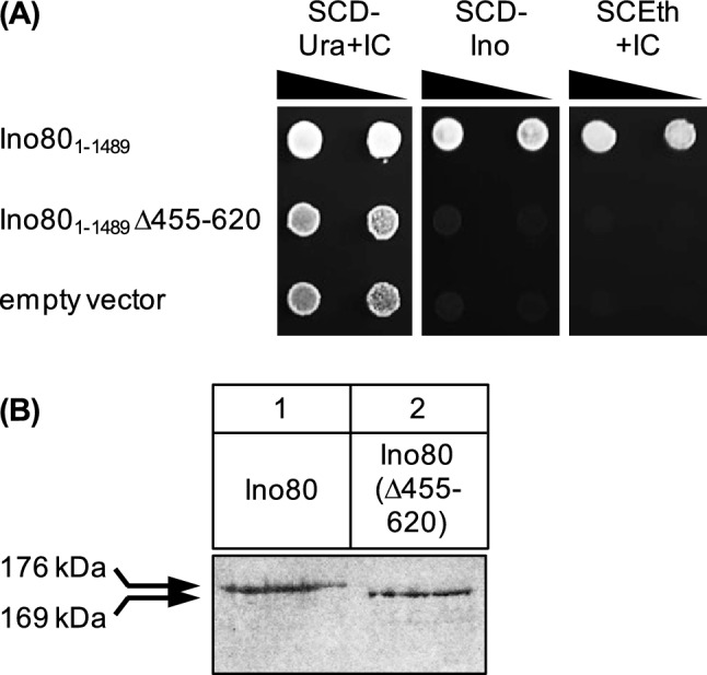
Fig. 9 Assay for functional complementation of an ino80 null mutation by INO80 variant lacking the activator binding domain (ABD). A S. cerevisiae strain YAB1 (relevant genotype: ura3 ino80Δ::kanMX) was transformed with plasmids pRE49 (ARS CEN URA3 INO80; wild-type), pECW74 (INO80Δ455-620) and YCplac33 (empty vector), respectively. Serial dilutions of transformants were spotted on the media shown and incubated for one (SCD-Ura + IC), two (SCD-Ino) or three days (SCEth + IC). IC, inositol + choline; Ino, inositol; Eth, ethanol. B Detection of HA-Ino80 variant in strain C13-ABY.S86, transformed with expression plasmids pRE82 (MET25-HA3-INO80) and pECW75 (MET25-HA3-INO80Δ455-620), respectively. In all lanes, 50 µg of total protein were analyzed
Fig. 9 Assay for functional complementation of an ino80 null mutation by INO80 variant lacking the activator binding domain (ABD). A S. cerevisiae strain YAB1 (relevant genotype: ura3 ino80Δ::kanMX) was transformed with plasmids pRE49 (ARS CEN URA3 INO80; wild-type), pECW74 (INO80Δ455-620) and YCplac33 (empty vector), respectively. Serial dilutions of transformants were spotted on the media shown and incubated for one (SCD-Ura + IC), two (SCD-Ino) or three days (SCEth + IC). IC, inositol + choline; Ino, inositol; Eth, ethanol. B Detection of HA-Ino80 variant in strain C13-ABY.S86, transformed with expression plasmids pRE82 (MET25-HA3-INO80) and pECW75 (MET25-HA3-INO80Δ455-620), respectively. In all lanes, 50 µg of total protein were analyzedCC BY · PubMed Central
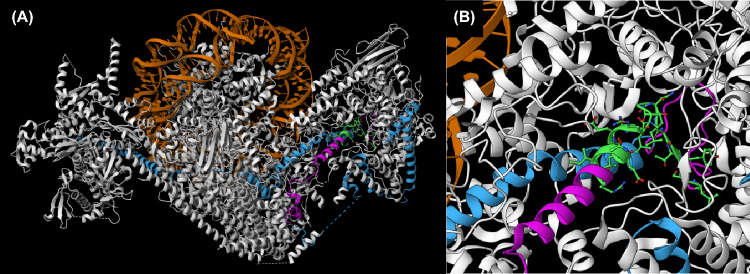
Fig. 10 Structural model of RSC based on cryo-EM analysis of Patel et al. (2019). A View of entire RSC complex (PBD: 6V92) with functional and structural domains of Sth1 depicted in different colours: DNA of the nucleosome (orange), Sth1 (blue), ABD as defined by in vitro interaction experiments (aa 160–232, purple), core region of ABD as defined by mutational analysis (aa 200–218, green). B Enlarged display of ABD and its core region
Fig. 10 Structural model of RSC based on cryo-EM analysis of Patel et al. (2019). A View of entire RSC complex (PBD: 6V92) with functional and structural domains of Sth1 depicted in different colours: DNA of the nucleosome (orange), Sth1 (blue), ABD as defined by in vitro interaction experiments (aa 160–232, purple), core region of ABD as defined by mutational analysis (aa 200–218, green). B Enlarged display of ABD and its core regionCC BY · PubMed Central

Verification

Claims the paper makes

The claims and statistics of this paper haven’t been checked yet.

Run the check — free, no account needed. It reads the open-access text and checks the sources it cites.

A few free checks a day · papers already verified open instantly · results are public

Self-check

Percentages, sums and reported differences re-computed from the paper’s own numbers.

The self-check needs the paper’s full text.

Citations

Cites

Show all 2 references

Research works printed but not linked: 57

  1. Bakshi R, Prakash T, Dash D, Brahmachari V (2004) In silico characterization of the INO80 subfamily of SWI2/SNF2 chromatin remodeling proteins. Biochem Biophys Res Commun 320:197–204. https://doi.org/10.1016/j.bbrc.2004.05.147 · DOI
  2. Bakshi R, Mehta AK, Sharma R, Maiti S, Pasha S, Brahmachari V (2006) Characterization of a human SWI2/SNF2 like protein hINO80: demonstration of catalytic and DNA binding activity. Biochem Biophys Res Commun 339:313–320. https://doi.org/10.1016/j.bbrc.2005.10.206 · DOI
  3. Bao Y, Shen X (2007) INO80 subfamily of chromatin remodeling complexes. Mutat Res 618:18–29. https://doi.org/10.1016/j.mrfmmm.2006.10.006 · DOI
  4. Biegel JA, Busse TM, Weissman BE (2014) SWI/SNF chromatin remodeling complexes and cancer. Am J Med Genet 166C:350–366. https://doi.org/10.1002/ajmg.c.31410 · DOI
  5. Brahma S, Ngubo M, Paul S, Udugama M, Bartholomew B (2018) The Arp8 and Arp4 module acts as a DNA sensor controlling INO80 chromatin remodeling. Nat Commun 9:3309. https://doi.org/10.1038/s41467-018-05710-7 · DOI
  6. Cairns BR, Lorch Y, Li Y, Zhang M, Lacomis L, Erdjument-Bromage H, Tempst P, Du J, Laurent B, Kornberg RD (1996) RSC, an essential, abundant chromatin-remodeling complex. Cell 87:1249–1260. https://doi.org/10.1016/s0092-8674(00)81820-6 · DOI
  7. Chou KY, Lee JY, Kim KB, Kim E, Lee HS, Ryu HY (2023) Histone modification in Saccharomyces cerevisiae: a review of the current status. Comput Struct Biotechnol J 21:1843–1850. https://doi.org/10.1016/j.csbj.2023.02.037 · DOI
  8. Clapier CR, Iwasa J, Cairns BR, Peterson CL (2017) Mechanisms of action and regulation of ATP-dependent chromatin-remodelling complexes. Nat Rev Mol Cell Biol 18:407–422. https://doi.org/10.1038/nrm.2017.26 · DOI · Paper page
  9. Cosma MP, Tanaka T, Nasmyth K (1999) Ordered recruitment of transcription and chromatin remodeling factors to a cell cycle- and developmentally regulated promoter. Cell 97:299–311. https://doi.org/10.1016/s0092-8674(00)80740-0 · DOI
  10. Dietz M, Heyken WT, Hoppen J, Geburtig S, Schüller HJ (2003) TFIIB and subunits of the SAGA complex are involved in transcriptional activation of phospholipid biosynthetic genes by the regulatory protein Ino2 in the yeast Saccharomyces cerevisiae. Mol Microbiol 48:1119–1130. https://doi.org/10.1046/j.1365-2958.2003.03501.x · DOI
  11. Ebbert R, Birkmann A, Schüller HJ (1999) The product of the SNF2/SWI2 paralogue INO80 of Saccharomyces cerevisiae required for efficient expression of various yeast structural genes is part of a high-molecular-weight protein complex. Mol Microbiol 32:741–751. https://doi.org/10.1046/j.1365-2958.1999.01390.x · DOI
  12. Engelhardt M, Hintze S, Wendegatz EC, Lettow J, Schüller HJ (2023) Ino2, activator of yeast phospholipid biosynthetic genes, interacts with basal transcription factors TFIIA and Bdf1. Curr Genet 69:289–300. https://doi.org/10.1007/s00294-023-01277-z · DOI
  13. Erijman A, Kozlowski L, Sohrabi-Jahromi S, Fishburn J, Warfield L, Schreiber J, Noble WS, Söding J, Hahn S (2020) A high-throughput screen for transcription activation domains reveals their sequence features and permits prediction by deep learning. Mol Cell 78:890–902. https://doi.org/10.1016/j.molcel.2020.04.020 · DOI
  14. Eustermann S, Patel AB, Hopfner KP, He Y, Korber P (2024) Energy-driven genome regulation by ATP-dependent chromatin remodellers. Nat Rev Mol Cell Biol 25:309–332. https://doi.org/10.1038/s41580-023-00683-y · DOI
  15. Ford J, Odeyale O, Eskandar A, Kouba N, Shen CH (2007) A SWI/SNF- and INO80-dependent nucleosome movement at the INO1 promoter. Biochem Biophys Res Commun 361:974–979. https://doi.org/10.1016/j.bbrc.2007.07.109 · DOI
  16. Ford J, Odeyale O, Shen CH (2008) Activator-dependent recruitment of SWI/SNF and INO80 during INO1 activation. Biochem Biophys Res Commun 373:602–606. https://doi.org/10.1016/j.bbrc.2008.06.079 · DOI
  17. Gietz RD, Sugino A (1988) New yeast-Escherichia coli shuttle vectors constructed with in vitro mutagenized yeast genes lacking six-base pair restriction sites. Gene 74:527–534. https://doi.org/10.1016/0378-1119(88)90185-0 · DOI
  18. Hahn S (2018) Phase separation, protein disorder, and enhancer function. Cell 175:1723–1725. https://doi.org/10.1016/j.cell.2018.11.034 · DOI
  19. Han Y, Reyes AA, Malik S, He Y (2020) Cryo-EM structure of SWI/SNF complex bound to a nucleosome. Nature 579:452–455. https://doi.org/10.1038/s41586-020-2087-1 · DOI
  20. Hartley PD, Madhani HD (2009) Mechanisms that specify promoter nucleosome location and identity. Cell 137:445–458. https://doi.org/10.1016/j.cell.2009.02.043 · DOI
  21. Hassan AH, Awad S, Al-Natour Z, Othman S, Mustafa F, Rizvi TA (2007) Selective recognition of acetylated histones by bromodomains in transcriptional co-activators. Biochem J 402:125–133. https://doi.org/10.1042/BJ20060907 · DOI
  22. Hintze S, Engelhardt M, van Diepen L, Witt E, Schüller HJ (2017) Multiple Taf subunits of TFIID interact with Ino2 activation domains and contribute to expression of genes required for yeast phospholipid biosynthesis. Mol Microbiol 106:876–890. https://doi.org/10.1111/mmi.13850 · DOI
  23. Hsieh LJ, Gourdet MA, Moore CM, Muñoz EN, Gamarra N, Ramani V, Narlikar GJ (2022) A hexasome is the preferred substrate for the INO80 chromatin remodeling complex, allowing versatility of function. Mol Cell 82:2098–2112. https://doi.org/10.1016/j.molcel.2022.04.026 · DOI
  24. Hu Z, Chen K, Li W, Tyler JK (2014) A matter of access. Nucleosome disassembly from gene promoters is the central goal of transcriptional activators. Transcription 5:e29355. https://doi.org/10.4161/trns.29355 · DOI
  25. Imai Y, Matsushima Y, Sugimura T, Terada M (1991) A simple and rapid method for generating a deletion by PCR. Nucleic Acids Res 19:2785. https://doi.org/10.1093/nar/19.10.2785 · DOI
  26. Kadosh D, Struhl K (1997) Repression by Ume6 involves recruitment of a complex containing Sin3 corepressor and Rpd3 histone deacetylase to target promoters. Cell 89:365–371. https://doi.org/10.1016/s0092-8674(00)80217-2 · DOI
  27. Kalpana GV, Marmon S, Wang W, Crabtree GR, Goff SP (1994) Binding and stimulation of HIV-1 integrase by a human homolog of yeast transcription factor SNF5. Science 266:2002–2006. https://doi.org/10.1126/science.7801128 · DOI
  28. Kim JH, Saraf A, Florens L, Washburn M, Workman JL (2010) Gcn5 regulates the dissociation of SWI/SNF from chromatin by acetylation of Swi2/Snf2. Genes Dev 24:2766–2771. https://doi.org/10.1101/gad.1979710 · DOI
  29. Knoll KR, Eustermann S, Niebauer V, Oberbeckmann E, Stoehr G, Schall K, Tosi A, Schwarz M, Buchfellner A, Korber P, Hopfner KP (2018) The nuclear actin-containing Arp8 module is a linker DNA sensor driving INO80 chromatin remodeling. Nat Struct Mol Biol 25:823–832. https://doi.org/10.1038/s41594-018-0115-8 · DOI
  30. Kunert F, Metzner FJ, Jung J, Höpfler M, Woike S, Schall K, Kostrewa D, Moldt M, Chen JX, Bantele S, Pfander B, Eustermann S, Hopfner KP (2022) Structural mechanism of extranucleosomal DNA readout by the INO80 complex. Sci Adv 8:eadd3189. https://doi.org/10.1126/sciadv.add3189 · DOI
  31. Lorch Y, Kornberg RD (2017) Chromatin-remodeling for transcription. Q Rev Biophys 50:e5. https://doi.org/10.1017/S003358351700004X · DOI
  32. Malik S, Roeder RG (2023) Regulation of the RNA polymerase II pre-initiation complex by its associated coactivators. Nat Rev Genet 24:767–782. https://doi.org/10.1038/s41576-023-00630-9 · DOI
  33. Mittal C, Lang O, Lai WKM, Pugh BF (2022) An integrated SAGA and TFIID PIC assembly pathway selective for poised and induced promoters. Genes Dev 36:985–1001. https://doi.org/10.1101/gad.350026.122 · DOI
  34. Mumberg D, Müller R, Funk M (1994) Regulatable promoters of Saccharomyces cerevisiae: comparison of transcriptional activity and their use for heterologous expression. Nucleic Acids Res 22:5767–5768. https://doi.org/10.1093/nar/22.25.5767 · DOI
  35. Neely KE, Hassan AH, Brown CE, Howe L, Workman JL (2002) Transcription activator interactions with multiple SWI/SNF subunits. Mol Cell Biol 22:1615–1625. https://doi.org/10.1128/MCB.22.6.1615-1625.2002 · DOI · Paper page
  36. Neigeborn L, Carlson M (1984) Genes affecting the regulation of SUC2 gene expression by glucose repression in Saccharomyces cerevisiae. Genetics 108:845–858. https://doi.org/10.1093/genetics/108.4.845 · DOI
  37. Papamichos-Chronakis M, Watanabe S, Rando OJ, Peterson CL (2011) Global regulation of H2A.Z localization by the INO80 chromatin-remodeling enzyme is essential for genome integrity. Cell 144:200–213. https://doi.org/10.1016/j.cell.2010.12.021 · DOI
  38. Patel AB, Moore CM, Greber BJ, Luo J, Zukin SA, Ranish J, Nogales E (2019) Architecture of the chromatin remodeler RSC and insights into its nucleosome engagement. Elife 8:e54449. https://doi.org/10.7554/eLife.54449 · DOI
  39. Peterson CL, Herskowitz I (1992) Characterization of the yeast SWI1, SWI2, and SWI3 genes, which encode a global activator of transcription. Cell 68:573–583. https://doi.org/10.1016/0092-8674(92)90192-f · DOI
  40. Peterson CL, Kruger W, Herskowitz I (1991) A functional interaction between the C-terminal domain of RNA polymerase II and the negative regulator SIN1. Cell 64:1135–1143. https://doi.org/10.1016/0092-8674(91)90268-4 · DOI
  41. Prochasson P, Neely KE, Hassan AH, Li B, Workman JL (2003) Targeting activity is required for SWI/SNF function in vivo and is accomplished through two partially redundant activator-interaction domains. Mol Cell 12:983–990. https://doi.org/10.1016/s1097-2765(03)00366-6 · DOI
  42. Schwank S, Ebbert R, Rautenstrauss K, Schweizer E, Schüller HJ (1995) Yeast transcriptional activator INO2 interacts as an Ino2p/Ino4p basic helix-loop-helix heteromeric complex with the inositol/choline-responsive element necessary for expression of phospholipid biosynthetic genes in Saccharomyces cerevisiae. Nucleic Acids Res 23:230–237. https://doi.org/10.1093/nar/23.2.230 · DOI
  43. Sen P, Ghosh S, Pugh BF, Bartholomew B (2011) A new, highly conserved domain in Swi2/Snf2 is required for SWI/SNF remodeling. Nucleic Acids Res 39:9155–9166. https://doi.org/10.1093/nar/gkr622 · DOI
  44. Shen X, Mizuguchi G, Hamiche A, Wu C (2000) A chromatin remodelling complex involved in transcription and DNA processing. Nature 406:541–544. https://doi.org/10.1038/35020123 · DOI
  45. Shen X, Ranallo R, Choi E, Wu C (2003) Involvement of actin-related proteins in ATP-dependent chromatin remodeling. Mol Cell 12:147–155. https://doi.org/10.1016/s1097-2765(03)00264-8 · DOI
  46. Sikorski RS, Boeke JD (1991) In vitro mutagenesis and plasmid shuffling: from cloned gene to mutant yeast. Methods Enzymol 194:302–318. https://doi.org/10.1016/0076-6879(91)94023-6 · DOI
  47. Stern M, Jensen R, Herskowitz I (1984) Five SWI genes are required for expression of the HO gene in yeast. J Mol Biol 178:853–868. https://doi.org/10.1016/0022-2836(84)90315-2 · DOI
  48. Sudarsanam P, Iyer VR, Brown PO, Winston F (2000) Whole-genome expression analysis of snf/swi mutants of Saccharomyces cerevisiae. Proc Natl Acad Sci USA 97:3364–3369. https://doi.org/10.1073/pnas.97.7.3364 · DOI · Paper page
  49. Szerlong H, Hinata K, Viswanathan R, Erdjument-Bromage H, Tempst P, Cairns BR (2008) The HSA domain binds nuclear actin-related proteins to regulate chromatin-remodeling ATPases. Nat Struct Mol Biol 15:469–476. https://doi.org/10.1038/nsmb.1403 · DOI
  50. Wagner FR, Dienemann C, Wang H, Stützer A, Tegunov D, Urlaub H, Cramer P (2020) Structure of SWI/SNF chromatin remodeller RSC bound to a nucleosome. Nature 579:448–451. https://doi.org/10.1038/s41586-020-2088-0 · DOI
  51. Wang T, Zhang J, Zhang X, Tu X (2012) Solution structure of SWI1 AT-rich interaction domain from Saccharomyces cerevisiae and its nonspecific binding to DNA. Proteins 80:1911–1917. https://doi.org/10.1002/prot.24091 · DOI
  52. Wang C, Guo Z, Zhan X, Yang F, Wu M, Zhang X (2020) Structure of the yeast Swi/Snf complex in a nucleosome free state. Nat Commun 11:3398. https://doi.org/10.1038/s41467-020-17229-x · DOI · Paper page
  53. Winston F, Carlson M (1992) Yeast SNF/SWI transcriptional activators and the SPT/SIN chromatin connection. Trends Genet 8:387–391. https://doi.org/10.1016/0168-9525(92)90300-s · DOI
  54. Ye Y, Wu H, Chen K, Clapier CR, Verma N, Zhang W, Deng H, Cairns BR, Gao N, Chen Z (2019) Structure of the RSC complex bound to the nucleosome. Science 366:838–843. https://doi.org/10.1126/science.aay0033 · DOI
  55. Yen K, Vinayachandran V, Batta K, Koerber RT, Pugh BF (2012) Genome-wide nucleosome specificity and directionality of chromatin remodelers. Cell 149:1461–1473. https://doi.org/10.1016/j.cell.2012.04.036 · DOI
  56. Yudkovsky N, Logie C, Hahn S, Peterson CL (1999) Recruitment of the SWI/SNF chromatin remodeling complex by transcriptional activators. Genes Dev 13:2369–2374. https://doi.org/10.1101/gad.13.18.2369 · DOI · Paper page
  57. Zhang M, Jungblut A, Kunert F, Hauptmann L, Hoffmann T, Kolesnikova O, Metzner F, Moldt M, Weis F, DiMaio F, Hopfner KP, Eustermann S (2023) Hexasome-INO80 complex reveals structural basis of noncanonical nucleosome remodeling. Science 381:313–319. https://doi.org/10.1126/science.adf6287 · DOI

Metadata

Authors
Wendegatz, E.-C., Engelhardt, M., & Schüller, H.-J.
Affiliations
Universität Greifswald (DE)
Corresponding author
Schüller, Hans-Joachim
Venue
Current Genetics
Published
2024-09-05
Volume
70
Issue
1
Pages
15-15
ISSN
  • 0172-8083 (ISSN-L)
  • 1432-0983
OpenAlex
W4402282790
Cited by
3
References
2
Citations by year
2026: 1 · 2025: 2
FWCI
0.41
Type
journal article
Language
en
Open access
Locations
  • Current Genetics (publishedVersion, cc-by)
  • Research Square (acceptedVersion, cc-by)
  • PubMed Central (publishedVersion, cc-by)
  • PubMed (publishedVersion)
Funders
Universität Greifswald
Topics
  • Life Sciences › Biochemistry, Genetics and Molecular Biology › Molecular Biology › Genomics and Chromatin Dynamics
  • Life Sciences › Biochemistry, Genetics and Molecular Biology › Molecular Biology › Chromatin Remodeling and Cancer
  • Life Sciences › Biochemistry, Genetics and Molecular Biology › Molecular Biology › Fungal and yeast genetics research
Keywords
chromatin remodeling complexes, SWI/SNF complex, RSC, INO80 complex, transactivation domain, ATPase subunits, Swi1, SMARCB1, INO2, nucleosome positioning, actin-related proteins, Saccharomyces cerevisiae
MeSH terms
Protein Domains, Adenosine Triphosphatases, Chromosomal Proteins, Non-Histone, DNA-Binding Proteins, Nuclear Proteins, Protein Binding, Saccharomyces cerevisiae, Transcription Factors, Transcriptional Activation, Gene Expression Regulation, Fungal, Cell Cycle Proteins, Saccharomyces cerevisiae Proteins, Chromatin Assembly and Disassembly, Basic Helix-Loop-Helix Proteins

About these checks

Registry, retraction, Cites and statistics results are computed, not judged, with one exception: a printed reference that no index lists is read once by a model before it counts as a citation error. Claims are extracted by a model and kept only when their quote appears verbatim in the text; each cited source is then checked, with a model’s help, for whether it says what the claim relies on. Those verdicts can be wrong — read the claim’s quote and the cited source before relying on one.

Statistics and arithmetic are recomputed, not judged.

Confidence bands are shown only for fields calibrated on our annotated test set. None is calibrated yet, so no band is shown.

What counts as a claim ›How we measure confidence ›How we check this ›

Are you an author, or do you see a problem with this page? Request a review

How to cite

Cite CoChat for the checks on this page, and the paper for its own findings.

This page
CoChat. (2026). Citation check: Transcriptional activation domains interact with ATPase subunits of yeast chromatin remodelling complexes SWI/SNF, RSC and INO80. https://app.cochat.ai/papers/10.1007/s00294-024-01300-x
The paper
Wendegatz, E.-C., Engelhardt, M., & Schüller, H.-J. (2024). Transcriptional activation domains interact with ATPase subunits of yeast chromatin remodelling complexes SWI/SNF, RSC and INO80. Current Genetics. https://doi.org/10.1007/s00294-024-01300-x