

The DOI matches its Crossref record; 80 of the 80 references it cites were found in a registry or an index.
Analyzing microglial phenotypes across neuropathologies: a practical guide
Open accessAbstract
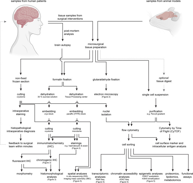
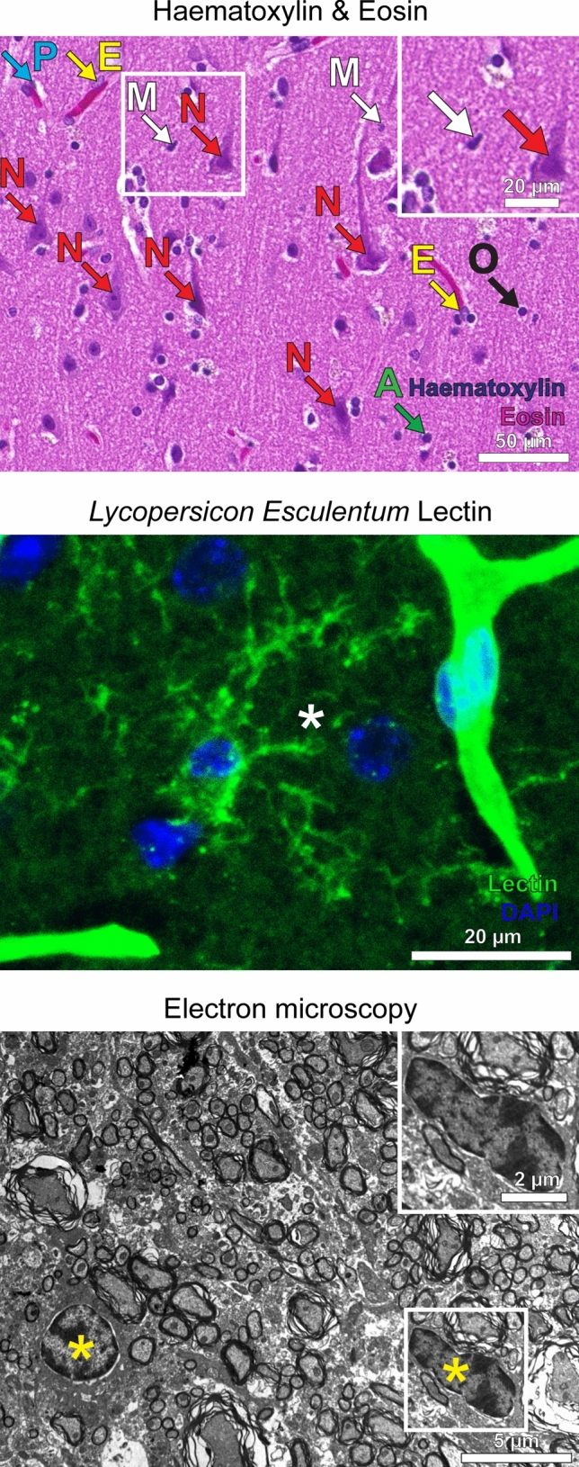
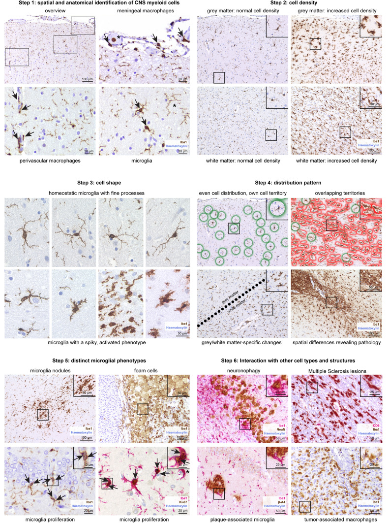
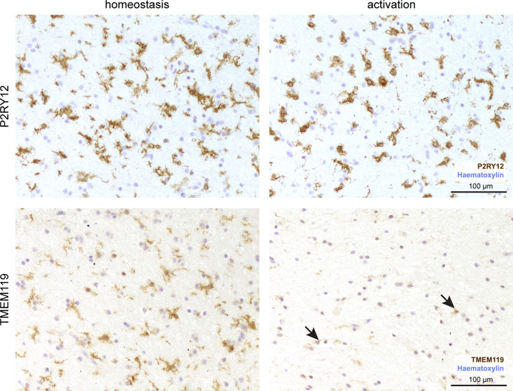
As extremely sensitive immune cells, microglia act as versatile watchdogs of the central nervous system (CNS) that tightly control tissue homeostasis. Therefore, microglial activation is an early and easily detectable hallmark of virtually all neuropsychiatric, neuro-oncological, neurodevelopmental, neurodegenerative and neuroinflammatory diseases. The recent introduction of novel high-throughput technologies and several single-cell methodologies as well as advances in epigenetic analyses helped to identify new microglia expression profiles, enhancer-landscapes and local signaling cues that defined diverse previously unappreciated microglia states in the healthy and diseased CNS. Here, we give an overview on the recent developments in the field of microglia biology and provide a practical guide to analyze disease-associated microglia phenotypes in both the murine and human CNS, on several morphological and molecular levels. Finally, technical limitations, potential pitfalls and data misinterpretations are discussed as well.
Overview
The claims and statistics of this paper haven’t been checked yet.
Run the check — free, no account needed. It reads the open-access text and checks the sources it cites.
Integrity signals
Registry
Cites
Statistics
Figures
Verification
Claims the paper makes
The claims and statistics of this paper haven’t been checked yet.
Run the check — free, no account needed. It reads the open-access text and checks the sources it cites.
Claims other papers make about this one
Each is checked against this paper’s own text: does it say what the citing paper says it does?
The cited source says what the claim attributes to it.
In the citing paper
“Histopathological examination of the hippocampal parenchyma revealed microgliosis17 in aged mice, which was reduced after STING inhibition (Fig. 2a).”
From cGAS–STING drives ageing-related inflammation and neurodegeneration (2023) · cites this paper as [17]
The cited source says what the claim attributes to it.
In the citing paper
“Homeostatic microglia present a ramified morphology with longer processes (Schwabenland et al., 2021), such as that observed in WT and CKO mice (Fig. 4, H, M, and N), while microglia from T mice display a hypertrophic morphology characterized by a significant reduction in process length and number of branches per cell (P < 0.0001).”
From Cholesterol 25-hydroxylase mediates neuroinflammation and neurodegeneration in a mouse model of tauopathy (2024) · cites this paper as Schwabenland et al., 2021
Self-check
Percentages, sums and reported differences re-computed from the paper’s own numbers.
The self-check needs the paper’s full text.
Citations
Cites
Polazzi, E., Mengoni, I., Caprini, M., Peña‐Altamira, E., Kurtys, E., & Monti, B. (2012) · Neurosignals · doi:10.1159/000337115 · Cited by 9,023
A Unique Microglia Type Associated with Restricting Development of Alzheimer’s Disease
Keren-Shaul, H., Spinrad, A., Weiner, A., Matcovitch-Natan, O., Dvir-Szternfeld, R., Ulland, T. K., David, E., Baruch, K., Lara-Astaiso, D., Toth, B., et al. (2017) · Cell · doi:10.1016/j.cell.2017.05.018 · Cited by 5,493
Synaptic Pruning by Microglia Is Necessary for Normal Brain Development
Paolicelli, R. C., Bolasco, G., Pagani, F., Maggi, L., Scianni, M., Panzanelli, P., Giustetto, M., Ferreira, T. A., Guiducci, E., Dumas, L., et al. (2011) · Science · doi:10.1126/science.1202529 · Cited by 4,244
Host microbiota constantly control maturation and function of microglia in the CNS
Erny, D., Hrabě de Angelis, A. L., Jaitin, D., Wieghofer, P., Staszewski, O., David, E., Keren-Shaul, H., Mahlakoiv, T., Jakobshagen, K., Buch, T., et al. (2015) · Nature Neuroscience · doi:10.1038/nn.4030 · Cited by 3,569
Self-citationATAC-seq: a method for assaying chromatin accessibility genome-wide
Buenrostro, J. D., Wu, B., Chang, H. Y., & Greenleaf, W. J. (2015) · Current Protocols in Molecular Biology · doi:10.1002/0471142727.mb2129s109 · Cited by 3,289
Identification of a unique TGF-β–dependent molecular and functional signature in microglia
Butovsky, O., Jedrychowski, M. P., Moore, C. S., Cialic, R., Lanser, A. J., Gabriely, G., Koeglsperger, T., Dake, B., Wu, P. M., Doykan, C. E., et al. (2014) · Nature Neuroscience · doi:10.1038/nn.3599 · Cited by 2,543
Slide-seq: A scalable technology for measuring genome-wide expression at high spatial resolution
Rodriques, S. G., Stickels, R. R., Goeva, A., Martin, C. A., Murray, E., Vanderburg, C. R., Welch, J., Chen, L. M., Chen, F., & Macosko, E. Z. (2019) · Science · doi:10.1126/science.aaw1219 · Cited by 2,424
Tissue-Resident Macrophage Enhancer Landscapes Are Shaped by the Local Microenvironment
Lavin, Y., Winter, D., Blecher-Gonen, R., David, E., Keren-Shaul, H., Merad, M., Jung, S., & Amit, I. (2014) · Cell · doi:10.1016/j.cell.2014.11.018 · Cited by 2,167
New tools for studying microglia in the mouse and human CNS
Bennett, M. L., Bennett, F. C., Liddelow, S. A., Ajami, B., Zamanian, J. L., Fernhoff, N. B., Mulinyawe, S. B., Bohlen, C. J., Adil, A., Tucker, A., et al. (2016) · Proceedings of the National Academy of Sciences · doi:10.1073/pnas.1525528113 · Cited by 1,804
The role of microglia and macrophages in glioma maintenance and progression
Hambardzumyan, D., Gutmann, D. H., & Kettenmann, H. (2016) · Nature Neuroscience · doi:10.1038/nn.4185 · Cited by 1,683
Cited by
Sorted by citations
cGAS–STING drives ageing-related inflammation and neurodegeneration
Gulen, M. F., Samson, N., Keller, A., Schwabenland, M., Liu, C., Glück, S., Thacker, V. V., Favre, L., Mangeat, B., Kroese, L. J., et al. (2023) · Nature · doi:10.1038/s41586-023-06373-1 · Cited by 1,104
Self-citationThe Impact of Systemic Inflammation on Alzheimer’s Disease Pathology
Xie, J., Van Hoecke, L., & Vandenbroucke, R. E. (2022) · Frontiers in Immunology · doi:10.3389/fimmu.2021.796867 · Cited by 324
Microglia in multiple sclerosis: protectors turn destroyers
Yong, V. W. (2022) · Neuron · doi:10.1016/j.neuron.2022.06.023 · Cited by 185
Detection of SARS-CoV-2 viral proteins and genomic sequences in human brainstem nuclei
Emmi, A., Rizzo, S., Barzon, L., Sandre, M., Carturan, E., Sinigaglia, A., Riccetti, S., Barbera, M. D., Rafael Boscolo-berto, A., Cocco, P., et al. (2023) · npj Parkinson s Disease · doi:10.1038/s41531-023-00467-3 · Cited by 75
CNS border-associated macrophages in the homeostatic and ischaemic brain
Gerganova, G., Riddell, A., & Miller, A. A. (2022) · Pharmacology & Therapeutics · doi:10.1016/j.pharmthera.2022.108220 · Cited by 72
Wang, M., Tang, G., Zhou, C., Guo, H., Hu, Z., Hu, Q., & Li, G. (2023) · Chemico-Biological Interactions · doi:10.1016/j.cbi.2023.110387 · Cited by 62
The niche matters: Origin, function and fate of CNS-associated macrophages during health and disease
Dalmau Gasull, A., Glavan, M., Samawar, S. K. R., Kapupara, K., Kelk, J., Rubio, M., Fumagalli, S., Sorokin, L., Vivien, D., & Prinz, M. (2024) · Acta Neuropathologica · doi:10.1007/s00401-023-02676-9 · Cited by 60
Self-citationLiu, R., Li, Y., Wang, Z., Chen, P., Xie, Y., Qu, W., Wang, M., Yu, Z., & Luo, X. (2023) · CNS Neuroscience & Therapeutics · doi:10.1111/cns.14161 · Cited by 58
Wei, W., Lattau, S. S. J., Xin, W., Pan, Y., Tatenhorst, L., Zhang, L., Graf, I., Kuang, Y., Zheng, X., Hao, Z., et al. (2024) · Advanced Science · doi:10.1002/advs.202306863 · Cited by 59
The contribution of neuropathology to multiple sclerosis research
Lassmann, H. (2022) · European Journal of Neurology · doi:10.1111/ene.15360 · Cited by 55
Self-citationZhang, C., Shao, W., Yuan, H., Xiao, R., Zhang, Y.-R., Wei, C., Ni, X., He, N., Chen, G., Gui, S., et al. (2024) · ACS Nano · doi:10.1021/acsnano.4c04674 · Cited by 53
Li, C., Ren, J., Zhang, M., Wang, H., Fang, Y., Wu, J., & Tang, Y. (2022) · Cellular and Molecular Life Sciences · doi:10.1007/s00018-022-04536-3 · Cited by 52
Ni, Y., Hu, Y., Zhu, L., Jiang, X., Zhang, H., Liu, J., & Zhao, Y. (2024) · Advanced Healthcare Materials · doi:10.1002/adhm.202304285 · Cited by 51
Toral-Rios, D., Long, J. M., Ulrich, J. D., Yu, J., Strickland, M. R., Han, X., Holtzman, D. M., Cashikar, A. G., & Paul, S. M. (2024) · Journal of Experimental Medicine · doi:10.1084/jem.20232000 · Cited by 47
Microglia–neuron crosstalk through Hex–GM2–MGL2 maintains brain homeostasis
Frosch, M., Shimizu, T., Wogram, E., Amann, L., Gruber, L., Groisman, A. I., Fliegauf, M., Schwabenland, M., Chhatbar, C., Zechel, S., et al. (2025) · Nature · doi:10.1038/s41586-025-09477-y · Cited by 56
Self-citationEndocannabinoid signaling in microglia
Marinelli, S., Marrone, M. C., Domenico, M. D., & Marinelli, S. (2022) · Glia · doi:10.1002/glia.24281 · Cited by 42
Claeys, W., Hoecke, L. V., Geerts, A. M., Vlierberghe, H. V., Lefere, S., Imschoot, G. V., Wonterghem, E. V., Ghesquière, B., Vandenbroucke, R. E., & Steenkiste, C. V. (2022) · Scientific Reports · doi:10.1038/s41598-022-22423-6 · Cited by 40
Teo, J. D., Marian, O. C., Spiteri, A. G., Nicholson, M., Song, H., Khor, J. X. Y., McEwen, H. P., Ge, A., Sen, M. K., Piccio, L. M., et al. (2022) · Glia · doi:10.1002/glia.24329 · Cited by 41
Astrocytic Neuroimmunological Roles Interacting with Microglial Cells in Neurodegenerative Diseases
Gotoh, M., Miyamoto, Y., & Ikeshima‐Kataoka, H. (2023) · International Journal of Molecular Sciences · doi:10.3390/ijms24021599 · Cited by 40
Tools for studying human microglia: In vitro and in vivo strategies
Warden, A. S., Han, C. Z., Hansen, E. A., Trescott, S., Nguyen, C. T., Kim, R. Y., Schafer, D. M., Johnson, A., Wright, M., Ramírez, G., et al. (2022) · Brain Behavior and Immunity · doi:10.1016/j.bbi.2022.10.008 · Cited by 38
USP25 Inhibits Neuroinflammatory Responses After Cerebral Ischemic Stroke by Deubiquitinating TAB2
Li, Z., Liu, B., Lambertsen, K. L., Clausen, B. H., Zhu, Z., Du, X., Xu, Y., Poulsen, F. R., Halle, B., Bonde, C., et al. (2023) · Advanced Science · doi:10.1002/advs.202301641 · Cited by 35
Vinnakota, J. M., Adams, R. C., Athanassopoulos, D., Schmidt, D., Biavasco, F., Zähringer, A., Erny, D., Schwabenland, M., Langenbach, M., Wenger, V., et al. (2024) · Science Translational Medicine · doi:10.1126/scitranslmed.adj9672 · Cited by 35
Self-citationLi, P., Wang, Y., Xu, Y., Feng, L., Jiang, N., Fang, Y., Song, G., Yu, L., Xu, L., Zhu, Z., et al. (2025) · Neurobiology of Disease · doi:10.1016/j.nbd.2025.107061 · Cited by 32
New Insights into Microglial Mechanisms of Memory Impairment in Alzheimer’s Disease
Li, N., Deng, M., Hu, G., Li, N., Yuan, H., & Zhou, Y. (2022) · Biomolecules · doi:10.3390/biom12111722 · Cited by 30
Wang, L., Ouyang, D., Li, L., Cao, Y., Wang, Y., Gu, N., Zhang, Z., Zhao, L., Tang, S., Tang, H. D., et al. (2025) · Journal of Neuroinflammation · doi:10.1186/s12974-025-03337-2 · Cited by 31
Schwabenland, M., Mossad, O., Sievert, A., Peres, A. G., Ringel, E., Baasch, S., Kolter, J., Cascone, G., Dokalis, N., Vlachos, A., et al. (2023) · Nature Communications · doi:10.1038/s41467-023-38373-0 · Cited by 26
Self-citationHase, Y., Ameen‐Ali, K. E., Waller, R., Simpson, J. E., Stafford, C., Mahesh, A., Ryan, L., Pickering, L., Bodman, C., Hase, M., et al. (2022) · Brain Pathology · doi:10.1111/bpa.13101 · Cited by 27
Exploring the Disease-Associated Microglia State in Amyotrophic Lateral Sclerosis
Jauregui, C., Blanco‐Luquin, I., Macías, M., Roldán, M., Caballero, C., Pagola, I., Mendióroz, M., & Jericó, I. (2023) · Biomedicines · doi:10.3390/biomedicines11112994 · Cited by 27
Wang, Y., Dong, L., Zhang, Y., Zhang, Y., Qin, G., Zhang, D., Chen, L., He, W., & Zhou, J. (2023) · Frontiers in Molecular Neuroscience · doi:10.3389/fnmol.2023.1177171 · Cited by 24
A transcriptomic microglia taxonomy across mouse and human pathologies
Chhatbar, C., Sankowski, R., Schulz, M., Shimizu, T., Schwabenland, M., Staszewski, O., Scheiwe, C. F., Nessler, S., Borst, K., Dumas, A. A., et al. (2026) · Nature Immunology · doi:10.1038/s41590-026-02472-z · Cited by 26
Self-citationMicroglia in brain aging: An overview of recent basic science and clinical research developments
Fan, H., Zhang, M., Wen, J., Wang, S., Yuan, M., Sun, H., Shu, L., Xu, Y., Pu, Y., & Cai, Z. (2023) · Journal of Biomedical Research · doi:10.7555/jbr.37.20220220 · Cited by 23
Tang, X., Cui, K., Lü, X., Wu, P., Yu, S., Yang, B., Xu, Y., & Liang, X. (2022) · Investigative Ophthalmology & Visual Science · doi:10.1167/iovs.63.6.13 · Cited by 19
Young COVID-19 Patients Show a Higher Degree of Microglial Activation When Compared to Controls
Matschke, J., Lahann, H., Krasemann, S., Altmeppen, H. C., Pfefferle, S., Galliciotti, G., Fitzek, A., Sperhake, J., Ondruschka, B., Busch, M., et al. (2022) · Frontiers in Neurology · doi:10.3389/fneur.2022.908081 · Cited by 19
Zhang, F., Zhang, Z., Alt, J., Kambhampati, S. P., Sharma, A., Singh, S., Nance, E. A., Thomas, A. G., Rojas, C. J., Rais, R., et al. (2023) · Journal of Controlled Release · doi:10.1016/j.jconrel.2023.04.017 · Cited by 21
Mass‐Guided Single‐Cell MALDI Imaging of Low‐Mass Metabolites Reveals Cellular Activation Markers
Cairns, J. L., Huber, J., Lewen, A., Jung, J., Maurer, S. J., Bausbacher, T., Schmidt, S., Levkin, P. A., Sévin, D. C., Göpfrich, K., et al. (2024) · Advanced Science · doi:10.1002/advs.202410506 · Cited by 21
A novel automated morphological analysis of Iba1+ microglia using a deep learning assisted model
Stetzik, L. A., Mercado, G., Smith, L., Sonia, G., Emmanuel, Q., Luda, K. M., Emily, S., Meyerdirk, L. K., Lindquist, A., Bergsma, A. L., et al. (2022) · Frontiers in Cellular Neuroscience · doi:10.3389/fncel.2022.944875 · Cited by 19
Microglia subtypes in acute, subacute, and chronic multiple sclerosis
Prineas, J. W., & Lee, S. (2023) · Journal of Neuropathology & Experimental Neurology · doi:10.1093/jnen/nlad046 · Cited by 16
Cacace, R., Zhou, L., de Craen, E. H. V., Buist, A., Hoogmartens, J., Sieben, A., Cras, P. P., Vandenberghe, R. R., Deyn, P. P. D., Oehlrich, D., et al. (2023) · Molecular Psychiatry · doi:10.1038/s41380-023-02166-0 · Cited by 16
Kessler, W., Thomas, C., & Kuhlmann, T. (2023) · Acta Neuropathologica · doi:10.1007/s00401-023-02645-2 · Cited by 17
Neuroimmune mechanisms in autism etiology - untangling a complex problem using human cellular models
Vacharasin, J. M., Ward, J. A., McCord, M. M., Cox, K., Imitola, J., & Lizarraga, S. B. (2024) · Oxford Open Neuroscience · doi:10.1093/oons/kvae003 · Cited by 17
Regulation of disease-associated microglia in the optic nerve by lipoxin B4 and ocular hypertension
Maurya, S., Lin, M., Karnam, S., Singh, T., Kumar, M., Ward, E., Sivak, J. M., Flanagan, J. G., & Gronert, K. (2024) · Molecular Neurodegeneration · doi:10.1186/s13024-024-00775-z · Cited by 17
Claeys, W., Geerts, A. M., Hoecke, L. V., Steenkiste, C. V., & Vandenbroucke, R. E. (2024) · Neural Regeneration Research · doi:10.4103/nrr.nrr-d-24-00600 · Cited by 17
Positron Emission Tomography of Neuroimmune Responses in Humans: Insights and Intricacies
Raval, N. R., Wetherill, R. R., Wiers, C. E., Dubroff, J. G., & Hillmer, A. T. (2022) · Seminars in Nuclear Medicine · doi:10.1053/j.semnuclmed.2022.08.008 · Cited by 15
Molecular and spatial heterogeneity of microglia in Rasmussen encephalitis
Westfall, J. J., Schwind, W. N., Sran, S., Navarro, J. B., Leonard, J. R., Pindrik, J., Pierson, C. R., Boué, D. R., Koboldt, D. C., Ostendorf, A. P., et al. (2022) · Acta Neuropathologica Communications · doi:10.1186/s40478-022-01472-y · Cited by 17
Microglia in Microbiota-Gut-Brain Axis: A Hub in Epilepsy
Liu, Y., Jia, N., Tang, C., Long, H., & Wang, J. (2024) · Molecular Neurobiology · doi:10.1007/s12035-024-04022-w · Cited by 15
The cGAS-STING axis: a comprehensive review from immune defense to disease pathogenesis
Sood, A. (2025) · Immunologic Research · doi:10.1007/s12026-025-09648-z · Cited by 14
Possible Implications of Obesity-Primed Microglia that Could Contribute to Stroke-Associated Damage
Carreto, R. J. R., Rodríguez-Cortés, Y. M., Guerrero, H. T., & Chavarría, A. (2023) · Cellular and Molecular Neurobiology · doi:10.1007/s10571-023-01329-5 · Cited by 15
Glial receptor PLXNB2 regulates schizophrenia-related stress perception via the amygdala
Xuan, F., Yan, L., Li, Y.-L., Fan, F., Deng, H.-H., Gou, M.-Z., Chithanathan, K., Heinla, I., Yuan, L., Seppa, K., et al. (2022) · Frontiers in Immunology · doi:10.3389/fimmu.2022.1005067 · Cited by 12
Cai, M., Xu, Z., Zhou, X., Li, L., Hua, X., Guo, S., & Yuan, X. (2022) · APL Bioengineering · doi:10.1063/5.0118311 · Cited by 14
Feng, L., Luo, G., Li, Y., Zhang, C., Liu, Y., Liu, Y., Chen, H., He, D., Zhu, Y., & Gan, L. (2023) · Veterinary Research · doi:10.1186/s13567-023-01149-x · Cited by 14
Xiong, M., Wu, Z., Zhao, Y., Zhao, D. B., Pan, Z., Wu, X., Liu, W. Q., & Hu, K. (2023) · Brain Research Bulletin · doi:10.1016/j.brainresbull.2023.04.008 · Cited by 13
Ma, Y., Liu, Z., Jiang, L., Wang, L., Li, Y., Liu, Y., Wang, Y., Yang, G., Ding, J., & Zhang, Z. (2022) · Journal of Cerebral Blood Flow & Metabolism · doi:10.1177/0271678x221135841 · Cited by 12
Zhu, Y., Yu, J., Chen, T., Liu, W., Huang, Y., Li, J., Zhang, B., Zhu, G., He, Z., Long, Y., et al. (2023) · European Journal of Medicinal Chemistry · doi:10.1016/j.ejmech.2023.115620 · Cited by 12
Abedrabbo, M., Kazemian, P., Connolly, C., & Leavitt, B. R. (2025) · Journal of Huntington s Disease · doi:10.1177/18796397251330144 · Cited by 12
Battis, K., Florio, J. B., Mante, M., Lana, A. J., Naumann, I., Gauer, C., Lambrecht, V., Müller, S. J., Cobo, I., Fixsen, B. R., et al. (2022) · Journal of Neuroscience · doi:10.1523/jneurosci.0417-22.2022 · Cited by 13
Midkine attenuates amyloid-β fibril assembly and plaque formation
Zaman, M., Yang, S., Huang, Y., Yarbro, J. M., Hao, Y., Wang, Z., Liu, D., Harper, K. E., Soliman, H., Hemphill, A., et al. (2025) · Nature Structural & Molecular Biology · doi:10.1038/s41594-025-01657-8 · Cited by 17
Szabó, M., Lajkó, N., Dulka, K., Barczánfalvi, G., Lőrinczi, B., Szatmári, I., Mihály, A., Vécsei, L., & Gulya, K. (2023) · Scientific Reports · doi:10.1038/s41598-023-38107-8 · Cited by 10
Victoriano, G. M., Zhu, Y., Middleton, F. A., Massa, P. T., Ajulu, K., Webster, M. J., & Weickert, C. S. (2024) · Psychiatry Research · doi:10.1016/j.psychres.2024.116141 · Cited by 11
Li, H., Zhou, B., Liao, P. S., Liao, D., Yang, L., Wang, J., Liu, J., Jiang, R., & Chen, L. (2023) · Frontiers in Neurology · doi:10.3389/fneur.2023.1142739 · Cited by 10
Teixeira-Santos, L., Veríssimo, E., Martins, S., Sousa, T., Albino‐Teixeira, A., & Pinho, D. (2023) · Biomedicines · doi:10.3390/biomedicines11020416 · Cited by 10
Shi, G., Zhang, C., Bai, X., Sun, J. P., Wang, K., Meng, Q., Li, Y., Hu, G., Hu, R., Cai, Q., et al. (2024) · Environmental Toxicology · doi:10.1002/tox.24053 · Cited by 9
From glial cells to pain pathways: ICAM-1 as a central player in neuroinflammation and neuropathy
Chaudhari, A., Padmar, J., Awathale, S. N., Goyal, S. N., Nakhate, K. T., & Sherikar, A. K. (2025) · Discover Neuroscience · doi:10.1186/s13064-025-00201-0 · Cited by 9
Fliegauf, M., Levard, D., Cardamone, F., Frosch, M., Kuhn, S., Sankowski, R., Provinciali, M., Dumas, A. A., Gasull, A. D., Aktories, P., et al. (2026) · Nature Immunology · doi:10.1038/s41590-026-02457-y · Cited by 12
Self-citationHigh throughput spatial immune mapping reveals an innate immune scar in post-COVID-19 brains
Schwabenland, M., Hasavci, D., Frase, S., Wolf, K., Deigendesch, N., Buescher, J. M., Mertz, K. D., Ondruschka, B., Altmeppen, H. C., Matschke, J., et al. (2024) · Acta Neuropathologica · doi:10.1007/s00401-024-02770-6 · Cited by 10
Self-citationMast, N., Li, Y., & Pikuleva, I. A. (2022) · International Journal of Molecular Sciences · doi:10.3390/ijms23147669 · Cited by 10
Microglia and Microbiome-Gut-Brain Axis
Filho, A. M. C., Gomes, N. S., Lós, D. B., Leite, I. B., Tremblay, M., & Macêdo, D. S. (2024) · Advances in neurobiology · doi:10.1007/978-3-031-55529-9_17 · Cited by 8
Mondal, V., Ross-Munro, E., Balasuriya, G. K., Kumari, R., Hossen, M. M., Ageeli, M., Firipis, K., Nisbet, D. R., King, G. F., Williams, R. J., et al. (2025) · Cells · doi:10.3390/cells14120910 · Cited by 11
Repopulation of microglia and its implications for CNS disorders: Insights from the single-cell era
Chen, C., Qiao, G., Tang, D., Xu, F., Dong, J., Zhang, J., & Jin, B. (2025) · Neurobiology of Disease · doi:10.1016/j.nbd.2025.107066 · Cited by 8
Kühl, B., Beyerbach, M., Baumgärtner, W., & Gerhauser, I. (2022) · Frontiers in Veterinary Science · doi:10.3389/fvets.2022.942967 · Cited by 8
Yang, X., Wu, J., Cheng, H., Chen, S., & Wang, J. (2023) · Shock · doi:10.1097/shk.0000000000002217 · Cited by 8
Pichi, F., Neri, P., Aljeneibi, S., Hay, S., Chaudhry, H., Saturno, M. C., & Carreño, E. (2023) · Ocular Immunology and Inflammation · doi:10.1080/09273948.2023.2254369 · Cited by 8
Targeting Cholesterol Homeostasis Improves Recovery in Experimental Optic Neuritis
Godwin, C. R., Anders, J. J., Cheng, L. Z., Elwood, B. W., Kardon, R. H., & Gramlich, O. W. (2022) · Biomolecules · doi:10.3390/biom12101437 · Cited by 7
Zaichick, S., & Caraveo, G. (2023) · Frontiers in Molecular Biosciences · doi:10.3389/fmolb.2023.1292555 · Cited by 7
Wakid, M. H., Alsulami, M. N., Farid, M., & Kholy, W. A. E. (2023) · Infection and Drug Resistance · doi:10.2147/idr.s443047 · Cited by 9
Cheng, W., Wang, Y., Zhang, L., Cheng, C., Chen, X., & Huang, W. S. (2023) · Neurochemical Research · doi:10.1007/s11064-023-04066-9 · Cited by 7
Improved gene regulatory network inference from single cell data with dropout augmentation
Zhu, H., & Slonim, D. K. (2025) · PLoS Computational Biology · doi:10.1371/journal.pcbi.1013603 · Cited by 8
Mukherjee, L., Sagar, M. A. K., Ouellette, J. N., Watters, J. J., & Eliceiri, K. W. (2022) · Frontiers in Neuroinformatics · doi:10.3389/fninf.2022.1040008 · Cited by 6
From shape to contents: heterogeneity of CNS glial cells
Chhatbar, C., & Prinz, M. (2021) · Acta Neuropathologica · doi:10.1007/s00401-021-02398-w · Cited by 6
Self-citationExpanding the neuroimmune research toolkit with in vivo brain organoid technologies
Tian, A., Bhattacharya, A., Muffat, J., & Li, Y. (2025) · Disease Models & Mechanisms · doi:10.1242/dmm.052200 · Cited by 6
Ido1 or Ido2 deficiency in myeloid-derived cells attenuates TMEV-induced ictogenesis
Kaswan, Z. A. M., Brooks, A. K., Hurtado, M., Chen, E. Y., Steelman, A. J., & McCusker, R. H. (2025) · Journal of Neuroimmunology · doi:10.1016/j.bbi.2025.07.017 · Cited by 7
Baker, A., Vasudeva Bhat, K., Moreira, D. C., & Qaddoumi, I. A. (2025) · Frontiers in Oncology · doi:10.3389/fonc.2025.1618469 · Cited by 5
Metabolic control of neuroinflammation: focus on itaconate and its derivatives in CNS disorders
Wang, Y. L., Liu, S., Zhu, W., Hao, P., Xu, J., Mai, D., Chen, R., Han, H., Bian, X., & Wang, B. (2026) · Frontiers in Immunology · doi:10.3389/fimmu.2026.1726379 · Cited by 5
Vidman, S. R., Ma, Y. H. E., Fullenkamp, N., & Plant, G. W. (2024) · Neural Regeneration Research · doi:10.4103/nrr.nrr-d-24-00901 · Cited by 6
Spinal cord injury regulates circular RNA expression in axons
Siddiq, M. M., Toro, C. A., Johnson, N. P., Hansen, J., Xiong, Y., Mellado, W., Tolentino, R. E., Johnson, K., Jayaraman, G., Suhail, Z., et al. (2023) · Frontiers in Molecular Neuroscience · doi:10.3389/fnmol.2023.1183315 · Cited by 6
Morera, H., Dave, P., Kolinko, Y., Alahmari, S. S., Anderson, A., Denham, G., Davis, C., Riaño, J. C., Goldgof, D. B., Hall, L., et al. (2024) · Neurotoxicology and Teratology · doi:10.1016/j.ntt.2024.107336 · Cited by 5
Chen, M., Zhang, X., Zeng, Z., Fan, C., Chen, S., Quan, C., Chen, J., You, M., & Xia, X. (2025) · Investigative Ophthalmology & Visual Science · doi:10.1167/iovs.66.12.63 · Cited by 4
Hsu, C., Hsu, Y.-Y., Chang, B.-M., Raffensperger, K. V., Kadden, M., Ton, H. T., Ette, E.-A., Lin, S., Brooks, J., Burke, M., et al. (2025) · Communications Biology · doi:10.1038/s42003-025-07926-y · Cited by 7
Koçancı, F. G., & Erol, A. Y. G. (2023) · Scandinavian Journal of Immunology · doi:10.1111/sji.13339 · Cited by 4
Metabolic control of microglia
Garcia-Segura, M. E., Pluchino, S., & Peruzzotti-Jametti, L. (2024) · Advances in Neurobiology · doi:10.1007/978-3-031-55529-9_34 · Cited by 4
Noguchi, H., Koyama, S., Yagita, K., Shijo, M., Matsuzono, K., Hamasaki, H., Kanemaru, T., Okamoto, T., Kai, K., Aishima, S., et al. (2022) · Journal of Neuropathology & Experimental Neurology · doi:10.1093/jnen/nlac098 · Cited by 5
Bortolin, V., Mansuroglu, Z., Conquet, L., Calcagno, G., Lambert, F., Marin‐Obando, J. P., Segrt, H., Savino, M., Menidjel, R., Souès, S., et al. (2024) · Glia · doi:10.1002/glia.24619 · Cited by 4
Maschio, C., Moreno, Ó., Wang, J., Maheshwari, U., Keller, A., Konietzko, U., Razansky, D., Nordberg, A. K., Rominger, A., Höck, C., et al. (2025) · Alzheimer s Research & Therapy · doi:10.1186/s13195-025-01916-2 · Cited by 9
Zeng, S., Wang, Y., Liu, X., Feng, H., Cao, F., Chu, B., Wu, C., Fan, W., Hu, K., Lei, F., et al. (2026) · Science Advances · doi:10.1126/sciadv.aeb3991 · Cited by 4
Victoriano, G. M., Zhu, Y., Kostoglou, Y., Walker, A. K., Middleton, F. A., Massa, P. T., Eggen, B. J. L., Webster, M. J., Sommer, I. E., & Weickert, C. S. (2026) · Brain Behavior and Immunity · doi:10.1016/j.bbi.2026.106276 · Cited by 5
Höfling, C., Donkersloot, P., Ulrich, L., Burghardt, S., Opitz, M., Geißler, S., Schilling, S., Cynis, H., Michalski, D., & Roßner, S. (2024) · Neurobiology of Disease · doi:10.1016/j.nbd.2024.106671 · Cited by 3
Raspeño-García, J. F., González-Granero, S., Herranz‐Pérez, V., Cozar-Cuesta, A., Artacho‐Pérula, E., Insausti, R., García‐Verdugo, J. M., & de la Rosa‐Prieto, C. (2025) · Tissue and Cell · doi:10.1016/j.tice.2025.102737 · Cited by 3
Jubran, L., Wannemacher, R., Ulianytska, A., Gerhauser, I., Baumgärtner, W., & Leitzen, E. (2025) · Scientific Reports · doi:10.1038/s41598-025-15639-9 · Cited by 3
Ji, W., Zhao, W., Xu, Y., Wang, Y., Meng, X., Xiao, H., Zhuang, X., Pan, C., Mo, T., Wang, B., et al. (2025) · Phytomedicine · doi:10.1016/j.phymed.2025.157547 · Cited by 3
Nexus of virus and brain: recent advances in flavivirus and enterovirus neuro-infection
Lee, C. Y., & Lum, F. (2025) · Current Opinion in Virology · doi:10.1016/j.coviro.2025.101487 · Cited by 2
Hypoxia or tobacco-smoke exposure induce region-specific microvascular remodeling in the brain
Demirtas, N. S., Loku, E., Porschen, Y., Schäffer, J., Wu, C.-Y., Rummel, C. D., Sommer, N., Hadžić, S., Weißmann, N., Acker, T., et al. (2026) · Scientific Reports · doi:10.1038/s41598-026-45975-3 · Cited by 2
Metadata
- Authors
- Schwabenland, M., Brück, W., Priller, J., Stadelmann, C., Lassmann, H., & Prinz, M.
- Affiliations
- University of Freiburg (DE)
- Universitätsmedizin Göttingen (DE)
- German Center for Neurodegenerative Diseases (DE)
- Medical University of Vienna (AT)
- Corresponding author
- Lassmann, Hans
- Prinz, Marco
- ORCID
- Venue
- Acta Neuropathologica
- Published
- 2021-10-08
- Volume
- 142
- Issue
- 6
- Pages
- 923-936
- ISSN
- 0001-6322 (ISSN-L)
- 1432-0533
- PMC
- PMC8498770
- OpenAlex
- W3201995754
- Cited by
- 149
- References
- 80
- Citations by year
- 2026: 21 · 2025: 29 · 2024: 25 · 2023: 36 · 2022: 37 · 2021: 1
- FWCI
- 2.89
- Type
- journal article
- Language
- en
- Open access
- hybrid
- doi.org
- Locations
- Acta Neuropathologica (publishedVersion, cc-by)
- Europe PMC (PubMed Central) (publishedVersion, other-oa)
- PubMed Central (publishedVersion, cc-by)
- PubMed (publishedVersion)
- Informationszentrum Lebenswissenschaften (ZB MED) (submittedVersion, cc-by)
- mediaTUM – the media and publications repository of the Technical University Munich (Technical University Munich) (submittedVersion)
- DZNE Pub (submittedVersion, cc-by)
- GoeScholar The Publication Server of the Georg-August-Universität Göttingen (Georg-August-Universität Göttingen) (submittedVersion, cc-by)
- Funders
- Universitätsklinikum Freiburg
- Topics
- Life Sciences › Neuroscience › Neurology › Neuroinflammation and Neurodegeneration Mechanisms
- Health Sciences › Medicine › Clinical Neurology › Multiple Sclerosis Research Studies
- Life Sciences › Biochemistry, Genetics and Molecular Biology › Molecular Biology › Single-cell and spatial transcriptomics
- Keywords
- microglia, microglial phenotype, microglial activation, neurodegenerative diseases, neuroinflammation, single-cell transcriptomics, enhancer landscape, epigenetics, microglial morphology, CNS homeostasis, mouse model
- MeSH terms
- Neuropathology, Animals, Central Nervous System Diseases, Humans, Phenotype, Microglia, Mice
About these checks
Registry, retraction, Cites and statistics results are computed, not judged, with one exception: a printed reference that no index lists is read once by a model before it counts as a citation error. Claims are extracted by a model and kept only when their quote appears verbatim in the text; each cited source is then checked, with a model’s help, for whether it says what the claim relies on. Those verdicts can be wrong — read the claim’s quote and the cited source before relying on one.
Statistics and arithmetic are recomputed, not judged.
Confidence bands are shown only for fields calibrated on our annotated test set. None is calibrated yet, so no band is shown.
What counts as a claim ›How we measure confidence ›How we check this ›
Are you an author, or do you see a problem with this page? Request a review
