

The DOI matches its Crossref record; 81 of the 81 references it cites were found in a registry or an index.
Diverse human astrocyte and microglial transcriptional responses to Alzheimer’s pathology
Open accessPublished version of a preprint
Abstract
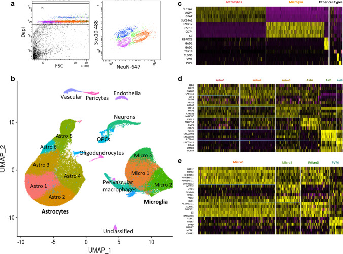
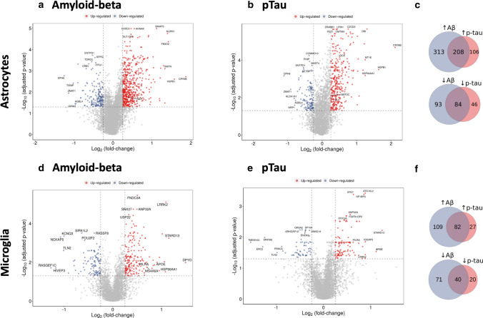
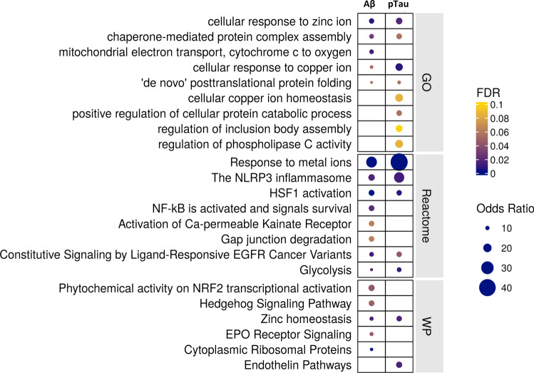
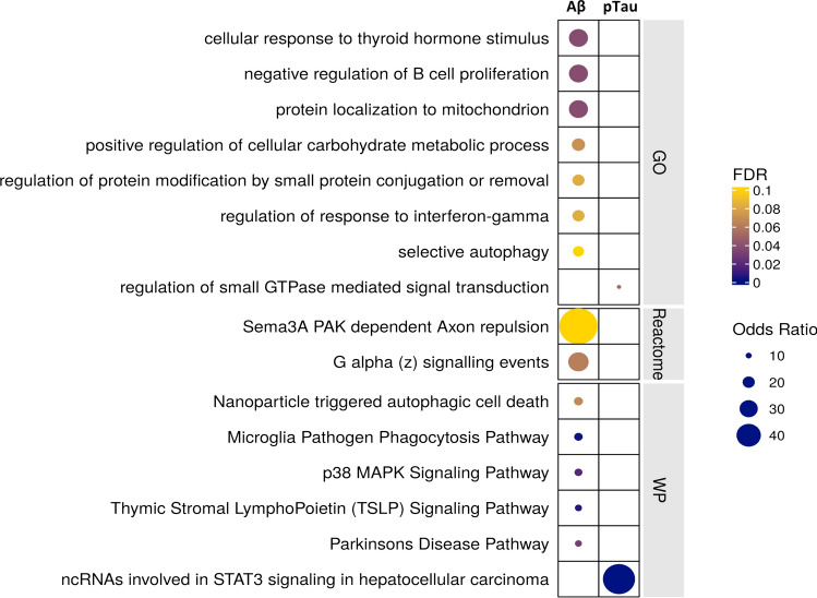
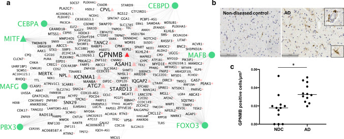
To better define roles that astrocytes and microglia play in Alzheimer’s disease (AD), we used single-nuclei RNA-sequencing to comprehensively characterise transcriptomes in astrocyte and microglia nuclei selectively enriched during isolation post-mortem from neuropathologically defined AD and control brains with a range of amyloid-beta and phospho-tau (pTau) pathology. Significant differences in glial gene expression (including AD risk genes expressed in both the astrocytes [ CLU , MEF2C , IQCK ] and microglia [ APOE , MS4A6A , PILRA ]) were correlated with tissue amyloid or pTau expression. The differentially expressed genes were distinct between with the two cell types and pathologies, although common (but cell-type specific) gene sets were enriched with both pathologies in each cell type. Astrocytes showed enrichment for proteostatic, inflammatory and metal ion homeostasis pathways. Pathways for phagocytosis, inflammation and proteostasis were enriched in microglia and perivascular macrophages with greater tissue amyloid, but IL1-related pathway enrichment was found specifically in association with pTau. We also found distinguishable sub-clusters in the astrocytes and microglia characterised by transcriptional signatures related to either homeostatic functions or disease pathology. Gene co-expression analyses revealed potential functional associations of soluble biomarkers of AD in astrocytes ( CLU ) and microglia ( GPNMB ). Our work highlights responses of both astrocytes and microglia for pathological protein clearance and inflammation, as well as glial transcriptional diversity in AD.
Overview
The claims and statistics of this paper haven’t been checked yet.
Run the check — free, no account needed. It reads the open-access text and checks the sources it cites.
Integrity signals
Registry
Cites
Statistics
Figures
Verification
Claims the paper makes
The claims and statistics of this paper haven’t been checked yet.
Run the check — free, no account needed. It reads the open-access text and checks the sources it cites.
Claims other papers make about this one
Each is checked against this paper’s own text: does it say what the citing paper says it does?
The cited source doesn’t support the claim as written.
In the citing paper
“resulting in over 100,000 astrocyte nuclei in each brain region with ≈6,000 average total number of reads and ≈2,500 average total number of genes detected, which is orders of magnitude higher than previous studies9–15”
From Astrocyte transcriptomic changes along the spatiotemporal progression of Alzheimer’s disease (2024) · cites this paper as [15]
Checked by a reasoning model — may be wrong.
Experimental — accuracy not yet measuredThe source backs part of the claim, not all of it — or backs it for a different population, condition or comparison.
In the citing paper
“may have confounded previous studies that compared only control and severe AD donors or grouped late- and end-stage donors (Braak stage V/VI) together9–15”
From Astrocyte transcriptomic changes along the spatiotemporal progression of Alzheimer’s disease (2024) · cites this paper as [15]
Experimental — accuracy not yet measuredThe cited source says what the claim attributes to it.
In the citing paper
“We found increased numbers of senescence genes upregulated in both microglia (YPEL3, IQGAP2, SQSTM1) and astrocytes (GLB1, SQSTM1, MRAS) with greater levels of tissue β-amyloid densities [66] (Supplementary Fig. 7a, b, Supplementary data S7).”
From Characterisation of premature cell senescence in Alzheimer's disease using single nuclear transcriptomics (2024) · cites this paper as [66]
The cited source says what the claim attributes to it.
In the citing paper
“Finally, we also explored senescence pathway expression in the six astroglial sub-populations that we described previously [66], but did not find that any of these sub-populations showed greater expression of the senescence gene set with AD (Fig. 6c, d, Supplementary Fig. 10c).”
From Characterisation of premature cell senescence in Alzheimer's disease using single nuclear transcriptomics (2024) · cites this paper as [66]
The cited source says what the claim attributes to it.
In the citing paper
“our sample sizes are similar to those reported in previous publications9–17”
From Astrocyte transcriptomic changes along the spatiotemporal progression of Alzheimer’s disease (2024) · cites this paper as [15]
In the citing paper
“The study found that microglial transcripts were most highly positively associated with tissue Aβ and tissue pTau density.”
From Microglia in neurodegenerative diseases: mechanism and potential therapeutic targets (2023) · cites this paper as [145]
Self-check
Percentages, sums and reported differences re-computed from the paper’s own numbers.
The self-check needs the paper’s full text.
Citations
Cites
Controlling the False Discovery Rate: A Practical and Powerful Approach to Multiple Testing
Benjamini, Y., & Hochberg, Y. (1995) · Journal of the Royal Statistical Society Series B: Statistical Methodology · doi:10.1111/j.2517-6161.1995.tb02031.x · Cited by 109,482
Cytoscape: A software Environment for integrated models of biomolecular interaction networks
Shannon, P., Markiel, A., Ozier, O., Baliga, N. S., Wang, J. T., Ramage, D., Amin, N., Schwikowski, B., & Ideker, T. (2003) · Genome Research · doi:10.1101/gr.1239303 · Cited by 51,888
limma powers differential expression analyses for RNA-sequencing and microarray studies
Ritchie, M. E., Phipson, B., Wu, D., Hu, Y., Law, C. W., Shi, W., & Smyth, G. K. (2015) · Nucleic Acids Research · doi:10.1093/nar/gkv007 · Cited by 40,458
Comprehensive Integration of Single-Cell Data
Stuart, T., Butler, A., Hoffman, P., Hafemeister, C., Papalexi, E., Mauck, W. M., Hao, Y., Stoeckius, M., Smibert, P., & Satija, R. (2019) · Cell · doi:10.1016/j.cell.2019.05.031 · Cited by 15,453
Inference and analysis of cell-cell communication using CellChat
Jin, S., Guerrero-Juarez, C. F., Zhang, L., Chang, I., Ramos, R., Kuan, C.-H., Myung, P., Plikus, M. V., & Nie, Q. (2021) · Nature Communications · doi:10.1038/s41467-021-21246-9 · Cited by 8,409
Neurotoxic reactive astrocytes are induced by activated microglia
Liddelow, S. A., Guttenplan, K. A., Clarke, L. E., Bennett, F. C., Bohlen, C. J., Schirmer, L., Bennett, M. L., Münch, A. E., Chung, W.-S., Peterson, T. C., et al. (2017) · Nature · doi:10.1038/nature21029 · Cited by 7,971
Enrichr: interactive and collaborative HTML5 gene list enrichment analysis tool
Chen, E. Y., Tan, C. M., Kou, Y., Duan, Q., Wang, Z., Meirelles, G. V., Clark, N. R., & Ma’ayan, A. (2013) · BMC Bioinformatics · doi:10.1186/1471-2105-14-128 · Cited by 7,814
Scenic: single-cell regulatory network inference and clustering.
Aibar, S., González-Blas, C. B., Moerman, T., Huynh-Thu, V. A., Imrichova, H., Hulselmans, G., Rambow, F., Marine, J.-C., Geurts, P., Aerts, J., et al. (2017) · Nature Methods · doi:10.1038/nmeth.4463 · Cited by 6,520
From Louvain to Leiden: guaranteeing well-connected communities
Traag, V. A., Waltman, L., & van Eck, N. J. (2019) · Scientific Reports · doi:10.1038/s41598-019-41695-z · Cited by 5,635 · cited 1×
“Sample integration was performed using Liger [72] and dimension reduction using PCA and UMAP, and finally, clustering was performed using the Leiden algorithm [65].”
MethodA Unique Microglia Type Associated with Restricting Development of Alzheimer’s Disease
Keren-Shaul, H., Spinrad, A., Weiner, A., Matcovitch-Natan, O., Dvir-Szternfeld, R., Ulland, T. K., David, E., Baruch, K., Lara-Astaiso, D., Toth, B., et al. (2017) · Cell · doi:10.1016/j.cell.2017.05.018 · Cited by 5,493 · cited 11×
“Our data show that this transcriptional functional diversity reflects distinguishable sub-types of human astrocytes and microglia enriched in gene sets related to (but distinct from) those previously described in transgenic amyloid or tau mouse models or with ageing [19, 27, 43, 48].”
BackgroundInfluential
Cited by
Sorted by citations
Microglia in neurodegenerative diseases: mechanism and potential therapeutic targets
Gao, C., Jiang, J., Tan, Y., & Chen, S. (2023) · Signal Transduction and Targeted Therapy · doi:10.1038/s41392-023-01588-0 · Cited by 1,616
“145 These results suggest that microglia respond to the pathology of AD.”
Neuroinflammation in Alzheimer disease
Heneka, M. T., van der Flier, W. M., Jessen, F., Hoozemanns, J., Thal, D. R., Boche, D., Brosseron, F., Teunissen, C., Zetterberg, H., Jacobs, A. H., et al. (2025) · Nature Reviews Immunology · doi:10.1038/s41577-024-01104-7 · Cited by 638
“Nevertheless, isolation of microglial cells from AD brains and subsequent analysis of the transcriptome has provided important insights into microglial cell activation states 220,222 .”
Nutma, E., Fancy, N. N., Weinert, M., Tsartsalis, S., Marzin, M. C., Muirhead, R. C. J., Falk, I. N., Breur, M., de Bruin, J., Hollaus, D., et al. (2023) · Nature Communications · doi:10.1038/s41467-023-40937-z · Cited by 204
“Quality control, sampleintegration, dimension reduction and clustering were performed using default parameters as previously described 41,83 .”
InfluentialSelf-citationDolan, M.-J., Therrien, M., Jereb, S., Kamath, T., Gazestani, V., Atkeson, T., Marsh, S. E., Goeva, A., Lojek, N. M., Murphy, S., et al. (2023) · Nature Immunology · doi:10.1038/s41590-023-01558-2 · Cited by 185
Human Microglia Show Unique Transcriptional Changes in Alzheimer's Disease
Prater, K. E., Green, K. J., Mamde, S., Sun, W., Cochoit, A., Smith, C. L., Chiou, K. L., Heath, L., Rose, S. E., Wiley, J., et al. (2023) · Nature Aging · doi:10.1038/s43587-023-00424-y · Cited by 164
Jie, F., Yang, X., Yang, B., Liu, Y., Wu, L., & Lu, B. (2022) · Biomedicine & Pharmacotherapy · doi:10.1016/j.biopha.2022.113317 · Cited by 161
Insights into Alzheimer’s disease from single-cell genomic approaches
Murdock, M. H., & Tsai, L.-H. (2023) · Nature Neuroscience · doi:10.1038/s41593-022-01222-2 · Cited by 148
“Smith et al.(134) GSE160936 12 Entorhinal and somatosensory cortex NEUN/SOX10 52,706 astrocytes and 27,592 microglia”
BackgroundAstrocyte transcriptomic changes along the spatiotemporal progression of Alzheimer’s disease
Serrano-Pozo, A., Li, H., Li, Z., Muñoz-Castro, C., Jaisa-aad, M., Healey, M. A., Welikovitch, L. A., Jayakumar, R., Bryant, A. G., Noori, A., et al. (2024) · Nature Neuroscience · doi:10.1038/s41593-024-01791-4 · Cited by 140
Inflammatory aspects of Alzheimer’s disease
Lucena, P. B., & Heneka, M. T. (2024) · Acta Neuropathologica · doi:10.1007/s00401-024-02790-2 · Cited by 152
Microglia-Astrocyte Communication in Alzheimer’s Disease
Wu, Y., & Eisel, U. L. M. (2023) · Journal of Alzheimer s Disease · doi:10.3233/jad-230199 · Cited by 125
“nuclei RNA-sequencing transcriptomics [167].”
Rodríguez-Giraldo, M., González-Reyes, R. E., Ramírez-Guerrero, S., Bonilla-Trilleras, C. E., Guardo-Maya, S., & Nava-Mesa, M. O. (2022) · International Journal of Molecular Sciences · doi:10.3390/ijms232113630 · Cited by 122
“Moreover, increased expression of CLU was associated with phosphorylated tau pathology in astrocytes [74].”
BackgroundInfluentialMa, Y., Deng, C., Zhou, Y., Zhang, Y., Qiu, F., Jiang, D., Zheng, G., Li, J., Shuai, J., Zhang, Y., et al. (2023) · Cell Genomics · doi:10.1016/j.xgen.2023.100383 · Cited by 113
Tsartsalis, S., Sleven, H., Fancy, N., Wessely, F., Smith, A. M., Willumsen, N., Cheung, T. K. D., Rokicki, M. J., Chau, V., Ifie, E., et al. (2024) · Nature Communications · doi:10.1038/s41467-024-46630-z · Cited by 95
“This downregulation could represent a partially adaptive response to chronically enhanced TLR signalling with AD 12 13,14 .”
Self-citationRevisiting the critical roles of reactive astrocytes in neurodegeneration
Qian, K., Jiang, X., Liu, Z., Zhang, J., Fu, P. F., Su, Y., Brazhe, N. A., Liu, D., & Zhu, L. (2023) · Molecular Psychiatry · doi:10.1038/s41380-023-02061-8 · Cited by 101
“Recently, the application of new methods, including snRNA-seq, has provided clues for astrocyte subset identi fi cation [135].”
MethodQian, Z., Qin, J., Lai, Y., Zhang, C., & Zhang, X. (2023) · Biomolecules · doi:10.3390/biom13040692 · Cited by 81
Seeker, L. A., Bestard-Cuche, N., Jäkel, S., Kazakou, N.-L., Bøstrand, S. M. K., Wagstaff, L. J., Cholewa-Waclaw, J., Kilpatrick, A. M., van Bruggen, D., Kabbe, M., et al. (2023) · Acta Neuropathologica Communications · doi:10.1186/s40478-023-01568-z · Cited by 72
“Human grey matter astrocytes are known to be transcriptionally heterogeneous [70, 90], but data for human white matter astrocytes is much sparser [33, 65].”
BackgroundNeurodegeneration cell per cell
Balusu, S., Praschberger, R., Lauwers, E., Strooper, B. D., & Verstreken, P. (2023) · Neuron · doi:10.1016/j.neuron.2023.01.016 · Cited by 53
Fancy, N. N., Smith, A. M., Caramello, A., Tsartsalis, S., Davey, K., Muirhead, R. C. J., McGarry, A., Jenkyns, M. H., Schneegans, E., Chau, V., et al. (2024) · Acta Neuropathologica · doi:10.1007/s00401-024-02727-9 · Cited by 48
Self-citationKuntić, M., Hahad, O., Münzel, T. F., & Daiber, A. (2024) · Antioxidants · doi:10.3390/antiox13030266 · Cited by 53
Kim, G. S., Harmon, E., Gutierrez, M. E. C., Kim, S., Vance, L., Burrous, H., Stephenson, J. M., Chauhan, A., Banerjee, A., Wise, Z., et al. (2025) · Nature Communications · doi:10.1038/s41467-025-56847-1 · Cited by 53
Alzheimer’s disease-related transcriptional sex differences in myeloid cells
Coales, I., Tsartsalis, S., Fancy, N., Weinert, M., Clode, D., Owen, D., & Matthews, P. M. (2022) · Journal of Neuroinflammation · doi:10.1186/s12974-022-02604-w · Cited by 49
“However, due to the relatively rarity of microglia in the brain, in this un-enriched population only a low resolution of microglia was be obtained (n = 1920 microglial nuclei compared to n = 27,592 in our enriched discovery data set [39] and n = 148,606 in the replication data set [41]).”
BackgroundMethodResultInfluentialSelf-citationRevealing cell vulnerability in Alzheimer’s disease by single-cell transcriptomics
Saura, C. A., Deprada-Fernandez, Á., Capilla-López, M. D., & Parra‐Damas, A. (2022) · Seminars in Cell and Developmental Biology · doi:10.1016/j.semcdb.2022.05.007 · Cited by 48
Microglia in Health and Diseases: Integrative Hubs of the Central Nervous System (CNS)
Sierra, A., Miron, V. E., Paolicelli, R. C., & Ransohoff, R. M. (2024) · Cold Spring Harbor Perspectives in Biology · doi:10.1101/cshperspect.a041366 · Cited by 47
Reactive astrocytes secrete the chaperone HSPB1 to mediate neuroprotection
Yang, F., Beltran-Lobo, P., Sung, K., Goldrick, C., Croft, C. L., Nishimura, A., Hedges, E., Mahiddine, F., Troakes, C., Golde, T. E., et al. (2024) · Science Advances · doi:10.1126/sciadv.adk9884 · Cited by 46
“…that, in AD brain, HSPB1 is predominantly expressed in astrocytes, in line with previous immunohistochemistry studies in AD and other tauopathies, where HSPB1 expression was mostly attributed to glial cells ( 24, 63 ), and with recent single-cell transcriptomic studies in human AD brain ( 64–66 ).”
ResultHaglund, A., Zuber, V., Abouzeid, M., Yang, Y., Ko, J. H., Wiemann, L., Otero‐Jimenez, M., Muhammed, L., Feleke, R., Nott, A., et al. (2025) · Nature Genetics · doi:10.1038/s41588-024-02050-9 · Cited by 46
“…‘Bryois_192’ (192 total, including 105 mixed disease and 87 control cases obtained from their recent publication 7 ) and ‘MATTHEWS’ (new samples generated in-house consisting of 65 individuals split into 38 AD cases and 27 controls), a subset of which have been used in published work 26,27 .”
Self-citationCholesterol Dysmetabolism in Alzheimer’s Disease: A Starring Role for Astrocytes?
Staurenghi, E., Giannelli, S., Testa, G., Sottero, B., Leonarduzzi, G., & Gamba, P. (2021) · Antioxidants · doi:10.3390/antiox10121890 · Cited by 44
“Recently, an enrichment in genes involved in inflammation also emerged from the transcriptomic analysis of astrocytes from human AD brains [75].”
BackgroundKoskeridis, F., Fancy, N. N., Tan, P. F., Meena, D., Εvangelou, Ε., Elliott, P., Wang, D. Y. Q., Matthews, P. M., Dehghan, A., & Tzoulaki, I. (2024) · Nature Communications · doi:10.1038/s41467-024-53452-6 · Cited by 46
“…to retrieve data for single cells of the left ventricle from 6 heart failure (HF) cases and 7 healthy controls 60 , left ventricular single nuclei from 13 dilated cardiomyopathy (dCM) cases and 25 healthy controls 61 , and post-mortem brain single nuclei from 9 AD cases and 8 healthy controls 62 .”
Self-citationGalectin-3 aggravates microglial activation and tau transmission in tauopathy
Siew, J. J., Chen, H.-M., Chiu, F., Lee, C., Chang, Y., Chen, H.-L., Nguyen, T. N. A., Liao, H.-T., Liu, M., Hagar, H. C., et al. (2023) · Journal of Clinical Investigation · doi:10.1172/jci165523 · Cited by 47
Synapses, Microglia, and Lipids in Alzheimer’s Disease
Paasila, P. J., Aramideh, J. A., Sutherland, G. T., & Graeber, M. B. (2022) · Frontiers in Neuroscience · doi:10.3389/fnins.2021.778822 · Cited by 40
“Indeed, single-nucleus RNAseq has demonstrated diverse astrocytic signatures in AD characterised by the enrichment of inflammatory, proteostatic genes, and metal ion homeostatic genes (Smith et al., 2021).”
BackgroundAquaporin 4 is differentially increased and dislocated in association with tau and amyloid-beta
Kecheliev, V., Boss, L., Maheshwari, U., Konietzko, U., Keller, A., Razansky, D., Nitsch, R. M., Klohs, J., & Ni, R. (2023) · Life Sciences · doi:10.1016/j.lfs.2023.121593 · Cited by 36
Lin, J.-P., Brake, A., Donadieu, M., Lee, A., Smith, G., Hu, K., Nair, G., Kawaguchi, R., Sati, P., Geschwind, D. H., et al. (2025) · Science · doi:10.1126/science.adp6325 · Cited by 35
Mosaic loss of Chromosome Y in aged human microgliaPreprint
Vermeulen, M., Pearse, R. V., Young‐Pearse, T. L., & Mostafavi, S. (2022) · Genome Research · doi:10.1101/gr.276409.121 · Cited by 35
“Smith et al. (2022) enriched for microglia and astrocyte nuclei from the ECs and SSCs of AD diagnosed and control donors, whereas Olah et al. (2020) and Mancuso et al. (2019) enriched for microglia from DLPFC and temporal cortex tissue, respectively.”
InfluentialLiu, Y., Tan, Y., Zhang, Z., Li, H., Yi, M., Zhang, Z., Hui, S., & Peng, W. (2022) · Ageing Research Reviews · doi:10.1016/j.arr.2022.101831 · Cited by 34
Sighencea, M. G., Popescu, R. Ș., & Trifu, S. C. (2024) · International Journal of Molecular Sciences · doi:10.3390/ijms252212311 · Cited by 33
“Indeed, astrocyte-mediated tauopathy risk loci, which encompass genes related to clustering, Myocyte Enhancer Factor 2C, and IQ Domain-Containing Protein K, have been identified in the postmortem brains of AD patients through single-nuclei RNA-sequencing transcriptomics [90].”
Lish, A. M., Ashour, N., Pearse, R. V., Galle, P. C., Orme, G. A., Heuer, S. E., Benoit, C. R., Alexander, K. D., Grogan, E. F. L., Terzioğlu, G., et al. (2025) · Cell Reports · doi:10.1016/j.celrep.2025.115777 · Cited by 35
“Given that AD mouse models and postmortem human AD brain tissues display upregulated DAM signatures, 4,61,62 we next investigated whether the DAM-like cluster in iMG TCs was altered in the presence of fAD neurons.”
Genetic insights into immune mechanisms of Alzheimer’s and Parkinson’s disease
Nott, A., & Holtman, I. R. (2023) · Frontiers in Immunology · doi:10.3389/fimmu.2023.1168539 · Cited by 33
Tarawneh, R. (2023) · Biomolecules · doi:10.3390/biom13050830 · Cited by 29
“Several AD risk genes are expressed in astrocytes (APOE4, WWOX, CLU, CDK2AP1, MEF2C, and IQCK) and are linked to both amyloid and tau metabolism [95,103].”
BackgroundSattarov, R., Havers, M., Orbjörn, C., Stomrud, E., Janelidze, S., Laurell, T., & Mattsson, N. (2024) · Scientific Reports · doi:10.1038/s41598-024-75406-0 · Cited by 29
“Microglial transcripts are associated with tissue Aβ and tau density 77 .”
BackgroundSt‐Pierre, M., VanderZwaag, J., Loewen, S. M., & Tremblay, M. (2022) · Frontiers in Cellular Neuroscience · doi:10.3389/fncel.2022.932572 · Cited by 27
Wang, Z., Ma, Y., Sun, Y., Tan, L., Wang, H.-F., Yu, J., & Initiative, A. D. N. (2022) · Journal of Neuroinflammation · doi:10.1186/s12974-022-02679-5 · Cited by 29
“However, excessive glial cell activation may be a direct pathway that causes Aβ deposition, tau pathology, and neuronal damage in AD [1, 6].”
BackgroundSt‐Pierre, M., Carrier, M., Ibáñez, F. G., Khakpour, M., Wallman, M., Parent, M., & Tremblay, M. (2023) · Journal of Neuroinflammation · doi:10.1186/s12974-023-02752-7 · Cited by 28
“This emerging role of astrocytes in AD is corroborated notably by genome-wide association studies highlighting several astrocytic gene variants connected with a higher risk of developing lateonset AD in humans [42, 43].”
ResultZou, D., Huang, X., Lan, Y., Pan, M., Xie, J., Huang, Q., Zeng, J., Zou, C., Pei, Z., Zou, C., et al. (2024) · Pharmacological Research · doi:10.1016/j.phrs.2024.107098 · Cited by 28
The cGAS−STING-mediated ROS and ferroptosis are involved in manganese neurotoxicity
Zhang, Z., Yang, J., Zhou, Q., Zhong, S., Liu, J., Zhang, X., Chang, X., & Wang, H. (2024) · Journal of Environmental Sciences · doi:10.1016/j.jes.2024.05.003 · Cited by 27
Neurodegenerative phagocytes mediate synaptic stripping in Neuro-HIV
Liberto, G. D., Égervári, K., Kreutzfeldt, M., Schürch, C. M., Hewer, E., Wagner, I., Pasquier, R. D., & Merkler, D. (2022) · Brain · doi:10.1093/brain/awac102 · Cited by 28
Microglia as a cellular target of diclofenac therapy in Alzheimer’s disease
Stopschinski, B. E., Weideman, R. A., McMahan, D., Jacob, D. A., Little, B. B., Chiang, H.-S., Saez Calveras, N., & Stuve, O. (2023) · Therapeutic Advances in Neurological Disorders · doi:10.1177/17562864231156674 · Cited by 26
“33,34 Taken together, further studies are needed to better understand the role of various astrocyte states in AD.”
BackgroundGlia as antigen-presenting cells in the central nervous system
Sutter, P. A., & Crocker, S. J. (2022) · Current Opinion in Neurobiology · doi:10.1016/j.conb.2022.102646 · Cited by 24
Hassan, H., Rawlinson, C., Lan, Y., Jenkins, S. I., & Chen, R. (2025) · Biomolecules · doi:10.3390/biom15111629 · Cited by 30
“However, prolonged exposure to these pathological proteins can convert microglia from a protective to a dysfunctional state, thereby contributing to AD progression [62].”
Xie, X., Hao, J.-J., Shi, J.-Z., Zhou, Y., Liu, P., Wang, F., Zheng, X.-M., Yu, X., Wang, C., Yan, Y., et al. (2023) · International Immunopharmacology · doi:10.1016/j.intimp.2023.109994 · Cited by 23
Plasticity of human microglia and brain perivascular macrophages in aging and Alzheimer’s disease
Lee, D., Vicari, J. M., Porras, C., Spencer, C., Pjanic, M., Wang, X., Kinrot, S., Weiler, P., Kosoy, R., Bendl, J. M., et al. (2026) · Nature Genetics · doi:10.1038/s41588-026-02716-6 · Cited by 32
“The MITF network is notably enriched with markers of phagocytic activity and has been recognized as a crucial regulator of homeostatic microglial functions, particularly in promoting autophagic states and enabling microglia to migrate, detect and clear Aβ/tau proteinopathies 22,23,69–71 .”
Asante, I., Louie, S. G., & Yassine, H. N. (2022) · Annals of the New York Academy of Sciences · doi:10.1111/nyas.14907 · Cited by 23
Luo, D., Li, J., Liu, H., Wang, J., Xia, Y., Qiu, W., Wang, N., Wang, X., Wang, X., Ma, C., et al. (2023) · Advanced Science · doi:10.1002/advs.202300876 · Cited by 20
Liu, M., Zhu, J., Zheng, J., Han, X., Jiang, L., Tong, X., Ke, Y., Guo, Z., Huang, W., Cong, J., et al. (2025) · Cell Reports · doi:10.1016/j.celrep.2025.115343 · Cited by 24
Ceyzériat, K., Zilli, T., Millet, P., Koutsouvelis, N., Dipasquale, G., Fossey, C., Cailly, T., Fabis, F., Frisoni, G. B., Garibotto, V., et al. (2022) · Journal of Neuroinflammation · doi:10.1186/s12974-022-02673-x · Cited by 22
Güvenek, A., Parikshak, N., Zamolodchikov, D., Gelfman, S., Moscati, A., Dobbyn, L., Stahl, E. A., Shuldiner, A. R., & Coppola, G. (2024) · Communications Biology · doi:10.1038/s42003-024-06684-7 · Cited by 23
Müller, M. A., Zweig, N., Spengler, B., Weinert, M., & Heiles, S. (2023) · Analytical Chemistry · doi:10.1021/acs.analchem.3c01533 · Cited by 19
Self-citationProteomics of the astrocyte secretome reveals changes in their response to soluble oligomeric Aβ
Matafora, V., Gorb, A., Yang, F., Noble, W. J., Bachi, A., Perez‐Nievas, B. G., & Jiménez-Sánchez, M. (2023) · Journal of Neurochemistry · doi:10.1111/jnc.15875 · Cited by 19
Minding the Gap: Exploring Neuroinflammatory and Microglial Sex Differences in Alzheimer’s Disease
Reed-Geaghan, E. G., & Keller-Norrell, P. R. (2023) · International Journal of Molecular Sciences · doi:10.3390/ijms242417377 · Cited by 19
Zhan, H., Cammann, D., Cummings, J. L., Dong, X., & Chen, J. (2025) · Journal of Translational Medicine · doi:10.1186/s12967-025-06317-5 · Cited by 17
“In addition, Smith et al. reported that the PILRA gene is upregulated in microglia isolated from neuropathologically defined AD brains and is correlated with Aβ and p-tau expression [96].”
Microglial apolipoprotein E particles contribute to neuronal senescence and synaptotoxicity
Wang, N., Cai, L., Pei, X., Lin, Z., Huang, L., Liang, C., Wei, M., Shao, L., Guo, T., Huang, F., et al. (2024) · iScience · doi:10.1016/j.isci.2024.110006 · Cited by 18
Chen, C., Lan, Z., Tang, X., Chen, W., Zhou, X. W., Su, H., Su, R., Chen, Z., Chen, H., Guo, Y., et al. (2024) · Annals of Neurology · doi:10.1002/ana.27001 · Cited by 16
Aghaizu, N. D., Jolly, S., Samra, S. K., Kalmár, B., Craessaerts, K., Greensmith, L., Salinas, P. C., Strooper, B. D., & Whiting, P. J. (2023) · eNeuro · doi:10.1523/eneuro.0306-22.2022 · Cited by 14
“In fact, numerous single-cell RNA-Seq analyses have identified gene expression signatures that differed between mouse and human DAM/ARM populations (Grubman et al., 2019; Mathys et al., 2019; Nguyen et al., 2020; Olah et al., 2020; Smith et al., 2022).”
BackgroundHe, Z., Chen, Q., Wang, K., Lin, J., Peng, Y., Zhang, J., Yan, X., & Yan, J. (2024) · European Journal of Neuroscience · doi:10.1111/ejn.16242 · Cited by 18
“Additionally, inflammatory pathways were significantly enriched in astrocytes, suggesting their proinflammatory role in AD progression (Smith et al., 2022).”
BackgroundSchauer, S. P., Tóth, B. I., Lee, J., Honigberg, L. A., Ramakrishnan, V., Jiang, J. Z., Kollmorgen, G., Bayfield, A., Wild, N., Hoffman, J., et al. (2024) · Alzheimer s & Dementia · doi:10.1002/alz.14346 · Cited by 17
“36,37 Together with the CSF YKL-40 observations, these findings were unexpected because semorinemab was developed on an IgG4 backbone, which has been demonstrated to have lower binding affinity to fragment crystallizable gamma receptor (FcR) and expected to have reduced immune effector function.”
Refining the interactions between microglia and astrocytes in Alzheimer’s disease pathology
Chen, J., Xu, S., Wang, L., Liu, X., Liu, G., Tan, Q., Li, W., Zhang, S., & Du, Y. (2025) · Neuroscience · doi:10.1016/j.neuroscience.2025.03.033 · Cited by 17
Mao, S., Qiao, R., Wang, Q., Shen, L., Li, D., Huo, X., Wang, J., Liu, K., Chen, W., Zhu, T., et al. (2025) · MedComm · doi:10.1002/mco2.70329 · Cited by 17
“Past studies have identified many AD-related risk genes expressed by astrocytes, such as APOE , SLC1A2 , CLU , MEF2C , IQCK , and so on [1, 51, 52].”
Kwon, S. H., Parthiban, S., Tippani, M., Divecha, H. R., Eagles, N. J., Lobana, J. S., Williams, S. R., Mak, M., Bharadwaj, R. A., Kleinman, J. E., et al. (2023) · GEN Biotechnology · doi:10.1089/genbio.2023.0019 · Cited by 14
Ma, Y., Xia, Y., Karako, K., Song, P., Tang, W., & Hu, X. (2025) · Molecular Neurobiology · doi:10.1007/s12035-025-04997-0 · Cited by 13
ApoE-calypse tau: ApoE–tau synergy in Alzheimer’s disease
Lennol, M. P., Bordier, C., Kamelher, L., Ulrich, J. D., Holtzman, D. M., & Gratuze, M. (2025) · The Journal of Experimental Medicine · doi:10.1084/jem.20250965 · Cited by 14
LRRK2 Kinase Inhibition Attenuates Astrocytic Activation in Response to Amyloid β1-42 Fibrils
Filippini, A., Salvi, V., Dattilo, V., Magri, C., Castrezzati, S., Veerhuis, R., Bosisio, D., Gennarelli, M., & Russo, I. (2023) · Biomolecules · doi:10.3390/biom13020307 · Cited by 13
“Given that multiple lines of evidence have indicated that amyloid‐β deposits trigger glia activation and inflammation [3,40], we investigated whether Aβ 1‐42 fibrils were able to activate primary astrocytes.”
BackgroundHu, Z., Yuan, Y.-Q., Tong, Z., Liao, M., Yuan, S., Jian, Y., Yang, J.-L., & Liu, W. (2023) · Molecular Neurobiology · doi:10.1007/s12035-023-03529-y · Cited by 13
MS4A superfamily molecules in tumors, Alzheimer’s and autoimmune diseases
Luo, X., Luo, B., Fei, L., Zhang, Q., Liang, X., Chen, Y., & Zhou, X. (2024) · Frontiers in Immunology · doi:10.3389/fimmu.2024.1481494 · Cited by 13
Liu, X., Wu, W., Li, X., Wang, C., Chai, K., Yuan, F., Zheng, H., Yao, Y., Li, C., Ye, Z., et al. (2024) · Neural Regeneration Research · doi:10.4103/nrr.nrr-d-23-01890 · Cited by 13
Potential of astrocytes in targeting therapy for Alzheimer’s disease
Bi, W., Lei, T., Cai, S., Zhang, X., Yang, Y., Xiao, Z., Wang, L., & Du, H. (2022) · International Immunopharmacology · doi:10.1016/j.intimp.2022.109368 · Cited by 13
Nutma, E., Fancy, N. N., Marzin, M. C., Tsartsalis, S., Muirhead, R. C. J., Falk, I. N., de Bruin, J., Hollaus, D., Pieterman, R., Anink, J. J., et al. (2022) · Research Square · doi:10.21203/rs.3.rs-1420033/v1 · Cited by 12
Self-citationTopalis, V., Voros, C., & Ziaka, M. (2025) · Journal of Geriatric Psychiatry and Neurology · doi:10.1177/08919887251361578 · Cited by 14
Nutma, E., Fancy, N. N., Weinert, M., Marzin, M. C., Tsartsalis, S., Muirhead, R. C. J., Falk, I. N., de Bruin, J., Hollaus, D., Pieterman, R., et al. (2022) · bioRxiv (Cold Spring Harbor Laboratory) · doi:10.1101/2022.05.11.491453 · Cited by 13
Self-citationJiao, H., Ge, Y., Huang, L.-Y., Liu, Y., Wu, B., Lian, P., Hao, Y., Han, S., Li, Y., Wu, K., et al. (2025) · Molecular Neurodegeneration · doi:10.1186/s13024-025-00887-0 · Cited by 11
Wickstead, E. S. (2023) · Biomedicines · doi:10.3390/biomedicines11082240 · Cited by 11
Yamakawa, M., & Rexach, J. E. (2024) · Biomedicines · doi:10.3390/biomedicines12020308 · Cited by 14
“The resulting microglial modules from human brain data captured reproducible states with distinct pathological associations and markers that have been validated in prior studies [9,10].”
BackgroundMethodInfluentialWei, J., Wang, M., Li, S., Han, R. F., Xu, W., Zhao, A., Yu, Q., Li, H., Li, M., & Chi, G. (2024) · Biomedicine & Pharmacotherapy · doi:10.1016/j.biopha.2024.116806 · Cited by 11
Associations of amyloid-β oligomers and plaques with neuropathology in the AppNL-G-F mouse
Tang, J., Huang, H. Y. R., Muirhead, R. C. J., Zhou, Y., Li, J., DeFelice, J., Kopanitsa, M. V., Serneels, L., Davey, K., Tilley, B. S., et al. (2024) · Brain Communications · doi:10.1093/braincomms/fcae218 · Cited by 11
“34 This emphasizes the protective functions of astrocyte activation.”
BackgroundSelf-citationLiu, H., Tan, A. Y. S., Mehrabi, N. F., Turner, C. P., Curtis, M. A., Faull, R. L. M., Dragunow, M., Singh‐Bains, M. K., & Smith, A. M. (2024) · Neurobiology of Disease · doi:10.1016/j.nbd.2024.106749 · Cited by 11
Self-citationInferring gene regulatory networks by hypergraph generative model
Su, G., Wang, H., Zhang, Y., Coster, A. C., Wilkins, M. R., Cañete, P. F., Yu, D., Yang, Y., & Zhang, W. (2025) · Cell Reports Methods · doi:10.1016/j.crmeth.2025.101026 · Cited by 17
HDAC Inhibitors recapitulate Human Disease-Associated Microglia Signatures in vitro
Haage, V. C., Tuddenham, J. F., Bautista, A. R., Garcia, F. G., White, C. C., Patel, R., Comandante-Lou, N., Marshe, V. S., Griffin, J. K., Zhou, Y., et al. (2024) · Brain Behavior and Immunity · doi:10.1016/j.bbi.2025.05.027 · Cited by 23
Maksour, S., & Ooi, L. (2023) · Journal of Neurochemistry · doi:10.1111/jnc.15885 · Cited by 12
TSPO contributes to neuropathology and cognitive deficits in Alzheimer’s disease
Ceyzériat, K., Badina, A. M., Abjean, L., Meyer, L., Bouteldja, F., Bałkota, M., Amossé, Q., Prudhomme, O., Middleton, R. J., Liu, G.-J., et al. (2025) · Journal of Neuroinflammation · doi:10.1186/s12974-025-03583-4 · Cited by 14
“Genome-wide association studies (GWAS) and single-cell RNA sequencing studies strongly suggested that glial cells are causally involved in the AD pathology [6, 105–109].”
Self-citationPenning, A., Snoeck, S., Garritsen, O., Tosoni, G., Hof, A., de Boer, F., van Hasenbroek, J., Zhang, L., Thrupp, N., Craessaerts, K., et al. (2024) · Scientific Reports · doi:10.1038/s41598-024-72096-6 · Cited by 11
“( E ) Re-plotting of data from Smith et al. 53 showing significantly increased NACC2 expression in astrocytes in human AD brain.”
BackgroundMethodAstrocytic proteostasis in the tale of aging and neurodegeneration
Cabral‐Miranda, F., Matias, I. C. P., & Gomes, F. C. A. (2024) · Ageing Research Reviews · doi:10.1016/j.arr.2024.102580 · Cited by 10
Common alterations to astrocytes across neurodegenerative disorders
Derevyanko, A., Tao, T., & Allen, N. J. (2025) · Current Opinion in Neurobiology · doi:10.1016/j.conb.2025.102970 · Cited by 10
Cell-death pathways and tau-associated neuronal vulnerability in Alzheimer’s disease
Lin, G., Chancellor, S. E., Kwon, T., Woodbury, M. E., Doering, A., Abdourahman, A., Bennett, R. E., Liao, F., Pastika, T., Tamm, J. A., et al. (2025) · Cell Reports · doi:10.1016/j.celrep.2025.115758 · Cited by 15
Nakatsuka, N. J., Adler, D. I., Jiang, L., Hartman, A., Cheng, E., Klann, E., & Satija, R. (2025) · Nature Communications · doi:10.1038/s41467-025-62579-z · Cited by 18
“…et al. 96 ), GSE157827 (Lau et al. 97 ), GSE163577 (Yang et al. 98 ), GSE183068 (Sayed et al. 99 ), GSE148822 (Gerrits et al. 100 ), GSE160936 (Smith et al. 101 ), GSE167494 (Sadick et al. 102 ), GSE157783 (Smajic et al. 10 ), GSE184950 (Wang et al. 15 ), GSE193688 (Adams et al. 14 ),…”
Astrocyte transcriptomic changes along the spatiotemporal progression of Alzheimer’s diseasePreprint
Serrano‐Pozo, A., Li, Z., Woodbury, M. E., Muñoz‐Castro, C., Wachter, A., Jayakumar, R., Bryant, A. G., Noori, A., Welikovitch, L. A., Hu, M., et al. (2022) · bioRxiv (Cold Spring Harbor Laboratory) · doi:10.1101/2022.12.03.518999 · Cited by 10
Pomeshchik, Y., Velásquez, E., Gil, J., Klementieva, O. L., Gidlöf, R., Sydoff, M., Bagnoli, S., Nacmias, B., Sorbi, S., Westergren‐Thorsson, G., et al. (2023) · Acta Neuropathologica Communications · doi:10.1186/s40478-023-01649-z · Cited by 10
ATP11B Modulates Microglial Lipid Metabolism and Alleviates Alzheimer's Disease Pathology
Zhang, Y., Zhang, S., Zhao, X., Wu, P., Ying, Y., Wu, L., Zhuang, J., Chen, Z., Chao, Y., Dong, X., et al. (2025) · MedComm · doi:10.1002/mco2.70139 · Cited by 10
“Additionally, there was a significant decrease in the expression of Atp11b of the brain tissue of AD patients (GSE160936) (Figure 1B) [29].”
Single-nucleus multi-omics identifies shared and distinct pathways in Pick’s and Alzheimer’s disease
Shi, Z., Das, S., Morabito, S., Stocksdale, J. T., Miyoshi, E., Srinivasan, S. S., Emerson, N., Shahin, A., Rahimzadeh, N., Cao, Z., et al. (2025) · Science Advances · doi:10.1126/sciadv.ads7973 · Cited by 9
Mechanisms of increased Alzheimer’s disease pathology with R47H and R62H TREM2 variants
Fancy, N. N., Willumsen, N., Chau, V. M. N., Boulger, S. L., Whitwell, H. J., Wang, W., Avot, B., Thomas, M., Talbot-Martin, J., Tsartsalis, S., et al. (2026) · Acta Neuropathologica · doi:10.1007/s00401-026-03036-z · Cited by 8
Self-citationTournier, B. B., Ceyzériat, K., Badina, A. M., Gloria, Y., Fall, A. B., Amossé, Q., Tsartsalis, S., & Millet, P. (2024) · NeuroImage · doi:10.1016/j.neuroimage.2024.120778 · Cited by 8
Self-citationTrivedi, M. R., Joshi, A. M., Shah, J., Readhead, B. P., Wilson, M. A., Su, Y., Reiman, E. M., Wu, T., & Wang, Q. (2025) · npj Aging · doi:10.1038/s41514-025-00258-5 · Cited by 9
Recent advances in Alzheimer’s disease: From molecular mechanisms to therapeutic strategies
Rudman, M. D., Ulrich, J. D., & Holtzman, D. M. (2026) · Cell · doi:10.1016/j.cell.2026.06.006 · Cited by 12
Gao, W., Kong, W., Wang, S., Wen, G., & Yu, Y. (2022) · Brain Sciences · doi:10.3390/brainsci12091196 · Cited by 9
“Therefore, to overcome these problems and to improve the expression of gene regulatory network (GRN) inference, the integration of other data is considered to be a modified approach [5,6].”
Background
Metadata
- Authors
- Smith, A. M., Davey, K., Tsartsalis, S., Khozoie, C., Fancy, N., Tang, S. S., Liaptsi, E., Weinert, M., McGarry, A., Muirhead, R. C. J., Gentleman, S., Owen, D. R., & Matthews, P. M.
- Affiliations
- Hammersmith Hospital (GB)
- University of Geneva (CH)
- Corresponding author
- Matthews, Paul M.
- ORCID
- Venue
- Acta Neuropathologica
- Published
- 2021-11-12
- Volume
- 143
- Issue
- 1
- Pages
- 75-91
- ISSN
- 0001-6322 (ISSN-L)
- 1432-0533
- PMC
- PMC8732962
- OpenAlex
- W3214651914
- Cited by
- 209
- References
- 81
- Citations by year
- 2026: 43 · 2025: 41 · 2024: 50 · 2023: 49 · 2022: 24 · 2021: 1
- FWCI
- 21.05
- Type
- journal article
- Language
- en
- Open access
- hybrid
- doi.org
- Locations
- Acta Neuropathologica (publishedVersion, cc-by)
- bioRxiv (Cold Spring Harbor Laboratory) (acceptedVersion, cc-by-nc-nd)
- PubMed Central (publishedVersion, cc-by)
- PubMed (publishedVersion)
- Archive ouverte UNIGE (University of Geneva) (publishedVersion, other-oa)
- Archive ouverte UNIGE (University of Geneva) (submittedVersion, cc-by)
- Funders
- National Science Foundation
- Alzheimer's Society
- National Institute for Health and Care Research (NIHR202376, NF-SI-0514-10022)
- Parkinson's UK
- Schweizerischer Nationalfonds zur Förderung der Wissenschaftlichen Forschung (191446)
- UK Dementia Research Institute
- Medical Research Council (MR/K501013/1, MR/N008219/1, MR/M024903/1, MR/T031891/1, MR/N026934/1, MC_PC_17114)
- NIHR Imperial Biomedical Research Centre
- Topics
- Life Sciences › Neuroscience › Cellular and Molecular Neuroscience › Alzheimer's disease research and treatments
- Life Sciences › Neuroscience › Neurology › Neuroinflammation and Neurodegeneration Mechanisms
- Health Sciences › Medicine › Psychiatry and Mental health › Dementia and Cognitive Impairment Research
- Keywords
- Alzheimer's disease, astrocytes, microglia, amyloid-beta, phosphorylated tau, single-nucleus RNA sequencing, postmortem brain, apolipoprotein E, clusterin, MEF2C, MS4A6A, proteostasis, inflammation, phagocytosis, metal homeostasis, perivascular macrophages, GPNMB, gene co-expression, disease biomarkers
- MeSH terms
- Aged, Aged, 80 and over, Alzheimer Disease, Astrocytes, Brain, Female, Humans, Male, Microglia, Transcriptome
About these checks
Registry, retraction, Cites and statistics results are computed, not judged, with one exception: a printed reference that no index lists is read once by a model before it counts as a citation error. Claims are extracted by a model and kept only when their quote appears verbatim in the text; each cited source is then checked, with a model’s help, for whether it says what the claim relies on. Those verdicts can be wrong — read the claim’s quote and the cited source before relying on one.
Statistics and arithmetic are recomputed, not judged.
Confidence bands are shown only for fields calibrated on our annotated test set. None is calibrated yet, so no band is shown.
What counts as a claim ›How we measure confidence ›How we check this ›
Are you an author, or do you see a problem with this page? Request a review
