

The DOI matches its Crossref record; 14 of the 14 references it cites were found in a registry or an index.
Pathologic and clinical correlates of region-specific brain GFAP in Alzheimer’s disease
Abstract
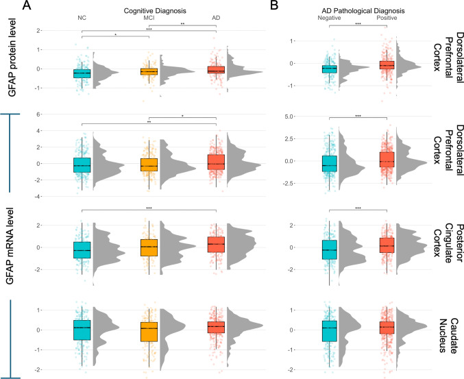
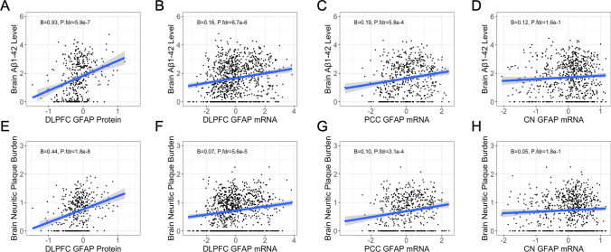
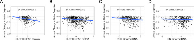
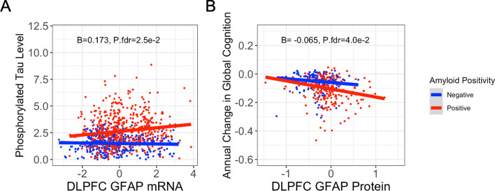
Plasma glial fibrillary acidic protein (GFAP) is an emerging biomarker of Alzheimer’s disease (AD), with higher blood GFAP levels linked to faster cognitive decline, particularly among individuals with high brain amyloid burden. However, few studies have examined brain GFAP expression to clarify if peripheral associations reflect brain changes. This study aimed to correlate region-specific GFAP mRNA expression ( n = 917) and protein abundance (n=386) with diverse neuropathological measures at autopsy in the Religious Orders Study and Rush Memory and Aging Project (ROS/MAP) and to characterize the interaction between brain GFAP and brain amyloid burden on downstream outcomes. We assessed GFAP gene expression in the dorsolateral prefrontal cortex, caudate nucleus, and posterior cingulate cortex with respect to core AD pathology (amyloid-β and tau), cerebrovascular (microinfarcts, macroinfarcts, and cerebral amyloid angiopathy [CAA]), proteinopathic (TDP-43, Lewy bodies), and cognitive outcomes. These associations were further examined at the protein level using tandem-mass tag proteomic measurements from the dorsolateral prefrontal cortex. We also assessed GFAP interactions with AD neuropathology on downstream outcomes. Cortical GFAP gene and protein expression were significantly upregulated in participants with a neuropathologically confirmed AD diagnosis at autopsy (all P FDR < 3.5e−4), but not in individuals positive for tau pathology and negative for amyloid pathology (all P FDR > 0.05). Higher cortical GFAP levels were associated with increased amyloid pathology, CAA pathology, and faster cognitive decline (all P FDR < 3.3e−3). GFAP’s associations with phosphorylated tau burden and cognition were influenced by amyloid burden, being most pronounced among amyloid-positive individuals, confirming previous in vivo biomarker observations. No associations were observed between GFAP gene expression and outcomes in the caudate nucleus. Our results support previous biomarker findings and suggest that higher brain GFAP levels are associated with higher brain amyloid burden and faster cognitive decline among amyloid-positive individuals.
Overview
The claims and statistics of this paper haven’t been checked yet.
Run the check — free, no account needed. It reads the open-access text and checks the sources it cites.
Integrity signals
Registry
Cites
Statistics
Figures
Verification
Claims the paper makes
The claims and statistics of this paper haven’t been checked yet.
Run the check — free, no account needed. It reads the open-access text and checks the sources it cites.
Claims other papers make about this one
Each is checked against this paper’s own text: does it say what the citing paper says it does?
In the citing paper
“Elevated levels of GFAP in plasma and serum are associated with the degree and rate of neurodegeneration, Aβ positivity and cognitive decline in individuals with AD [27, 28].”
Self-check
Percentages, sums and reported differences re-computed from the paper’s own numbers.
The self-check needs the paper’s full text.
Citations
Cites
Neurotoxic reactive astrocytes are induced by activated microglia
Liddelow, S. A., Guttenplan, K. A., Clarke, L. E., Bennett, F. C., Bohlen, C. J., Schirmer, L., Bennett, M. L., Münch, A. E., Chung, W.-S., Peterson, T. C., et al. (2017) · Nature · doi:10.1038/nature21029 · Cited by 7,971
Reactive astrocyte nomenclature, definitions, and future directions
Escartin, C., Galea, E., Lakatos, A., O’Callaghan, J. P., Petzold, G. C., Serrano-Pozo, A., Steinhäuser, C., Volterra, A., Carmignoto, G., Agarwal, A., et al. (2021) · Nature Neuroscience · doi:10.1038/s41593-020-00783-4 · Cited by 2,513
Religious Orders Study and Rush Memory and Aging Project
Bennett, D. A., Buchman, A. S., Boyle, P. A., Barnes, L. L., Wilson, R. S., & Schneider, J. A. (2018) · Journal of Alzheimer's Disease · doi:10.3233/jad-179939 · Cited by 1,260
Self-citationMassively parallel single-nucleus RNA-seq with DroNc-seq
Habib, N., Avraham-Davidi, I., Basu, A., Burks, T., Shekhar, K., Hofree, M., Choudhury, S. R., Aguet, F., Gelfand, E., Ardlie, K., et al. (2017) · Nature Methods · doi:10.1038/nmeth.4407 · Cited by 1,069
Sofroniew, M. V. (2015) · Cold Spring Harbor Perspectives in Biology · doi:10.1101/cshperspect.a020420 · Cited by 695
Benedet, A. L., Milà-Alomà, M., Vrillon, A., Ashton, N. J., Pascoal, T. A., Lussier, F., Karikari, T. K., Hourregue, C., Cognat, E., Dumurgier, J., et al. (2021) · JAMA Neurology · doi:10.1001/jamaneurol.2021.3671 · Cited by 638
Reactive Glia not only Associates with Plaques but also Parallels Tangles in Alzheimer’s Disease
Serrano-Pozo, A., Mielke, M. L., Gómez-Isla, T., Betensky, R. A., Growdon, J. H., Frosch, M. P., & Hyman, B. T. (2011) · The American Journal of Pathology · doi:10.1016/j.ajpath.2011.05.047 · Cited by 488
Attenuating astrocyte activation accelerates plaque pathogenesis in APP/PS1 mice
Kraft, A. W., Hu, X., Yoon, H., Yan, P., Xiao, Q., Wang, Y., Gil, S. C., Brown, J., Wilhelmsson, U., Restivo, J. L., et al. (2013) · The FASEB Journal · doi:10.1096/fj.12-208660 · Cited by 324
Ceyzériat, K., Ben Haim, L., Denizot, A., Pommier, D., Matos, M., Guillemaud, O., Palomares, M.-A., Abjean, L., Petit, F., Gipchtein, P., et al. (2018) · Acta Neuropathologica Communications · doi:10.1186/s40478-018-0606-1 · Cited by 256
Specific associations between plasma biomarkers and postmortem amyloid plaque and tau tangle loads
Salvadó, G., Ossenkoppele, R., Ashton, N. J., Beach, T. G., Serrano, G. E., Reiman, E. M., Zetterberg, H., Mattsson‐Carlgren, N., Janelidze, S., Blennow, K., et al. (2023) · EMBO Molecular Medicine · doi:10.15252/emmm.202217123 · Cited by 205
Research works printed but not linked: 38
- Ally M, Sugarman MA, Zetterberg H, Blennow K, Ashton NJ, Karikari TK et al (2023) Cross-sectional and longitudinal evaluation of plasma glial fibrillary acidic protein to detect and predict clinical syndromes of Alzheimer’s disease. Alzheimers Dement 15:e12492. https://doi.org/10.1002/dad2.12492 · DOI
- Arvanitakis Z, Leurgans SE, Barnes LL, Bennett DA, Schneider JA (2011) Microinfarct pathology, dementia, and cognitive systems. Stroke 42:722–727. https://doi.org/10.1161/STROKEAHA.110.595082 · DOI · Paper page
- Bellaver B, Povala G, Ferreira PCL, Ferrari-Souza JP, Leffa DT, Lussier FZ et al (2023) Astrocyte reactivity influences amyloid-β effects on tau pathology in preclinical Alzheimer’s disease. Nat Med 29:1775–1781. https://doi.org/10.1038/s41591-023-02380-x · DOI · Paper page
- Bennett DA, Schneider JA, Buchman AS, Barnes LL, Boyle PA, Wilson RS (2012) Overview and Findings from the rush memory and aging project. Curr Alzheimer Res 9:646–663. https://doi.org/10.2174/156720512801322663 · DOI
- Blasko I, Veerhuis R, Stampfer-Kountchev M, Saurwein-Teissl M, Eikelenboom P, Grubeck-Loebenstein B (2000) Costimulatory effects of interferon-γ and interleukin-1β or tumor necrosis factor α on the synthesis of Aβ1-40 and Aβ1-42 by human astrocytes. Neurobiology of Disease 7:682–689. https://doi.org/10.1006/nbdi.2000.0321 · DOI · Paper page
- Boyle PA, Yu L, Nag S, Leurgans S, Wilson RS, Bennett DA et al (2015) Cerebral amyloid angiopathy and cognitive outcomes in community-based older persons. Neurology 85:1930–1936. https://doi.org/10.1212/WNL.0000000000002175 · DOI · Paper page
- Chatterjee P, Pedrini S, Stoops E, Goozee K, Villemagne VL, Asih PR et al (2021) Plasma glial fibrillary acidic protein is elevated in cognitively normal older adults at risk of Alzheimer’s disease. Transl Psychiatry 11:27. https://doi.org/10.1038/s41398-020-01137-1 · DOI · Paper page
- Cousins KAQ, Irwin DJ, Chen-Plotkin A, Shaw LM, Arezoumandan S, Lee EB et al (2023) Plasma GFAP associates with secondary Alzheimer’s pathology in Lewy body disease. Ann Clin Transl Neurol 10:802–813. https://doi.org/10.1002/acn3.51768 · DOI
- Dai DL, Li M, Lee EB (2023) Human Alzheimer’s disease reactive astrocytes exhibit a loss of homeostastic gene expression. Acta Neuropathol Commun 11:127. https://doi.org/10.1186/s40478-023-01624-8 · DOI · Paper page
- Dark HE, Paterson C, Daya GN, Peng Z, Duggan MR, Bilgel M et al (2024) Proteomic indicators of health predict Alzheimer’s disease biomarker levels and dementia risk. Ann Neurol 95:260–273. https://doi.org/10.1002/ana.26817 · DOI
- Ingannato A, Bagnoli S, Mazzeo S, Giacomucci G, Bessi V, Ferrari C et al (2024) Plasma GFAP, NfL and pTau 181 detect preclinical stages of dementia. Front Endocrinol 15:1375302. https://doi.org/10.3389/fendo.2024.1375302 · DOI
- Johnson ECB, Dammer EB, Duong DM, Ping L, Zhou M, Yin L et al (2020) Large-scale proteomic analysis of Alzheimer’s disease brain and cerebrospinal fluid reveals early changes in energy metabolism associated with microglia and astrocyte activation. Nat Med 26:769–780. https://doi.org/10.1038/s41591-020-0815-6 · DOI · Paper page
- Kim J-H, Rahman MH, Park D, Jo M, Kim H-J, Suk K (2021) Identification of genetic modifiers of TDP-43: inflammatory activation of astrocytes for neuroinflammation. Cells 10:676. https://doi.org/10.3390/cells10030676 · DOI
- Koenigsknecht-Talboo J, Landreth GE (2005) Microglial phagocytosis induced by Fibrillar β-Amyloid and IgGs are differentially regulated by proinflammatory cytokines. J Neurosci 25:8240–8249. https://doi.org/10.1523/JNEUROSCI.1808-05.2005 · DOI · Paper page
- Koob AO, Paulino AD, Masliah E (2010) GFAP reactivity, apolipoprotein E redistribution and cholesterol reduction in human astrocytes treated with α-synuclein. Neurosci Lett 469:11–14. https://doi.org/10.1016/j.neulet.2009.11.034 · DOI
- Liddelow SA, Olsen ML, Sofroniew MV (2024) Reactive astrocytes and emerging roles in central nervous system (CNS) disorders. Cold Spring Harb Perspect Biol. https://doi.org/10.1101/cshperspect.a041356 · DOI · Paper page
- Mathys H, Davila-Velderrain J, Peng Z, Gao F, Mohammadi S, Young JZ, Menon M, He L, Abdurrob F, Jiang X, Martorell AJ, Ransohoff RM, Hafler BP, Bennett DA, Kellis M, Tsai L-H (2019) Single-cell transcriptomic analysis of Alzheimer’s disease. Nature 570:332–337. https://doi.org/10.1038/s41586-019-1195-2 · DOI · Paper page
- Nag S, Yu L, Boyle PA, Leurgans SE, Bennett DA, Schneider JA (2018) TDP-43 pathology in anterior temporal pole cortex in aging and Alzheimer’s disease. Acta Neuropathol Commun 6:33. https://doi.org/10.1186/s40478-018-0531-3 · DOI
- O’Leary LA, Davoli MA, Belliveau C, Tanti A, Ma JC, Farmer WT et al (2020) Characterization of vimentin-immunoreactive astrocytes in the human brain. Front Neuroanat 14:31. https://doi.org/10.3389/fnana.2020.00031 · DOI
- Oveisgharan S, Buchman AS, Yu L, Farfel J, Hachinski V, Gaiteri C et al (2018) APOE ε2ε4 genotype, incident AD and MCI, cognitive decline, and AD pathology in older adults. Neurology. https://doi.org/10.1212/WNL.0000000000005677 · DOI · Paper page
- Pais MV, Forlenza OV, Diniz BS (2023) Plasma biomarkers of Alzheimer’s disease: a review of available assays, recent developments, and implications for clinical practice. J Alzheimer’s Dis Rep 7:355–380. https://doi.org/10.3233/ADR-230029 · DOI
- Pereira JB, Janelidze S, Smith R, Mattsson-Carlgren N, Palmqvist S, Teunissen CE et al (2021) Plasma GFAP is an early marker of amyloid-β but not tau pathology in Alzheimer’s disease. Brain 144:3505–3516. https://doi.org/10.1093/brain/awab223 · DOI · Paper page
- Puspitasari V, Gunawan PY, Wiradarma HD, Hartoyo V (2019) Glial fibrillary acidic protein serum level as a predictor of clinical outcome in ischemic stroke. Open Access Maced J Med Sci 7:1471–1474. https://doi.org/10.3889/oamjms.2019.326 · DOI
- Rodríguez-Giraldo M, González-Reyes RE, Ramírez-Guerrero S, Bonilla-Trilleras CE, Guardo-Maya S, Nava-Mesa MO (2022) Astrocytes as a therapeutic target in Alzheimer’s disease-comprehensive review and recent developments. IJMS 23:13630. https://doi.org/10.3390/ijms232113630 · DOI
- Sánchez-Juan P, Valeriano-Lorenzo E, Ruiz-González A, Pastor AB, Rodrigo Lara H, López-González F et al (2024) Serum GFAP levels correlate with astrocyte reactivity, post-mortem brain atrophy and neurofibrillary tangles. Brain 147:1667–1679. https://doi.org/10.1093/brain/awae035 · DOI
- Schneider JA, Bienias JL, Wilson RS, Berry-Kravis E, Evans DA, Bennett DA (2005) The apolipoprotein E ε4 allele increases the odds of chronic cerebral infarction detected at autopsy in older persons. Stroke 36:954–959. https://doi.org/10.1161/01.STR.0000160747.27470.2a · DOI
- Schneider JA, Arvanitakis Z, Yu L, Boyle PA, Leurgans SE, Bennett DA (2012) Cognitive impairment, decline and fluctuations in older community-dwelling subjects with Lewy bodies. Brain 135:3005–3014. https://doi.org/10.1093/brain/aws234 · DOI · Paper page
- Seto M, Weiner RL, Dumitrescu L, Mahoney ER, Hansen SL, Janve V et al (2022) RNASE6 is a novel modifier of APOE-ε4 effects on cognition. Neurobiol Aging 118:66–76. https://doi.org/10.1016/j.neurobiolaging.2022.06.011 · DOI
- Shen X-Y, Gao Z-K, Han Y, Yuan M, Guo Y-S, Bi X (2021) Activation and role of astrocytes in ischemic stroke. Front Cell Neurosci 15:755955. https://doi.org/10.3389/fncel.2021.755955 · DOI
- Shir D, Graff-Radford J, Hofrenning EI, Lesnick TG, Przybelski SA, Lowe VJ et al (2022) Association of plasma glial fibrillary acidic protein (GFAP) with neuroimaging of Alzheimer’s disease and vascular pathology. Alzheimers Dement 14:e12291. https://doi.org/10.1002/dad2.12291 · DOI
- Smethurst P, Risse E, Tyzack GE, Mitchell JS, Taha DM, Chen Y-R et al (2020) Distinct responses of neurons and astrocytes to TDP-43 proteinopathy in amyotrophic lateral sclerosis. Brain 143:430–440. https://doi.org/10.1093/brain/awz419 · DOI
- Sofroniew MV (2020) Astrocyte reactivity: subtypes, states, and functions in CNS innate immunity. Trends Immunol 41:758–770. https://doi.org/10.1016/j.it.2020.07.004 · DOI · Paper page
- Thal DR, Rüb U, Orantes M, Braak H (2002) Phases of Aβ-deposition in the human brain and its relevance for the development of AD. Neurology 58:1791–1800. https://doi.org/10.1212/WNL.58.12.1791 · DOI · Paper page
- Verberk IMW, Laarhuis MB, Van Den Bosch KA, Ebenau JL, Van Leeuwenstijn M, Prins ND et al (2021) Serum markers glial fibrillary acidic protein and neurofilament light for prognosis and monitoring in cognitively normal older people: a prospective memory clinic-based cohort study. Lancet Healthy Longev 2:e87–e95. https://doi.org/10.1016/S2666-7568(20)30061-1 · DOI
- Wang X, Shi Z, Qiu Y, Sun D, Zhou H (2024) Peripheral GFAP and NfL as early biomarkers for dementia: longitudinal insights from the UK Biobank. BMC Med 22:192. https://doi.org/10.1186/s12916-024-03418-8 · DOI
- Winfree RL, Seto M, Dumitrescu L, Menon V, De Jager P, Wang Y et al (2023) TREM2 gene expression associations with Alzheimer’s disease neuropathology are region-specific: implications for cortical versus subcortical microglia. Acta Neuropathol 145:733–747. https://doi.org/10.1007/s00401-023-02564-2 · DOI
- Wunderlich MT, Wallesch CW, Goertler M (2006) Release of glial fibrillary acidic protein is related to the neurovascular status in acute ischemic stroke. Eur J Neurol 13:1118–1123. https://doi.org/10.1111/j.1468-1331.2006.01435.x · DOI
- Yang Z, Wang KKW (2015) Glial fibrillary acidic protein: from intermediate filament assembly and gliosis to neurobiomarker. Trends Neurosci 38:364–374. https://doi.org/10.1016/j.tins.2015.04.003 · DOI · Paper page
Cited by
Sorted by citations
Di Caro, V., Cho, E., Thiel, J., Lizama, B. N., Koel-Simmelink, M. J. A., Pandey, K., Duong, D., Seyfried, N. T., Grundman, M., Teunissen, C. E., et al. (2026) · Alzheimer's Research & Therapy · doi:10.1186/s13195-026-02025-4 · Cited by 1
Metadata
- Authors
- Phillips, J. M., Winfree, R. L., Seto, M., Schneider, J. A., Bennett, D. A., Dumitrescu, L. C., & Hohman, T. J.
- Venue
- Acta Neuropathologica
- Published
- 2024-11-24
- Volume
- 148
- Issue
- 1
- ISSN
- 1432-0533
- PMC
- PMC11586308
- Cited by
- 25
- References
- 14
- Language
- en
- Funders
- National Institute on Aging (F31 AG085980, U24 AG074855, R01 AG22018)
About these checks
Registry, retraction, Cites and statistics results are computed, not judged, with one exception: a printed reference that no index lists is read once by a model before it counts as a citation error. Claims are extracted by a model and kept only when their quote appears verbatim in the text; each cited source is then checked, with a model’s help, for whether it says what the claim relies on. Those verdicts can be wrong — read the claim’s quote and the cited source before relying on one.
Statistics and arithmetic are recomputed, not judged.
Confidence bands are shown only for fields calibrated on our annotated test set. None is calibrated yet, so no band is shown.
What counts as a claim ›How we measure confidence ›How we check this ›
Are you an author, or do you see a problem with this page? Request a review
