

The DOI matches its Crossref record; 176 of the 177 references it cites were found in a registry or an index.
DNA glycosylases: in DNA repair and beyond
Open accessAbstract
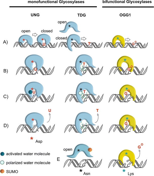
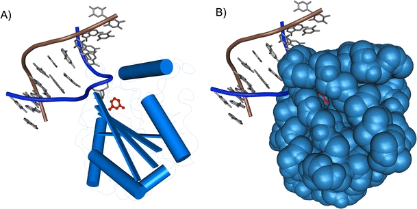
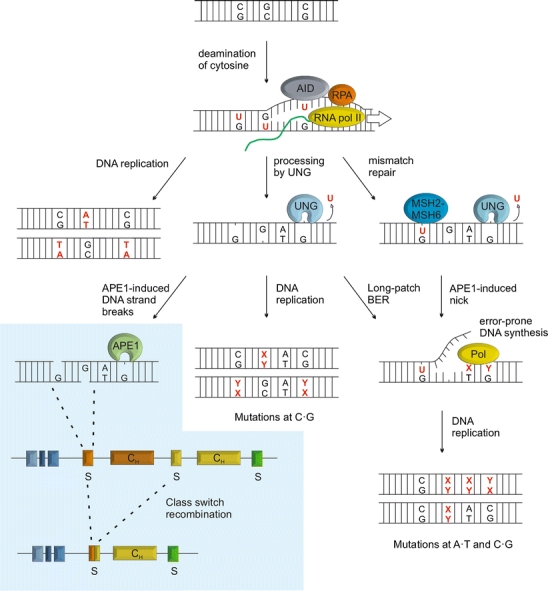
The base excision repair machinery protects DNA in cells from the damaging effects of oxidation, alkylation, and deamination; it is specialized to fix single-base damage in the form of small chemical modifications. Base modifications can be mutagenic and/or cytotoxic, depending on how they interfere with the template function of the DNA during replication and transcription. DNA glycosylases play a key role in the elimination of such DNA lesions; they recognize and excise damaged bases, thereby initiating a repair process that restores the regular DNA structure with high accuracy. All glycosylases share a common mode of action for damage recognition; they flip bases out of the DNA helix into a selective active site pocket, the architecture of which permits a sensitive detection of even minor base irregularities. Within the past few years, it has become clear that nature has exploited this ability to read the chemical structure of DNA bases for purposes other than canonical DNA repair. DNA glycosylases have been brought into context with molecular processes relating to innate and adaptive immunity as well as to the control of DNA methylation and epigenetic stability. Here, we summarize the key structural and mechanistic features of DNA glycosylases with a special focus on the mammalian enzymes, and then review the evidence for the newly emerging biological functions beyond the protection of genome integrity.
Overview
The claims and statistics of this paper haven’t been checked yet.
Run the check — free, no account needed. It reads the open-access text and checks the sources it cites.
Integrity signals
Registry
Cites
Statistics
Figures
Verification
Claims the paper makes
The claims and statistics of this paper haven’t been checked yet.
Run the check — free, no account needed. It reads the open-access text and checks the sources it cites.
Claims other papers make about this one
Each is checked against this paper’s own text: does it say what the citing paper says it does?
In the citing paper
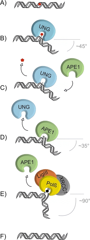
“The ligation process is carried by DNA ligase III working together with its cofactor X-ray repair cross-complementing protein 1(XRCC1) [111,112,113].”
From Focus on UV-Induced DNA Damage and Repair—Disease Relevance and Protective Strategies (2020) · cites this paper as [112]
Self-check
Percentages, sums and reported differences re-computed from the paper’s own numbers.
The self-check needs the paper’s full text.
Citations
Cites
1 retracted
DNA demethylation in hormone-induced transcriptional derepression
Kim, M. S., Kondo, T., Takada, I., Youn, M., Yamamoto, Y., Takahashi, S., Μatsumoto, T., Fujiyama, S., Shirode, Y., Yamaoka, I., et al. (2009) · Nature · doi:10.1038/nature08456 · Cited by 242
RetractedConversion of 5-Methylcytosine to 5-Hydroxymethylcytosine in Mammalian DNA by MLL Partner TET1
Tahiliani, M., Koh, K. P., Shen, Y., Pastor, W. A., Bandukwala, H., Brudno, Y., Agarwal, S., Iyer, L. M., Liu, D. R., Aravind, L., et al. (2009) · Science · doi:10.1126/science.1170116 · Cited by 5,836
Friedberg, E. C., Walker, G. C., Siede, W., Wood, R. D., Schultz, R. A., & Ellenberger, T. (2005) · doi:10.1128/9781555816704 · Cited by 5,683
Muramatsu, M., Kinoshita, K., Fagarasan, S., Yamada, S., Shinkai, Y., & Honjo, T. (2000) · Cell · doi:10.1016/s0092-8674(00)00078-7 · Cited by 3,475
Tet proteins can convert 5-methylcytosine to 5-formylcytosine and 5-carboxylcytosine
Ito, S., Shen, L., Dai, Q., Wu, S. C., Collins, L. B., Swenberg, J. A., He, C., & Zhang, Y. (2011) · Science · doi:10.1126/science.1210597 · Cited by 3,451
Tet-Mediated Formation of 5-Carboxylcytosine and Its Excision by TDG in Mammalian DNA
He, Y., Li, B.-Z., Li, Z., Liu, P., Wang, Y., Tang, Q., Ding, J., Jia, Y., Chen, Z., Li, L., et al. (2011) · Science · doi:10.1126/science.1210944 · Cited by 2,776
Ito, S., D’Alessio, A. C., Taranova, O., Hong, K., Sowers, L. C., & Zhang, Y. (2010) · Nature · doi:10.1038/nature09303 · Cited by 2,628
Highly Integrated Single-Base Resolution Maps of the Epigenome in Arabidopsis
Lister, R., O’Malley, R. C., Tonti‐Filippini, J., Gregory, B. D., Berry, C. C., Millar, A. H., & Ecker, J. R. (2008) · Cell · doi:10.1016/j.cell.2008.03.029 · Cited by 2,424
Lindahl, T. L., & Wood, R. D. (1999) · Science · doi:10.1126/science.286.5446.1897 · Cited by 1,521
Inherited variants of MYH associated with somatic G:C→T:A mutations in colorectal tumors
Tassan, N. A. A., Chmiel, N. H., Maynard, J. H., Fleming, N. I., Livingston, A. L., Williams, G. T., Hodges, A., Davies, D. R., David, S. S., Sampson, J. R., et al. (2002) · Nature Genetics · doi:10.1038/ng828 · Cited by 1,408
Cited by
Sorted by citations
Items 301–400 of 410
Sun, J., Wang, J., & Chen, X. (2023) · ACS Applied Materials & Interfaces · doi:10.1021/acsami.2c19257 · Cited by 2
The Base Excision Repair (BER) Pathway
Parsons, J. L., & Grundy, G. J. (2022) · Elsevier eBooks · doi:10.1016/b978-0-12-821618-7.00100-0 · Cited by 2
DNA Tile Self‐Assembly Guided by Base Excision Repair Enzymes
Farag, N., Ercolani, G., Grosso, E. D., & Ricci, F. (2022) · Angewandte Chemie · doi:10.1002/ange.202208367 · Cited by 2
Romano, A., Feola, A., Morgera, V., Tramontano, A., Messina, S., Gackowski, D., Zarakowska, E., Oliński, R., Avvedimento, V. E., Zuchegna, C., et al. (2025) · Redox Biology · doi:10.1016/j.redox.2025.103842 · Cited by 1
Ling, A., Jin, Y., Xia, Y., Jiang, S., Shu, J., Hu, X., Du, K., Zhang, P., & Kang, X. (2026) · Plant Biotechnology Journal · doi:10.1111/pbi.70543 · Cited by 1
Ji, T., Xie, N., Zhu, Y. Z., Xiong, J., Gu, Y.-H., Guo, X., Zhang, S., Gang, F., Hu, Z., Liu, Y., et al. (2026) · Analytical Chemistry · doi:10.1021/acs.analchem.6c00913 · Cited by 1
Structural basis for uracil removal from DNA by human SMUG1
Ludäscher, J. M., Scaletti, E. R., Vila-Julià, G., Jemth, A., Shahid, S., Wiita, E., de Vaca, I. C., Pach, S., Gajdos, L., Aggarwal, S., et al. (2026) · Nature Communications · doi:10.1038/s41467-026-72937-0 · Cited by 1
PARP1 inhibition in naïve mouse embryonic stem cells induces viral mimicry
Xu, J., Schwarz, S. D., Gunasekera, K., Steinacher, R., Kuśnierczyk, A., Hottiger, M. O., & Schär, P. (2026) · Nucleic Acids Research · doi:10.1093/nar/gkag537 · Cited by 1
Self-citationA Function-Centric Framework for Mitochondrial Quality Control
Ozcan, F., Vujovic, F., & Farahani, R. H. (2026) · Biomolecules · doi:10.3390/biom16081116 · Cited by 1
Antiviral defence is a conserved function of diverse bacterial DNA glycosylases
Getz, L. J., Qian, A. L., Liu, Y., Fairburn, S. R., Butt, M. S., Lee, Y., Weigele, P., & Maxwell, K. L. (2026) · Nature Microbiology · doi:10.1038/s41564-026-02441-0 · Cited by 1
The Biology and Genomic Localization of Cytosine Modifications
McInroy, G. R., Bell, N. M., Ficz, G., Balasubramanian, S., Reik, W., & Raiber, E. (2014) · Epigenetics and human health · doi:10.1007/978-3-642-31974-7_8 · Cited by 1
Simultaneous In Vitro Characterisation of DNA Deaminase Function and Associated DNA Repair Pathways
Franchini, D., Incorvaia, E., Gopinath, R., Coker, H. A., & Petersen‐Mahrt, S. K. (2013) · PLoS ONE · doi:10.1371/journal.pone.0082097 · Cited by 1
Insights into the substrate discrimination mechanisms of methyl-CpG-binding domain 4
Ouzon‐Shubeita, H., Schmaltz, L. F., & Lee, S. (2021) · Biochemical Journal · doi:10.1042/bcj20210017 · Cited by 2
Moysés, D. D. A., Galucio, N. C. D. R., Barbosa, W. D. S., Rodrigues, I. D. L., Santos, J. L. B., Vale, V. S., Terra, I. A., & Correa, R. M. D. S. (2023) · RECIMA21 - Revista Científica Multidisciplinar · doi:10.47820/recima21.v4i12.4536 · Cited by 1
Structural insights into the catalytic mechanism of the AP endonuclease AtARP
Guo, W., Wu, W., Wen, Y., Gao, Y., Zhuang, S., Meng, C., Chen, H., Zhao, Z., Hu, K., & Wu, B. (2024) · Structure · doi:10.1016/j.str.2024.02.014 · Cited by 3
Karwowski, B. T. (2024) · Molecules · doi:10.3390/molecules29122754 · Cited by 1
Structure of a DNA Glycosylase Bound to a Nicked T:G Mismatch-Containing DNA
Ouzon‐Shubeita, H., Barnes, R., Schmaltz, L. F., & Lee, S. (2025) · Molecules · doi:10.3390/molecules30092083 · Cited by 1
Barry, C., Shahi, A., & Kidane, D. (2025) · Scientific Reports · doi:10.1038/s41598-025-00393-9 · Cited by 1
Iakovlev, D. A., Alekseeva, I. V., Kuznetsov, N. A., & Fedorova, O. S. (2020) · Biochemistry (Moscow) · doi:10.1134/s0006297920050089 · Cited by 1
ROLES OF INOSITOL DIPHOSPHATES IN DNA REPAIR AND EFFECTS OF ASPIRIN ANALOGUES ON OESOPHAGEAL CANCER
Kilari, R. S. (2014) · Wolverhampton Intellectual Repository and E-Theses (University of Wolverhampton) · Cited by 1
Assessment of UVR-Induced DNA Damage and Repair in Nuclear Genome versus Mitochondrial Genome
Alrumaihi, F. (2016) · Figshare · Cited by 1
Design Aspects of Ruthenium Complexes as DNA Probes and Therapeutic Agents
Beer, M. D., & Swavey, S. (2017) · doi:10.1002/9783527695225.ch9 · Cited by 1
Rancourt, A., Sato, S., & Satoh, M. S. (2018) · bioRxiv (Cold Spring Harbor Laboratory) · doi:10.1101/319525 · Cited by 1
Introduction of cytosine-5 DNA methylation sensitizes cells to oxidative damagePreprint
Krwawicz, J., Sheeba, C. J., Hains, K., McMahon, T. C., Zhang, Y., Kriaučionis, S., & Sarkies, P. (2024) · eLife · doi:10.7554/elife.103432 · Cited by 1
Introduction of cytosine-5 DNA methylation sensitizes cells to oxidative damagePreprint
Krwawicz, J., Sheeba, C. J., Hains, K., McMahon, T. C., Zhang, Y., Kriaučionis, S., & Sarkies, P. (2024) · eLife · doi:10.7554/elife.103432.1 · Cited by 1
Characterizing Ku’s Role as an AP lyase in Nonhomologous End Joining
Strande, N. T. (2019) · Carolina Digital Repository (University of North Carolina at Chapel Hill) · doi:10.17615/jhtj-pn93
Implications of BRCA1 mutations in basal-like breast cancer development and treatment
Gu, Y. (2015) · Työväentutkimus Vuosikirja
Epigenetic Control of Seed Gene Imprinting
Ibarra, C. A., Frost, J. M., Shin, J., Hsieh, T., & Fischer, R. L. (2013) · doi:10.1002/9781118525524.ch4
Epigenomics – Understanding the Epigenetic Landscape of Cells
Eckhardt, F., & Walter, J. (2014) · Elsevier eBooks · doi:10.1016/b978-0-12-801238-3.00009-x
Khojah, S. M. (2015) · Enlighten: Theses (The University of Glasgow)
SURVEY AND SUMMARY Mammalian Base Excision Repair: the Forgotten Archangel
Dianov, G. L. (2012)
Active DNA Demethylation in the Arabidopsis Endosperm
Ibarra, C. A. (2012) · eScholarship (California Digital Library)
Krokeide, S. Z. (2013) · NORA - Norwegian Open Research Archives
Constitutive OGG1 variant together with BRCA mutations display accelerated telomere shortening
Ferreira, S. (2015) · Portuguese National Funding Agency for Science, Research and Technology (RCAAP Project by FCT)
Neil3 DNA Glycosylase in Maintenance and Repair of the Mammalian Brain
Sejersted, Y. (2012) · NORA - Norwegian Open Research Archives
Mechanisms and Dynamics of Oxidative DNA Damage Repair in Nucleosomes
Cannan, W. J. (2016) · ScholarWorks -A service of University of Vermont Libraries (University of Vermont) · doi:10.13140/rg.2.2.10290.20165
The combined role of NTH and MUTY DNA glycosylases in mycobacterium smegmatis
Nthambeleni, G. (2016)
Base Excision Repair and Epigenetic Regulation
Weber, A. R., & Schuermann, D. (2016) · WORLD SCIENTIFIC eBooks · doi:10.1142/9789814719735_0011
Investigating The Role Of Dna Polymerase Beta In The Aging Phenotype Of Down Syndrome
Ahmed, A. A. (2016) · Human Biology
The Role of Nej1-Lif1 in Double Strand Break Repair Pathway Choice and Non-Homologous End-Joining
Sorenson, K. (2017) · Libraries and Cultural Resources (University of Calgary) · doi:10.11575/prism/28337
Plant genome stability—General mechanisms
Bilichak, A., & Yadav, N. S. (2021) · Elsevier eBooks · doi:10.1016/b978-0-323-85679-9.00012-x
Genome Instability in Bacteria and Archaea
Messling, J., & Williams, A. B. (2016) · Elsevier eBooks · doi:10.1016/b978-0-12-803309-8.00004-5 · Cited by 1
Hecox-Lea (2017) · Digital Repository Service (Northeastern University) · doi:10.17760/d20261161
Wicker, C. A. (2018) · UKnowledge (University of Kentucky) · doi:10.13023/etd.2018.001
Estudo do papel de mTOR na regulação da atividade de reparo do DNA mitocondrial humano
Faria, C. M. P. B. (2018) · Digital Library of Theses and Dissertations (Universidade de São Paulo) · doi:10.11606/d.46.2018.tde-08052018-151734
Regulation of the Base Excision Repair Pathway by Ubiquitination
Carter, R. J., & Parsons, J. L. (2018) · InTech eBooks · doi:10.5772/intechopen.70733
Decoding the mutational history of human stem cells : From patterns to mechanisms
Blokzijl, F. (2018) · Utrecht University Repository (Utrecht University)
Trinucleotide Repeat Instability Modulated by DNA Repair Enzymes and Cofactors
Ren, Y. (2018) · FIU - Digital Commons (Florida International University) · doi:10.25148/etd.fidc006873
Caracterização de sistemas de reparo de lesões oxidativas e fotoprodutos de Caulobacter crescentus
Fernández‐Silva, F. S. (2019) · Digital Library of Theses and Dissertations (Universidade de São Paulo) · doi:10.11606/t.42.2019.tde-16102020-014935
Dodd, T. (2022) · Digital Archive @ GSU · doi:10.57709/20345880
DNA damage and repair in aging
Kawale, A. S., & Sung, P. (2021) · Elsevier eBooks · doi:10.1016/b978-0-12-815962-0.00002-0
DNA Base Excision Repair Modulates DNA Repeat Instability and Non-B Form DNA Structures
Laverde, E. E. (2020) · FIU - Digital Commons (Florida International University) · doi:10.25148/etd.fidc008956
Natural and Synthetic Strategies for DNA Maintenance and Recording
Gharaei, N. (2019) · Digital Access to Scholarship at Harvard (DASH) (Harvard University)
Algorithmic Problems in Bioinformatics
Szalkai, B. (2018) · Eötvös Loránd Tudományegyetem · doi:10.15476/elte.2018.077
Genome instability in bacteria and archaea: Strategies for maintaining genome stability
Golubov, A. (2021) · Elsevier eBooks · doi:10.1016/b978-0-323-85679-9.00004-0
Intra and extracellular responses to DNA damage
Fisher, M. (2021) · Queensland University of Technology · doi:10.5204/thesis.eprints.214106
Dawn of a new era in sponge biotechnology
Hesp, K. (2021) · doi:10.18174/540280
Uracil Repair - A Source of DNA Glycosylase Dependent Genome InstabilityPreprint
Krawczyk, C., Bentele, M., Latypov, V., Woolcock, K., Noreen, F., Fritsch, O., & Schär, P. (2023) · bioRxiv (Cold Spring Harbor Laboratory) · doi:10.1101/2023.03.07.530818
Self-citationCho, E. (2023) · Carleton University's Institutional Repository (MacOdrum Library, Carleton University) · doi:10.22215/etd/2023-15419
Genetic etiology of DNA-mismatch repair deficiency in cancer
Rosa, R. C. A. (2022) · Digital Library of Theses and Dissertations (Universidade de São Paulo) · doi:10.11606/t.17.2022.tde-10042023-085050
Costa, S., Pires, J., & Gomes, A. (2023) · Elsevier eBooks · doi:10.1016/b978-0-12-824315-2.01072-1
Li, L., Li, S., Luo, J., Chen, T., Xi, Q., Zhang, Y., & Sun, J. (2023) · Figshare · doi:10.6084/m9.figshare.24208967
Li, L., Li, S., Luo, J., Chen, T., Xi, Q., Zhang, Y., & Sun, J. (2023) · Figshare · doi:10.6084/m9.figshare.24208973
Li, L., Li, S., Luo, J., Chen, T., Xi, Q., Zhang, Y., & Sun, J. (2023) · Figshare · doi:10.6084/m9.figshare.24208964
Li, L., Li, S., Luo, J., Chen, T., Xi, Q., Zhang, Y., & Sun, J. (2023) · Figshare · doi:10.6084/m9.figshare.24208970
Kladova, O. A., Tyugashev, T. E., Miroshnikov, A. A., Новопашина, Д. С., Kuznetsov, N. A., & Кузнецова, А. А. (2024) · Biomolecules · doi:10.3390/biom14050547
Repair Mechanisms and Initiation in Carcinogenesis
Rebok, A., & Spratt, T. E. (2024) · Elsevier eBooks · doi:10.1016/b978-0-323-95488-4.00089-9
Introduction of cytosine-5 DNA methylation sensitizes cells to oxidative damagePreprint
Krwawicz, J., Sheeba, C. J., Hains, K., McMahon, T. C., Zhang, Y., Kriaučionis, S., & Sarkies, P. (2024) · bioRxiv (Cold Spring Harbor Laboratory) · doi:10.1101/2024.09.13.612259
Structural Basis for Udg Identifying DNA Damage in the NucleosomePreprint
Ghediri, S., Blossey, R., & Cleri, F. (2025) · SSRN Electronic Journal · doi:10.2139/ssrn.5222808
Structural basis for UDG identifying DNA damage in the nucleosomePreprint
Ghediri, S., Blossey, R., & Cleri, F. (2025) · bioRxiv (Cold Spring Harbor Laboratory) · doi:10.1101/2025.04.17.649289
Introduction of cytosine-5 DNA methylation sensitizes cells to oxidative damagePreprint
Krwawicz, J., Sheeba, C. J., Hains, K., McMahon, T. C., Zhang, Y., Kriaučionis, S., & Sarkies, P. (2025) · eLife · doi:10.7554/elife.103432.2
Overcoming brain cancer treatment resistance: mechanisms and strategies
Mosoane, B., Marutha, T., Nonxuba, M., Sengupta, S., & Dlamini, Z. (2025) · Elsevier eBooks · doi:10.1016/b978-0-443-45495-0.00004-8
Introduction of cytosine-5 DNA methylation sensitizes cells to oxidative damagePreprint
Krwawicz, J., Sheeba, C. J., Hains, K., McMahon, T. C., Zhang, Y., Kriaučionis, S., & Sarkies, P. (2025) · eLife · doi:10.7554/elife.103432.3
Thorve, M., & Kishore, N. (2025) · Journal of Biomolecular Structure and Dynamics · doi:10.1080/07391102.2025.2549781
Osuji, E., Wright, J. L., Brar, T., Scott, S. S., & Smith, S. A. (2025) · Oxidative stress in applied basic research and clinical practice · doi:10.1007/978-3-031-97517-2_6
Karwowski, B. T. (2025) · Cells · doi:10.3390/cells14211665
Purification and characterization of an unusual DNA glycosylase in diatoms
Andersen, M. V. (2012) · BIBSYS Brage (BIBSYS (Norway))
Li, L., Li, S., Luo, J., Chen, T., Xi, Q., Zhang, Y., & Sun, J. (2023) · Figshare · doi:10.6084/m9.figshare.24208961
Role of alphaOGG1 in the Maintenance of Mitochondrial Physiology
Lia, D. (2018) · theses.fr (ABES) · doi:10.70675/679004d9z93c1z48b3zbba9zf7828ee3d37a
Bagacean, C. (2018) · theses.fr (ABES) · doi:10.70675/4409fbafz6d37z497cza3b9zf76d4e09d242
APOBEC3A cytidine deaminase, a potent mutator enzyme leading to nuclear DNA damages
Li, X. (2018) · theses.fr (ABES) · doi:10.70675/c6c54ef9za374z4871zb0e0z9d11e8ea3d84
APOBEC1 cytidine deaminases induce DNA mutagenesis in different animal species
Jiao, W. (2019) · theses.fr (ABES) · doi:10.70675/1c94d7f4zed6fz4958zbfdazfd2f9fea884b
The role and therapeutic potential of DNA glycosylases in Alzheimer's disease
Zhao, X., Li, Y., Meng, Z., Xu, X., Chen, L., Yu, Q., Wang, R., Ren, Y., Chen, F., Ai, Q., et al. (2026) · Ageing Research Reviews · doi:10.1016/j.arr.2026.103129
Vessel co-option and therapy resistance in glioblastoma
Pettiwala, A. M. (2022) · theses.fr (ABES) · doi:10.70675/15693d9dzd047z44d6za761z3f8f6cb4721a
The role of NEIL3 DNA glycosylase in the resistance to cisplatin
Aliyaskarova, U. (2022) · theses.fr (ABES) · doi:10.70675/30a458baz068az4324zad6az39ec2d9d0493
How DNA methylation helps specify and maintain centromere identity and function
Salinas‐Luypaert, C. (2023) · theses.fr (ABES) · doi:10.70675/a93709d4z9314z49a4z9c09z676f84888eca
Manapkyzy, D. (2024) · theses.fr (ABES) · doi:10.70675/f4d1f783z2b22z4817z9efdze580c3bb548c
Mechanisms of search for specific sites in DNA by DNA-binding proteins
Lukina, M. V., Fedorova, O. S., Chernonosov, A. A., & Koval, V. V. (2026) · Biochemistry and Biophysics Reports · doi:10.1016/j.bbrep.2026.102604
Choi, D., & Lee, G. (2026) · Current Opinion in Structural Biology · doi:10.1016/j.sbi.2026.103282
A SMUG1 Inhibitor Modulates the Excision of Pyrimidine DNA Damage
Thacker, P. S., Gao, Y., McPherson, L. A., Lee, A., Averill, A. M., Pratihar, S., Doublié, S., Ford, J. M., & Kool, E. T. (2026) · ACS Medicinal Chemistry Letters · doi:10.1021/acsmedchemlett.6c00051
Therapeutic targeting of DNA repair pathway dysregulation in aging, cancer, and neurodegeneration
Kumar, V., Kashif, M., Singh, V., Rawat, R. S., Khan, A., Sarkar, G. C., & Majood, M. (2026) · Expert Opinion on Therapeutic Targets · doi:10.1080/14728222.2026.2683686
Genetic polymorphisms of DNA repair pathways in pancreatic cancer progression and treatment
Saraf, F., Purohit, M. R., Lakshmisundaram, R., Verma, H. K., & Bhaskar, L. V. K. S. (2026) · Elsevier eBooks · doi:10.1016/b978-0-443-43976-6.00017-x
Metadata
- Authors
- Jacobs, A. L., & Schär, P.
- Affiliations
- University of Basel (CH)
- Corresponding author
- Schär, Primo
- ORCID
- Schär, Primo
- Venue
- Chromosoma
- Published
- 2011-11-03
- Volume
- 121
- Issue
- 1
- Pages
- 1-20
- ISSN
- 0009-5915 (ISSN-L)
- 1432-0886
- PMC
- PMC3260424
- OpenAlex
- W2052679982
- Cited by
- 407
- References
- 177
- Citations by year
- 2026: 24 · 2025: 30 · 2024: 32 · 2023: 28 · 2022: 35 · 2021: 34 · 2020: 35 · 2019: 28 · 2018: 33 · 2017: 29 · 2016: 29 · 2015: 18 · 2014: 22 · 2013: 14 · 2012: 16
- FWCI
- 1.51
- Type
- journal article
- Language
- en
- Open access
- hybrid
- doi.org
- Licence
- cc-by-nc
- Locations
- Chromosoma (publishedVersion, cc-by-nc)
- PubMed Central (publishedVersion, cc-by-nc)
- PubMed (publishedVersion)
- reroDoc Digital Library (submittedVersion)
- Funders
- National Science Foundation
- Schweizerischer Nationalfonds zur Förderung der Wissenschaftlichen Forschung (122574, 31003A)
- Topics
- Life Sciences › Biochemistry, Genetics and Molecular Biology › Molecular Biology › DNA Repair Mechanisms
- Life Sciences › Biochemistry, Genetics and Molecular Biology › Molecular Biology › DNA and Nucleic Acid Chemistry
- Life Sciences › Biochemistry, Genetics and Molecular Biology › Molecular Biology › Epigenetics and DNA Methylation
- Keywords
- DNA glycosylase, base excision repair, DNA damage recognition, base flipping, DNA oxidation, alkylation, deamination, mutagenesis, innate immunity, adaptive immunity, DNA methylation, epigenetic stability, genome stability
- MeSH terms
- Animals, Base Sequence, DNA Repair, Humans, Models, Biological, Models, Molecular, Molecular Sequence Data, Structure-Activity Relationship, Genomic Instability, DNA Glycosylases
About these checks
Registry, retraction, Cites and statistics results are computed, not judged, with one exception: a printed reference that no index lists is read once by a model before it counts as a citation error. Claims are extracted by a model and kept only when their quote appears verbatim in the text; each cited source is then checked, with a model’s help, for whether it says what the claim relies on. Those verdicts can be wrong — read the claim’s quote and the cited source before relying on one.
Statistics and arithmetic are recomputed, not judged.
Confidence bands are shown only for fields calibrated on our annotated test set. None is calibrated yet, so no band is shown.
What counts as a claim ›How we measure confidence ›How we check this ›
Are you an author, or do you see a problem with this page? Request a review
