

The DOI matches its Crossref record; 65 of the 65 references it cites were found in a registry or an index.
Pyroglutamate Abeta pathology in APP/PS1KI mice, sporadic and familial Alzheimer’s disease cases
Open accessAbstract
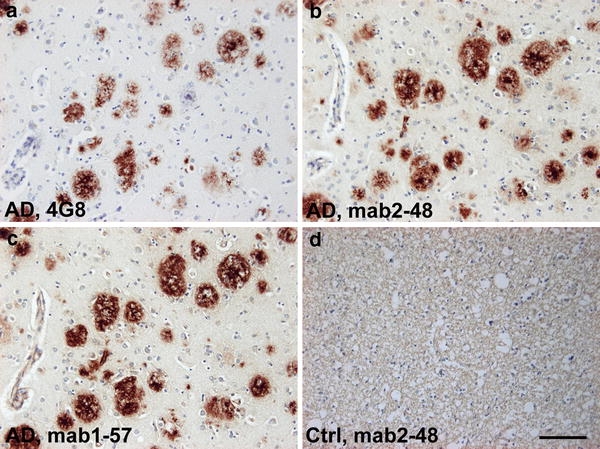
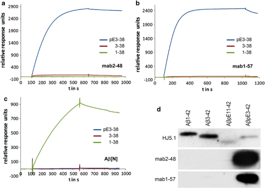
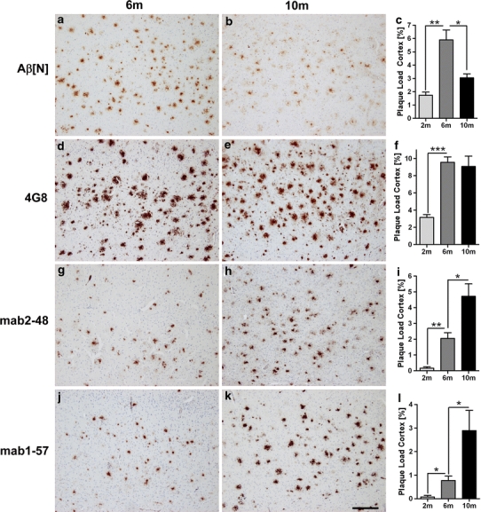
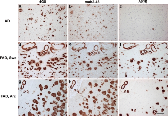
The presence of Abeta(pE3) (N-terminal truncated Abeta starting with pyroglutamate) in Alzheimer's disease (AD) has received considerable attention since the discovery that this peptide represents a dominant fraction of Abeta peptides in senile plaques of AD brains. This was later confirmed by other reports investigating AD and Down's syndrome postmortem brain tissue. Importantly, Abeta(pE3) has a higher aggregation propensity, and stability, and shows an increased toxicity compared to full-length Abeta. We have recently shown that intraneuronal accumulation of Abeta(pE3) peptides induces a severe neuron loss and an associated neurological phenotype in the TBA2 mouse model for AD. Given the increasing interest in Abeta(pE3), we have generated two novel monoclonal antibodies which were characterized as highly specific for Abeta(pE3) peptides and herein used to analyze plaque deposition in APP/PS1KI mice, an AD model with severe neuron loss and learning deficits. This was compared with the plaque pattern present in brain tissue from sporadic and familial AD cases. Abundant plaques positive for Abeta(pE3) were present in patients with sporadic AD and familial AD including those carrying mutations in APP (arctic and Swedish) and PS1. Interestingly, in APP/PS1KI mice we observed a continuous increase in Abeta(pE3) plaque load with increasing age, while the density for Abeta(1-x ) plaques declined with aging. We therefore assume that, in particular, the peptides starting with position 1 of Abeta are N-truncated as disease progresses, and that, Abeta(pE3) positive plaques are resistant to age-dependent degradation likely due to their high stability and propensity to aggregate.
Overview
The claims and statistics of this paper haven’t been checked yet.
Run the check — free, no account needed. It reads the open-access text and checks the sources it cites.
Integrity signals
Registry
Cites
Statistics
Figures
Verification
Claims the paper makes
The claims and statistics of this paper haven’t been checked yet.
Run the check — free, no account needed. It reads the open-access text and checks the sources it cites.
Claims other papers make about this one
Each is checked against this paper’s own text: does it say what the citing paper says it does?
The source backs part of the claim, not all of it — or backs it for a different population, condition or comparison.
In the citing paper
“N-terminal Aβ1-x/βCTF-specific antibodies also fail to label N-truncated Aβ42 peptides that appear to be particularly important in AD [60, 98, 157, 158, 206].”
From Intraneuronal β-amyloid accumulation and synapse pathology in Alzheimer’s disease (2010) · cites this paper as [206]
Experimental — accuracy not yet measured
Self-check
Percentages, sums and reported differences re-computed from the paper’s own numbers.
The self-check needs the paper’s full text.
Citations
Cites
Alzheimer's Disease: Genes, Proteins, and Therapy
Selkoe, D. J. (2001) · Physiological Reviews · doi:10.1152/physrev.2001.81.2.741 · Cited by 6,063
Amyloid plaque core protein in Alzheimer disease and Down syndrome.
Masters, C. L., Simms, G., Weinman, N. A., Multhaup, G., McDonald, B. L., & Beyreuther, K. (1985) · Proceedings of the National Academy of Sciences · doi:10.1073/pnas.82.12.4245 · Cited by 4,431
Self-citationOakley, H., Cole, S. L., Logan, S., Maus, E., Shao, P., Craft, J., Guillozet-Bongaarts, A., Ohno, M., Disterhoft, J., Van Eldik, L., et al. (2006) · The Journal of Neuroscience · doi:10.1523/jneurosci.1202-06.2006 · Cited by 3,530 · cited 1×
“Although learning deficits (Billings et al. 2005; Holcomb et al. 1998; Oakley et al. 2006; Puolivali et al. 2002) were evident in various APP models, the extent of Ab-amyloid deposition did not correlate with the behavioral phenotype (Holcomb et al.”
BackgroundHolcomb, L., Gordon, M. N., McGowan, E., Yu, X., Benkovic, S., Jantzen, P., Wright, K., Saad, I., Mueller, R., Morgan, D., et al. (1998) · Nature Medicine · doi:10.1038/nm0198-097 · Cited by 1,386
Billings, L. M., Oddo, S., Green, K. N., McGaugh, J. L., & LaFerla, F. M. (2005) · Neuron · doi:10.1016/j.neuron.2005.01.040 · Cited by 1,294
Correlation Between Elevated Levels of Amyloid β-Peptide in the Brain and Cognitive Decline
Näslund, J. (2000) · JAMA · doi:10.1001/jama.283.12.1571 · Cited by 1,268
Plaque-independent disruption of neural circuits in Alzheimer’s disease mouse models
Hsia, A. Y., Masliah, E., McConlogue, L., Yu, G.-Q., Tatsuno, G., Hu, K., Kholodenko, D., Malenka, R. C., Nicoll, R. A., & Mucke, L. (1999) · Proceedings of the National Academy of Sciences · doi:10.1073/pnas.96.6.3228 · Cited by 1,123
Masters, C. L., Multhaup, G., Simms, G., Pottgiesser, J., Martins, R. N., & Beyreuther, K. T. (1985) · The EMBO Journal · doi:10.1002/j.1460-2075.1985.tb04000.x · Cited by 1,048
Self-citationIntraneuronal Aβ42 Accumulation in Human Brain
Gouras, G. K., Tsai, J., Naslund, J., Vincent, B., Edgar, M., Checler, F., Greenfield, J. P., Haroutunian, V., Buxbaum, J. D., Xu, H., et al. (2000) · The American Journal of Pathology · doi:10.1016/s0002-9440(10)64700-1 · Cited by 991
Age, Neuropathology, and Dementia
Savva, G. M., Wharton, S. B., Ince, P. G., Forster, G., Matthews, F. E., & Brayne, C. (2009) · New England Journal of Medicine · doi:10.1056/nejmoa0806142 · Cited by 900
Cited by
Sorted by citations
Items 101–107 of 107
Henniges, P. (2023) · eDiss (Georg-August-Universität Göttingen) · doi:10.53846/goediss-9694
Développement de formes vectorisables innovantes dans le cadre de maladies liées à l'âge
Iskandar, S. (2019) · theses.fr (ABES) · doi:10.70675/1d771122z75d0z4ba8z8ee6zbd1a858ffc38
Valverde, A. (2020) · theses.fr (ABES) · doi:10.70675/f1b70734zf4e3z4870z8fbdz950d831642a7
BindingDB (2024) · UC San Diego · doi:10.7270/q2sn0f5x
Saul, A. (2013) · eDiss (Georg-August-Universität Göttingen) · doi:10.53846/goediss-3915
Metadata
- Authors
- Wirths, O., Bethge, T., Marcello, A., Harmeier, A., Jawhar, S., Lucassen, P. J., Multhaup, G., Brody, D. L., Esparza, T., Ingelsson, M., Kalimo, H., Lannfelt, L., & Bayer, T. A.
- Affiliations
- University of Göttingen (DE)
- Freie Universität Berlin (DE)
- University of Amsterdam (NL)
- Washington University in St. Louis (US)
- Uppsala University (SE)
- Corresponding author
- Bayer, Thomas A.
- ORCID
- Venue
- Journal of Neural Transmission
- Published
- 2009-10-13
- Volume
- 117
- Issue
- 1
- Pages
- 85-96
- ISSN
- 0300-9564 (ISSN-L)
- 1435-1463
- PMC
- PMC2789212
- OpenAlex
- W2097338951
- Cited by
- 106
- References
- 65
- Citations by year
- 2024: 1 · 2023: 3 · 2022: 4 · 2021: 3 · 2020: 3 · 2019: 6 · 2018: 4 · 2017: 6 · 2016: 7 · 2015: 13 · 2014: 7 · 2013: 10 · 2012: 14
- FWCI
- 8.61
- Type
- journal article
- Language
- en
- Open access
- hybrid
- doi.org
- Licence
- cc-by-nc
- Locations
- Journal of Neural Transmission (publishedVersion, cc-by-nc)
- Europe PMC (PubMed Central) (publishedVersion, cc-by-nc)
- PubMed Central (publishedVersion, cc-by-nc)
- PubMed (publishedVersion)
- UvA-DARE (University of Amsterdam) (publishedVersion, other-oa)
- GoeScholar The Publication Server of the Georg-August-Universität Göttingen (Georg-August-Universität Göttingen) (submittedVersion, cc-by)
- Data Archiving and Networked Services (DANS) (submittedVersion, other-oa)
- Funders
- European Commission (MEST-CT-2005-020013)
- Topics
- Life Sciences › Neuroscience › Cellular and Molecular Neuroscience › Alzheimer's disease research and treatments
- Health Sciences › Medicine › Psychiatry and Mental health › Dementia and Cognitive Impairment Research
- Life Sciences › Neuroscience › Neurology › Neuroinflammation and Neurodegeneration Mechanisms
- Keywords
- pyroglutamate amyloid-beta, Alzheimer's disease, amyloid plaques, amyloid-beta aggregation, amyloid beta degradation, APP mutations, PS1 mutations, monoclonal antibodies, neuronal loss, aging, familial Alzheimer's disease, sporadic Alzheimer's disease
- MeSH terms
- Aged, Aged, 80 and over, Aging, Alzheimer Disease, Animals, Brain, Disease Models, Animal, Female, Humans, Male, Mice, Transgenic, Middle Aged, Mutation, Peptide Fragments, Pyrrolidonecarboxylic Acid, Receptors, Cell Surface, Amyloid beta-Peptides, Amyloid beta-Protein Precursor, Mice, Presenilin-1, Plaque, Amyloid, Protease Nexins
About these checks
Registry, retraction, Cites and statistics results are computed, not judged, with one exception: a printed reference that no index lists is read once by a model before it counts as a citation error. Claims are extracted by a model and kept only when their quote appears verbatim in the text; each cited source is then checked, with a model’s help, for whether it says what the claim relies on. Those verdicts can be wrong — read the claim’s quote and the cited source before relying on one.
Statistics and arithmetic are recomputed, not judged.
Confidence bands are shown only for fields calibrated on our annotated test set. None is calibrated yet, so no band is shown.
What counts as a claim ›How we measure confidence ›How we check this ›
Are you an author, or do you see a problem with this page? Request a review
