

The DOI matches its Crossref record; 65 of the 65 references it cites were found in a registry or an index.
Pyroglutamate Abeta pathology in APP/PS1KI mice, sporadic and familial Alzheimer’s disease cases
Open accessAbstract
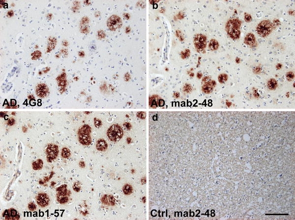
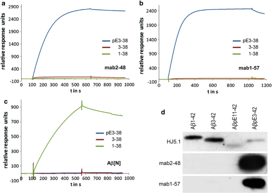
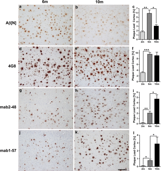
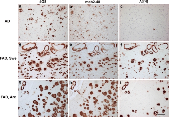
The presence of Abeta(pE3) (N-terminal truncated Abeta starting with pyroglutamate) in Alzheimer's disease (AD) has received considerable attention since the discovery that this peptide represents a dominant fraction of Abeta peptides in senile plaques of AD brains. This was later confirmed by other reports investigating AD and Down's syndrome postmortem brain tissue. Importantly, Abeta(pE3) has a higher aggregation propensity, and stability, and shows an increased toxicity compared to full-length Abeta. We have recently shown that intraneuronal accumulation of Abeta(pE3) peptides induces a severe neuron loss and an associated neurological phenotype in the TBA2 mouse model for AD. Given the increasing interest in Abeta(pE3), we have generated two novel monoclonal antibodies which were characterized as highly specific for Abeta(pE3) peptides and herein used to analyze plaque deposition in APP/PS1KI mice, an AD model with severe neuron loss and learning deficits. This was compared with the plaque pattern present in brain tissue from sporadic and familial AD cases. Abundant plaques positive for Abeta(pE3) were present in patients with sporadic AD and familial AD including those carrying mutations in APP (arctic and Swedish) and PS1. Interestingly, in APP/PS1KI mice we observed a continuous increase in Abeta(pE3) plaque load with increasing age, while the density for Abeta(1-x ) plaques declined with aging. We therefore assume that, in particular, the peptides starting with position 1 of Abeta are N-truncated as disease progresses, and that, Abeta(pE3) positive plaques are resistant to age-dependent degradation likely due to their high stability and propensity to aggregate.
Overview
The claims and statistics of this paper haven’t been checked yet.
Run the check — free, no account needed. It reads the open-access text and checks the sources it cites.
Integrity signals
Registry
Cites
Statistics
Figures
Verification
Claims the paper makes
The claims and statistics of this paper haven’t been checked yet.
Run the check — free, no account needed. It reads the open-access text and checks the sources it cites.
Claims other papers make about this one
Each is checked against this paper’s own text: does it say what the citing paper says it does?
The source backs part of the claim, not all of it — or backs it for a different population, condition or comparison.
In the citing paper
“N-terminal Aβ1-x/βCTF-specific antibodies also fail to label N-truncated Aβ42 peptides that appear to be particularly important in AD [60, 98, 157, 158, 206].”
From Intraneuronal β-amyloid accumulation and synapse pathology in Alzheimer’s disease (2010) · cites this paper as [206]
Experimental — accuracy not yet measured
Self-check
Percentages, sums and reported differences re-computed from the paper’s own numbers.
The self-check needs the paper’s full text.
Citations
Cites
Alzheimer's Disease: Genes, Proteins, and Therapy
Selkoe, D. J. (2001) · Physiological Reviews · doi:10.1152/physrev.2001.81.2.741 · Cited by 6,063
Amyloid plaque core protein in Alzheimer disease and Down syndrome.
Masters, C. L., Simms, G., Weinman, N. A., Multhaup, G., McDonald, B. L., & Beyreuther, K. (1985) · Proceedings of the National Academy of Sciences · doi:10.1073/pnas.82.12.4245 · Cited by 4,431
Self-citationOakley, H., Cole, S. L., Logan, S., Maus, E., Shao, P., Craft, J., Guillozet-Bongaarts, A., Ohno, M., Disterhoft, J., Van Eldik, L., et al. (2006) · The Journal of Neuroscience · doi:10.1523/jneurosci.1202-06.2006 · Cited by 3,530 · cited 1×
“Although learning deficits (Billings et al. 2005; Holcomb et al. 1998; Oakley et al. 2006; Puolivali et al. 2002) were evident in various APP models, the extent of Ab-amyloid deposition did not correlate with the behavioral phenotype (Holcomb et al.”
BackgroundHolcomb, L., Gordon, M. N., McGowan, E., Yu, X., Benkovic, S., Jantzen, P., Wright, K., Saad, I., Mueller, R., Morgan, D., et al. (1998) · Nature Medicine · doi:10.1038/nm0198-097 · Cited by 1,386
Billings, L. M., Oddo, S., Green, K. N., McGaugh, J. L., & LaFerla, F. M. (2005) · Neuron · doi:10.1016/j.neuron.2005.01.040 · Cited by 1,294
Correlation Between Elevated Levels of Amyloid β-Peptide in the Brain and Cognitive Decline
Näslund, J. (2000) · JAMA · doi:10.1001/jama.283.12.1571 · Cited by 1,268
Plaque-independent disruption of neural circuits in Alzheimer’s disease mouse models
Hsia, A. Y., Masliah, E., McConlogue, L., Yu, G.-Q., Tatsuno, G., Hu, K., Kholodenko, D., Malenka, R. C., Nicoll, R. A., & Mucke, L. (1999) · Proceedings of the National Academy of Sciences · doi:10.1073/pnas.96.6.3228 · Cited by 1,123
Masters, C. L., Multhaup, G., Simms, G., Pottgiesser, J., Martins, R. N., & Beyreuther, K. T. (1985) · The EMBO Journal · doi:10.1002/j.1460-2075.1985.tb04000.x · Cited by 1,048
Self-citationIntraneuronal Aβ42 Accumulation in Human Brain
Gouras, G. K., Tsai, J., Naslund, J., Vincent, B., Edgar, M., Checler, F., Greenfield, J. P., Haroutunian, V., Buxbaum, J. D., Xu, H., et al. (2000) · The American Journal of Pathology · doi:10.1016/s0002-9440(10)64700-1 · Cited by 991
Age, Neuropathology, and Dementia
Savva, G. M., Wharton, S. B., Ince, P. G., Forster, G., Matthews, F. E., & Brayne, C. (2009) · New England Journal of Medicine · doi:10.1056/nejmoa0806142 · Cited by 900
Cited by
Sorted by citations
Jawhar, S., Trawicka, A., Jenneckens, C., Bayer, T. A., & Wirths, O. (2012) · Neurobiology of Aging · doi:10.1016/j.neurobiolaging.2010.05.027 · Cited by 623
“The following antibodies were used: 4G8 (A 17-14, :10,000, Covance, Princeton, USA), NF200 (200 kDa-neuofilament, 1:1,000, Millipore, Schwalbach, Germany), 2–48 A 3pE-x, 1:100, Synaptic Systems, Göttingen, Germany, (Wirths et al., 2010)), A [N] (1:2,000, IBL, Hamburg, Gerany), 692 against A (1:500) and 23850 against h…”
InfluentialSelf-citationBiochemistry of Amyloid -Protein and Amyloid Deposits in Alzheimer DiseasePreprint
Masters, C. L., & Selkoe, D. J. (2012) · Cold Spring Harbor Perspectives in Medicine · doi:10.1101/cshperspect.a006262 · Cited by 554
Endothelial LRP1 transports amyloid-β1–42 across the blood-brain barrier
Storck, S. E., Meister, S., Nahrath, J., Meißner, J. N., Schubert, N., Di Spiezio, A., Baches, S., Vandenbroucke, R. E., Bouter, Y., Prikulis, I., et al. (2015) · Journal of Clinical Investigation · doi:10.1172/jci81108 · Cited by 477
Self-citationIntraneuronal β-amyloid accumulation and synapse pathology in Alzheimer’s disease
Gouras, G. K., Tampellini, D., Takahashi, R. H., & Capetillo-Zarate, E. (2010) · Acta Neuropathologica · doi:10.1007/s00401-010-0679-9 · Cited by 372
Pyroglutamate Amyloid-β (Aβ): A Hatchet Man in Alzheimer Disease
Jawhar, S., Wirths, O., & Bayer, T. A. (2011) · Journal of Biological Chemistry · doi:10.1074/jbc.r111.288308 · Cited by 220
Self-citationBouter, Y., Dietrich, K., Wittnam, J. L., Rezaei-Ghaleh, N., Pillot, T., Papot-Couturier, S., Lefebvre, T., Sprenger, F., Wirths, O., Zweckstetter, M., et al. (2013) · Acta Neuropathologica · doi:10.1007/s00401-013-1129-2 · Cited by 175
Self-citationBayer, T. A., & Wirths, O. (2014) · Acta Neuropathologica · doi:10.1007/s00401-014-1287-x · Cited by 170
Self-citationHureau, C., & Dorlet, P. (2012) · Coordination Chemistry Reviews · doi:10.1016/j.ccr.2012.03.034 · Cited by 148
Richard, B. C., Kurdakova, A., Baches, S., Bayer, T. A., Weggen, S., & Wirths, O. (2015) · Journal of Alzheimer’s Disease · doi:10.3233/jad-143120 · Cited by 142
Self-citationFrost, J. L., Le, K. X., Cynis, H., Ekpo, E., Kleinschmidt, M., Palmour, R. M., Ervin, F. R., Snigdha, S., Cotman, C. W., Saido, T. C., et al. (2013) · American Journal Of Pathology · doi:10.1016/j.ajpath.2013.05.005 · Cited by 135
Morawski, M., Schilling, S., Kreuzberger, M., Waniek, A., Jäger, C., Koch, B. M., Cynis, H., Kehlen, A., Arendt, T., Hartlage‐Rübsamen, M., et al. (2014) · Journal of Alzheimer s Disease · doi:10.3233/jad-131535 · Cited by 114
Luccarini, I., Grossi, C., Rigacci, S., Coppi, E., Pugliese, A. M., Pantano, D., la Marca, G., Dami, T. E., Berti, A., Stefani, M., et al. (2014) · Neurobiology of Aging · doi:10.1016/j.neurobiolaging.2014.08.029 · Cited by 113
Jawhar, S., Wirths, O., Schilling, S., Graubner, S., Demuth, H., & Bayer, T. A. (2010) · Journal of Biological Chemistry · doi:10.1074/jbc.m110.185819 · Cited by 105
Self-citationIdentification of Low Molecular Weight Pyroglutamate Aβ Oligomers in Alzheimer Disease
Wirths, O., Erck, C., Martens, H., Harmeier, A., Geumann, C., Jawhar, S., Kumar, S., Multhaup, G., Walter, J., Ingelsson, M., et al. (2010) · Journal of Biological Chemistry · doi:10.1074/jbc.m110.178707 · Cited by 103
Self-citationAlexandru, A., Jagla, W., Graubner, S., Becker, A., Bäuscher, C., Kohlmann, S., Sedlmeier, R., Raber, K. A., Cynis, H., Rönicke, R., et al. (2011) · The Journal of Neuroscience · doi:10.1523/jneurosci.1794-11.2011 · Cited by 102
Pyroglutamate Aβ cascade as drug target in Alzheimer’s disease
Bayer, T. A. (2021) · Molecular Psychiatry · doi:10.1038/s41380-021-01409-2 · Cited by 102
Self-citationFrost, J. L., Liu, B., Kleinschmidt, M., Schilling, S., Demuth, H., & Lemere, C. A. (2012) · Neurodegenerative Diseases · doi:10.1159/000335913 · Cited by 82
Copper(II) Coordination to Amyloid β: Murine versus Human Peptide
Eury, H., Bijani, C., Faller, P., & Hureau, C. (2010) · Angewandte Chemie International Edition · doi:10.1002/anie.201005838 · Cited by 79
Aβ truncated species: Implications for brain clearance mechanisms and amyloid plaque deposition
Cabrera, E., Mathews, P. M., Mezhericher, E., Beach, T. G., Deng, J., Neubert, T. A., Rostagno, A. A., & Ghiso, J. A. (2017) · Biochimica et Biophysica Acta (BBA) - Molecular Basis of Disease · doi:10.1016/j.bbadis.2017.07.005 · Cited by 78
Wittnam, J. L., Portelius, E., Zetterberg, H., Gustavsson, M. K., Schilling, S., Koch, B. C. P., Demuth, H., Blennow, K., Wirths, O., & Bayer, T. A. (2012) · Journal of Biological Chemistry · doi:10.1074/jbc.m111.308601 · Cited by 77
Self-citationMichno, W., Nyström, S., Wehrli, P. M., Lashley, T., Brinkmalm, G., Guérard, L., Syvänen, S., Sehlin, D., Kaya, I., Brinet, D., et al. (2019) · Journal of Biological Chemistry · doi:10.1074/jbc.ra118.006604 · Cited by 75
Bouter, Y., López‐Noguerola, J. S., Tucholla, P., Crespi, G. A. N., Parker, M. W., Wiltfang, J., Miles, L. A., & Bayer, T. A. (2015) · Acta Neuropathologica · doi:10.1007/s00401-015-1489-x · Cited by 74
Self-citationSpencer, B. J., Marr, R. A., Gindi, R., Potkar, R., Michael, S. A., Adame, A., Rockenstein, E. M., Verma, I. M., & Masliah, E. (2011) · PLoS ONE · doi:10.1371/journal.pone.0016575 · Cited by 69
Hartlage‐Rübsamen, M., Morawski, M., Waniek, A., Jäger, C., Zeitschel, U., Koch, B. C. P., Cynis, H., Schilling, S., Schliebs, R., Demuth, H., et al. (2011) · Acta Neuropathologica · doi:10.1007/s00401-011-0806-2 · Cited by 66
Pyroglutamate-Modified Amyloid Beta Peptides: Emerging Targets for Alzheimer´s Disease Immunotherapy
Pérez-Garmendia, R., & Gevorkian, G. (2013) · Current Neuropharmacology · doi:10.2174/1570159x11311050004 · Cited by 65
Wirths, O., Walter, S., Kraus, I., Klafki, H., Stazi, M., Oberstein, T. J., Ghiso, J. A., Wiltfang, J., Bayer, T. A., & Weggen, S. (2017) · Alzheimer s Research & Therapy · doi:10.1186/s13195-017-0309-z · Cited by 66
Self-citationAn anti-pyroglutamate-3 Aβ vaccine reduces plaques and improves cognition in APPswe/PS1ΔE9 mice
Frost, J. L., Liu, B., Rahfeld, J., Kleinschmidt, M., O’Nuallain, B., Le, K. X., Lues, I., Caldarone, B. J., Schilling, S., Demuth, H., et al. (2015) · Neurobiology of Aging · doi:10.1016/j.neurobiolaging.2015.08.021 · Cited by 64
Targets for AD treatment: conflicting messages from γ-secretase inhibitors
Sambamurti, K., Greig, N. H., Utsuki, T., Barnwell, E., Sharma, E., Mazell, C., Bhat, N. R., Kindy, M. S., Lahiri, D. K., & Pappolla, M. A. (2011) · Journal of Neurochemistry · doi:10.1111/j.1471-4159.2011.07213.x · Cited by 62
Lues, I., Weber, F., Meyer, A., Uli, B., Hoffmann, T., Kühn‐Wache, K., Manhart, S., Heiser, U. F., Pokorny, R., Chiesa, J., et al. (2015) · Alzheimer s & Dementia Translational Research & Clinical Interventions · doi:10.1016/j.trci.2015.08.002 · Cited by 62
Alzheimer therapy with an antibody against N-terminal Abeta 4-X and pyroglutamate Abeta 3-X
Antonios, G., Borgers, H., Richard, B. C., Brauß, A., Meißner, J., Weggen, S., Pena, V., Pillot, T., Davies, S. L., Bakrania, P., et al. (2015) · Scientific Reports · doi:10.1038/srep17338 · Cited by 61
Self-citationMoon, M., Hong, H.-S., Nam, D. W., Baik, S. H., Song, H., Kook, S.-Y., Kim, Y. S., Lee, J., & Mook-Jung, I. (2012) · Journal of Alzheimer's Disease · doi:10.3233/jad-2011-111778 · Cited by 60
Daulatzai, M. A. (2013) · Neurotoxicity Research · doi:10.1007/s12640-013-9407-2 · Cited by 56
Protein amino-terminal modifications and proteomic approaches for N-terminal profiling
Lai, Z. W., Petrera, A., & Schilling, O. (2014) · Current Opinion in Chemical Biology · doi:10.1016/j.cbpa.2014.10.026 · Cited by 55
Antonios, G., Saiepour, N., Bouter, Y., Richard, B. C., Paetau, A., Verkkoniemi-Ahola, A., Lannfelt, L., Ingelsson, M., Kovacs, G. G., Pillot, T., et al. (2013) · Acta Neuropathologica Communications · doi:10.1186/2051-5960-1-56 · Cited by 53
Self-citationKalimo, H., Lalowski, M. M., Bogdanović, N., Philipson, O., Bird, T. D., Nochlin, D., Schellenberg, G. D., Brundin, R., Olofsson, T., Soliymani, R., et al. (2013) · Acta Neuropathologica Communications · doi:10.1186/2051-5960-1-60 · Cited by 52
Self-citationCu II Binding to Various Forms of Amyloid‐β Peptides: Are They Friends or Foes?
Borghesani, V., Aliès, B., & Hureau, C. (2017) · European Journal of Inorganic Chemistry · doi:10.1002/ejic.201700776 · Cited by 50
Alzheimer’s Disease-Related Protein Expression in the Retina of Octodon degus
Du, L. Y., Chang, L., Ardiles, Á. O., Tapia‐Rojas, C., Araya, J., Inestrosa, N. C., Palacios, A. G., & Acosta, M. L. (2015) · PLoS ONE · doi:10.1371/journal.pone.0135499 · Cited by 51
Sullivan, C. P., Berg, E. A., Elliott-Bryant, R., Fishman, J. B., McKee, A. C., Morin, P. J., Shia, M. A., & Fine, R. E. (2011) · Neuroscience Letters · doi:10.1016/j.neulet.2011.09.071 · Cited by 46
Jakobsen, J. E., Johansen, M., Schmidt, M. H., Liu, Y., Li, R., Callesen, H., Melnikova, M., Habekost, M., Matrone, C., Bouter, Y., et al. (2016) · Journal of Alzheimer s Disease · doi:10.3233/jad-160408 · Cited by 45
Self-citationAhlemeyer, B., Halupczok, S., Rodenberg-Frank, E., Valerius, K.-P., & Baumgart‐Vogt, E. (2018) · Journal of Alzheimer s Disease · doi:10.3233/jad-170923 · Cited by 49
Aliès, B., Bijani, C., Sayen, S., Guillon, E., Faller, P., & Hureau, C. (2012) · Inorganic Chemistry · doi:10.1021/ic302097d · Cited by 44
Emerging roles of N- and C-terminally truncated Aβ species in Alzheimer’s disease
Wirths, O., & Zampar, S. (2019) · Expert Opinion on Therapeutic Targets · doi:10.1080/14728222.2019.1702972 · Cited by 44
Self-citationHuman glutaminyl cyclase: Structure, function, inhibitors and involvement in Alzheimer’s disease
Dileep, K. V., & Zhang, K. Y. J. (2019) · Pharmacological Research · doi:10.1016/j.phrs.2019.104342 · Cited by 43
Drew, S. C., Masters, C. L., & Barnham, K. J. (2010) · PLoS ONE · doi:10.1371/journal.pone.0015875 · Cited by 42
Lemmens, M. A. M., Sierksma, A. S. R., Rutten, B. P. F., Dennissen, F., Steinbusch, H. W. M., Lucassen, P. J., & Schmitz, C. (2011) · Brain Structure and Function · doi:10.1007/s00429-011-0305-1 · Cited by 37
Self-citationpGluAβ increases accumulation of Aβ in vivo and exacerbates its toxicity
Sofola-Adesakin, O., Khericha, M., Snoeren, I., Tsuda, L., & Partridge, L. (2016) · Acta Neuropathologica Communications · doi:10.1186/s40478-016-0380-x · Cited by 37
Philipson, O., Lord, A., Lalowski, M. M., Soliymani, R., Baumann, M., Thyberg, J., Bogdanović, N., Olofsson, T., Tjernberg, L. O., Ingelsson, M., et al. (2011) · Neurobiology of Aging · doi:10.1016/j.neurobiolaging.2011.10.022 · Cited by 40
Self-citationMorawski, M., Hartlage‐Rübsamen, M., Jäger, C., Waniek, A., Schilling, S., Schwab, C., McGeer, P. L., Arendt, T., Demuth, H., & Roßner, S. (2010) · Acta Neuropathologica · doi:10.1007/s00401-010-0685-y · Cited by 37
Glutaminyl Cyclase, Diseases, and Development of Glutaminyl Cyclase Inhibitors
Xu, C., Wang, Y., & Wu, H. (2021) · Journal of Medicinal Chemistry · doi:10.1021/acs.jmedchem.1c00325 · Cited by 36
Role of amyloid-β CSF levels in cognitive deficit in MS
Gentile, A., Mori, F., Bernardini, S., & Centonze, D. (2015) · Clinica Chimica Acta · doi:10.1016/j.cca.2015.01.035 · Cited by 32
A vaccine against Alzheimer`s disease: anything left but faith?
Bachmann, M. F., Jennings, G. T., & Vogel, M. (2018) · Expert Opinion on Biological Therapy · doi:10.1080/14712598.2019.1554646 · Cited by 31
Wu, G., Miller, R. A., Connolly, B., Marcus, J., Renger, J. J., & Savage, M. J. (2013) · Neurodegenerative Diseases · doi:10.1159/000353634 · Cited by 31
Härtig, W., Goldhammer, S., Bauer, U. U. I., Wegner, F., Wirths, O., Bayer, T. A., & Grosche, J. (2010) · Journal of Chemical Neuroanatomy · doi:10.1016/j.jchemneu.2010.03.006 · Cited by 29
Self-citationMatsumoto, Y., Niimi, N., & Kohyama, K. (2013) · PLoS ONE · doi:10.1371/journal.pone.0075203 · Cited by 28
Bakrania, P., Hall, G., Bouter, Y., Bouter, C., Beindorff, N., Cowan, R. H., Davies, S. L., Price, J., Mpamhanga, C., Love, E. A., et al. (2021) · Molecular Psychiatry · doi:10.1038/s41380-021-01385-7 · Cited by 31
Self-citationPiechotta, A., Parthier, C., Kleinschmidt, M., Gnoth, K., Pillot, T., Lues, I., Demuth, H., Schilling, S., Rahfeld, J., & Stubbs, M. T. (2017) · Journal of Biological Chemistry · doi:10.1074/jbc.m117.777839 · Cited by 28
Amyloid-β: The seeds of darkness
Fodero‐Tavoletti, M. T., Villemagne, V. L., Rowe, C. C., Masters, C. L., Barnham, K. J., & Cappai, R. (2011) · The International Journal of Biochemistry & Cell Biology · doi:10.1016/j.biocel.2011.05.001 · Cited by 30
AβPP Accumulation and/or Intraneuronal Amyloid-β Accumulation? The 3xTg-AD Mouse Model Revisited
Wirths, O., Dins, A., & Bayer, T. A. (2012) · Journal of Alzheimer s Disease · doi:10.3233/jad-2011-111529 · Cited by 26
Self-citationPyroglutamate-Modified Amyloid-β(3–42) Shows α-Helical Intermediates before Amyloid Formation
Dammers, C., Reiß, K., Gremer, L., Lecher, J., Ziehm, T., Stoldt, M., Schwarten, M., & Willbold, D. (2017) · Biophysical Journal · doi:10.1016/j.bpj.2017.03.007 · Cited by 25
Bouter, Y., Liekefeld, H., Pichlo, S., Westhoff, A. C., Fenn, L., Bakrania, P., & Bayer, T. A. (2022) · Acta Neuropathologica · doi:10.1007/s00401-022-02418-3 · Cited by 26
Self-citationN-terminally truncated Aβ4-x proteoforms and their relevance for Alzheimer’s pathophysiology
Rostagno, A. A., Cabrera, E., Lashley, T., & Ghiso, J. A. (2022) · Translational Neurodegeneration · doi:10.1186/s40035-022-00303-3 · Cited by 23
Brain Region-Specific Differences in Amyloid-β Plaque Composition in 5XFAD Mice
Bader, A. S., Gnädig, M.-U., Fricke, M., Büschgens, L., Berger, L. J., Klafki, H., Meyer, T., Jahn, O., Weggen, S., & Wirths, O. (2023) · Life · doi:10.3390/life13041053 · Cited by 26
Self-citationSun, N., Hartmann, R., Lecher, J., Stoldt, M., Funke, S. A., Gremer, L., Ludwig, H., Demuth, H., Kleinschmidt, M., & Willbold, D. (2012) · Journal of Peptide Science · doi:10.1002/psc.2456 · Cited by 22
Gene Expression Profiling in the APP/PS1KI Mouse Model of Familial Alzheimer’s Disease
Weißmann, R., Hüttenrauch, M., Kacprowski, T., Bouter, Y., Pradier, L., Bayer, T. A., Kuss, A. W., & Wirths, O. (2016) · Journal of Alzheimer s Disease · doi:10.3233/jad-150745 · Cited by 21
Self-citationWirths, O., Hillmann, A., Pradier, L., Härtig, W., & Bayer, T. A. (2013) · Journal of Alzheimer s Disease · doi:10.3233/jad-121945 · Cited by 21
Self-citationThe Hidden Role of Non-Canonical Amyloid β Isoforms in Alzheimer’s Disease
Busch, L., Eggert, S., Endres, K., & Bufe, B. (2022) · Cells · doi:10.3390/cells11213421 · Cited by 20
Saul, A., Lashley, T., Révész, T., Holton, J. L., Ghiso, J. A., Coomaraswamy, J., & Wirths, O. (2012) · Neurobiology of Aging · doi:10.1016/j.neurobiolaging.2012.11.014 · Cited by 23
Self-citationVenkataramani, V., Wirths, O., Budka, H., Härtig, W., Kovács, G. G., & Bayer, T. A. (2012) · Journal of Alzheimer s Disease · doi:10.3233/jad-2011-111379 · Cited by 17
Self-citationAβ Peptide Fibrillar Architectures Controlled by Conformational Constraints of the Monomer
Brännström, K., Öhman, A., & Olofsson, A. (2011) · PLoS ONE · doi:10.1371/journal.pone.0025157 · Cited by 17
Meißner, J. N., Bouter, Y., & Bayer, T. A. (2015) · Journal of Alzheimer s Disease · doi:10.3233/jad-142868 · Cited by 16
Self-citationBerger–Sieczkowski, E., Milenković, I., Venkataramani, V., Giera, R., Ströbel, T., Höftberger, R., Liberski, P. P., Auff, E., Wirths, O., Bayer, T. A., et al. (2015) · Journal of Alzheimer s Disease · doi:10.3233/jad-141524 · Cited by 16
Self-citationCopper(II) Coordination to Amyloid β: Murine versus Human Peptide
Eury, H., Bijani, C., Faller, P., & Hureau, C. (2010) · Angewandte Chemie · doi:10.1002/ange.201005838 · Cited by 15
Todd, K. L., Fossati, S., Ghiso, J. A., & Rostagno, A. A. (2014) · Biochimica et Biophysica Acta (BBA) - Molecular Basis of Disease · doi:10.1016/j.bbadis.2014.09.010 · Cited by 15
Schrempel, S., Kottwitz, A. K., Piechotta, A., Gnoth, K., Büschgens, L., Hartlage‐Rübsamen, M., Morawski, M., Schenk, M., Kleinschmidt, M., Serrano, G. E., et al. (2024) · Acta Neuropathologica · doi:10.1007/s00401-024-02824-9 · Cited by 14
Self-citationPérez-Garmendia, R., Ibarra-Bracamontes, V., Vasilevko, V., Luna‐Muñoz, J., Mena, R., Govezensky, T., Acero, G., Manoutcharian, K., Cribbs, D. H., & Gevorkian, G. (2010) · Journal of Neuroimmunology · doi:10.1016/j.jneuroim.2010.08.020 · Cited by 14
Plasma pyroglutamate‐modified amyloid beta differentiates amyloid pathology
Wang, P., Lin, K., Liu, H., Andréasson, U., Blennow, K., Zetterberg, H., & Yang, S. (2020) · Alzheimer s & Dementia Diagnosis Assessment & Disease Monitoring · doi:10.1002/dad2.12029 · Cited by 11
Unveiling Brain Aβ Heterogeneity Through Targeted Proteomic Analysis
Rostagno, A. A., Neubert, T. A., & Ghiso, J. A. (2018) · Methods in molecular biology · doi:10.1007/978-1-4939-7816-8_3 · Cited by 11
N-Terminally Truncated Aß Peptide Variants in Alzheimer’s Disease
Wirths, O., Zampar, S., & Weggen, S. (2019) · Codon Publications eBooks · doi:10.15586/alzheimersdisease.2019.ch7 · Cited by 13
Self-citationHernández-Zimbrón, L. F., Gorostieta-Salas, E., Díaz-Hung, M.-L., Pérez-Garmendia, R., Gevorkian, G., & Quiroz-Mercado, H. (2016) · InTech eBooks · doi:10.5772/64744 · Cited by 10
The Domestic Dog as a Model for Human Brain Aging and Alzheimer’s Disease
Schütt, T., Pedersen, J. T., & Berendt, M. (2018) · Elsevier eBooks · doi:10.1016/b978-0-12-811353-0.00015-4 · Cited by 9
Waniek, A., Hartlage‐Rübsamen, M., Höfling, C., Kehlen, A., Schilling, S., Demuth, H., & Roßner, S. (2014) · Biochimica et Biophysica Acta (BBA) - Molecular Basis of Disease · doi:10.1016/j.bbadis.2014.11.011 · Cited by 9
Li, Y., Ma, Y., Zong, L., Xing, X., Sha, S., & Cao, Y. (2011) · Neuroscience Letters · doi:10.1016/j.neulet.2011.10.005 · Cited by 6
The contribution of N-terminally modified amyloid beta to the etiology of Alzheimer's disease
Wittnam, J. L. (2012) · eDiss (Georg-August-Universität Göttingen) · doi:10.53846/goediss-3182 · Cited by 5
The role of amyloid beta 4-42 in the etiology of Alzheimer's disease
Bouter, Y. (2015) · eDiss (Georg-August-Universität Göttingen) · doi:10.53846/goediss-4849 · Cited by 5
Immunotherapy Against N-Truncated Amyloid-β Oligomers
Bayer, T. A., & Wirths, O. (2016) · Methods in pharmacology and toxicology · doi:10.1007/978-1-4939-3560-4_3 · Cited by 4
Self-citationHigh yield expression and purification of full-length Neurotensin with pyroglutamate modification
Asadollahi, K., Huang, K., Yan, F., de Zhang, L. A., Scott, D. J., & Gooley, P. R. (2022) · Protein Expression and Purification · doi:10.1016/j.pep.2022.106227 · Cited by 4
One or more β-amyloid(s)? New insights into the prion-like nature of Alzheimer's disease
Catania, M., & Fede, G. D. (2020) · Progress in molecular biology and translational science · doi:10.1016/bs.pmbts.2020.07.003 · Cited by 4
Amyloid Imaging PET Ligands as Biomarkers for Alzheimer’s Disease, Preclinical Evaluation
Svedberg, M. M., Hellström‐Lindahl, E., Rahman, O., & Hall, H. (2012) · InTech eBooks · doi:10.5772/31813 · Cited by 1
Schieb, H. (2011) · DuEPublico (University of Duisburg-Essen)
The 5XFAD mouse model: a tool for genetic modulation of Alzheime's disease pathology
Jawhar, S. (2012) · eDiss (Georg-August-Universität Göttingen) · doi:10.53846/goediss-3224
Self-citationSPECIFIC AMYLOID-BETA OLIGOMERS IN HUMAN CEREBROSPINAL FLUID
Handoko, M. (2012) · University of Minnesota Digital Conservancy (University of Minnesota)
Lues, I., Weber, F., Meyer, A., Uli, B., Hoffmann, T., Manhart, S., Heiser, U. F., Pokorny, R., Chiesa, J., & Glund, K. (2015)
Lucassen, P. J., & Schmitz, C. (2011)
Self-citationKurdakova, A. (2016) · eDiss (Georg-August-Universität Göttingen) · doi:10.53846/goediss-5882
In Vitro and In Vivo Studies on Antibodies - N-terminally Truncated Abeta in the 5XFAD Mouse Model
Richard, B. C. (2015) · eDiss (Georg-August-Universität Göttingen) · doi:10.53846/goediss-5098
Borgers, H. (2017) · eDiss (Georg-August-Universität Göttingen) · doi:10.53846/goediss-6363
Studie zur Rolle N-trunkierter Amyloid-β-Peptide bei der Alzheimer-Krankheit
Gießen, N. M.-E. (2019) · eDiss (Georg-August-Universität Göttingen) · doi:10.53846/goediss-7657
Studies on the Involvement of the Immune system in Alzheimer's disease
Marcello, A. (2010) · doi:10.53846/goediss-3277
Self-citation
Metadata
- Authors
- Wirths, O., Bethge, T., Marcello, A., Harmeier, A., Jawhar, S., Lucassen, P. J., Multhaup, G., Brody, D. L., Esparza, T., Ingelsson, M., Kalimo, H., Lannfelt, L., & Bayer, T. A.
- Affiliations
- University of Göttingen (DE)
- Freie Universität Berlin (DE)
- University of Amsterdam (NL)
- Washington University in St. Louis (US)
- Uppsala University (SE)
- Corresponding author
- Bayer, Thomas A.
- ORCID
- Venue
- Journal of Neural Transmission
- Published
- 2009-10-13
- Volume
- 117
- Issue
- 1
- Pages
- 85-96
- ISSN
- 0300-9564 (ISSN-L)
- 1435-1463
- PMC
- PMC2789212
- OpenAlex
- W2097338951
- Cited by
- 106
- References
- 65
- Citations by year
- 2024: 1 · 2023: 3 · 2022: 4 · 2021: 3 · 2020: 3 · 2019: 6 · 2018: 4 · 2017: 6 · 2016: 7 · 2015: 13 · 2014: 7 · 2013: 10 · 2012: 14
- FWCI
- 8.61
- Type
- journal article
- Language
- en
- Open access
- hybrid
- doi.org
- Licence
- cc-by-nc
- Locations
- Journal of Neural Transmission (publishedVersion, cc-by-nc)
- Europe PMC (PubMed Central) (publishedVersion, cc-by-nc)
- PubMed Central (publishedVersion, cc-by-nc)
- PubMed (publishedVersion)
- UvA-DARE (University of Amsterdam) (publishedVersion, other-oa)
- GoeScholar The Publication Server of the Georg-August-Universität Göttingen (Georg-August-Universität Göttingen) (submittedVersion, cc-by)
- Data Archiving and Networked Services (DANS) (submittedVersion, other-oa)
- Funders
- European Commission (MEST-CT-2005-020013)
- Topics
- Life Sciences › Neuroscience › Cellular and Molecular Neuroscience › Alzheimer's disease research and treatments
- Health Sciences › Medicine › Psychiatry and Mental health › Dementia and Cognitive Impairment Research
- Life Sciences › Neuroscience › Neurology › Neuroinflammation and Neurodegeneration Mechanisms
- Keywords
- pyroglutamate amyloid-beta, Alzheimer's disease, amyloid plaques, amyloid-beta aggregation, amyloid beta degradation, APP mutations, PS1 mutations, monoclonal antibodies, neuronal loss, aging, familial Alzheimer's disease, sporadic Alzheimer's disease
- MeSH terms
- Aged, Aged, 80 and over, Aging, Alzheimer Disease, Animals, Brain, Disease Models, Animal, Female, Humans, Male, Mice, Transgenic, Middle Aged, Mutation, Peptide Fragments, Pyrrolidonecarboxylic Acid, Receptors, Cell Surface, Amyloid beta-Peptides, Amyloid beta-Protein Precursor, Mice, Presenilin-1, Plaque, Amyloid, Protease Nexins
About these checks
Registry, retraction, Cites and statistics results are computed, not judged, with one exception: a printed reference that no index lists is read once by a model before it counts as a citation error. Claims are extracted by a model and kept only when their quote appears verbatim in the text; each cited source is then checked, with a model’s help, for whether it says what the claim relies on. Those verdicts can be wrong — read the claim’s quote and the cited source before relying on one.
Statistics and arithmetic are recomputed, not judged.
Confidence bands are shown only for fields calibrated on our annotated test set. None is calibrated yet, so no band is shown.
What counts as a claim ›How we measure confidence ›How we check this ›
Are you an author, or do you see a problem with this page? Request a review
