

The DOI matches its Crossref record; 344 of the 352 references it cites were found in a registry or an index; none of the 233 sources its claims cite is checked for support yet.
Peroxisomes in brain development and function
Open accessAbstract
Peroxisomes contain numerous enzymatic activities that are important for mammalian physiology. Patients lacking either all peroxisomal functions or a single enzyme or transporter function typically develop severe neurological deficits, which originate from aberrant development of the brain, demyelination and loss of axonal integrity, neuroinflammation or other neurodegenerative processes. Whilst correlating peroxisomal properties with a compilation of pathologies observed in human patients and mouse models lacking all or individual peroxisomal functions, we discuss the importance of peroxisomal metabolites and tissue- and cell type-specific contributions to the observed brain pathologies. This enables us to deconstruct the local and systemic contribution of individual metabolic pathways to specific brain functions. We also review the recently discovered variability of pathological symptoms in cases with unexpectedly mild presentation of peroxisome biogenesis disorders. Finally, we explore the emerging evidence linking peroxisomes to more common neurological disorders such as Alzheimer's disease, autism and amyotrophic lateral sclerosis.
Overview
AI snapshot
Written from the paper’s full text.
TL;DR
This review synthesizes how peroxisomal metabolic pathways contribute to brain development and function, using inherited peroxisomal disorders and mouse models to link specific pathways to neuronal migration, myelination, axonal integrity, neuroinflammation, and neurodegeneration. It also discusses emerging evidence connecting peroxisomes to more common neurological disorders such as Alzheimer’s disease, autism, amyotrophic lateral sclerosis, and schizophrenia.
Key findings
- Peroxisome biogenesis disorders (PBD) and single peroxisomal enzyme/transporter deficiencies cause major brain pathology grouped into neuronal migration/differentiation defects, white matter formation/maintenance defects, and post-developmental neuronal degeneration. Zellweger syndrome features cortical malformations such as centrosylvian pachygyria and polymicrogyria, heterotopic Purkinje cells, and white matter abnormalities; in a study of 19 patients aged 16–35 years with mild Zellweger spectrum disorder, MRI showed white matter abnormalities in 9 patients, restricted to the cerebellar hilus of the dentate nucleus and/or peridentate region.
- Cell-type-specific mouse studies indicate that in the murine brain peroxisomes are most crucial for oligodendrocytes and the myelin compartment: oligodendrocyte-selective Pex5 knockout caused progressive subcortical demyelination, axonal degeneration, neuroinflammation, impaired motor function, and premature death starting at 2–6 months, whereas Pex5 deletion in projection neurons or astrocytes had no obvious deleterious effect on brain development or function despite VLCFA accumulation and reduced plasmalogen levels in astrocytes.
- X-linked adrenoleukodystrophy (X-ALD) is the most common peroxisomal disorder, with estimated combined male and female incidence between 1:16,800 and 1:30,000; about 60% of male X-ALD patients develop cerebral ALD, and adrenal insufficiency affects 80% of male patients before adulthood. ABCD1 deficiency causes VLCFA accumulation, AMN with distal dying-back axonopathy, and in CALD an inflammatory demyelinating process with blood–brain barrier permeability, macrophage/T-cell infiltration, and loss of oligodendrocytes.
- Ether phospholipid deficiency in RCDP causes rhizomelia, chondrodysplasia punctata, cataracts, growth/developmental retardation, variable brain involvement including white matter abnormalities, delayed myelination, cerebellar atrophy with Purkinje cell loss, and peripheral neuropathy; RCDP incidence is estimated at about 1:100,000. Peroxisomal links are also reported for autism (PEX7 mutations, reduced plasmalogens), Alzheimer’s disease (plasmalogen depletion, altered VLCFA, catalase, DHA, peroxisome distribution), ALS (DAO mutation, D-serine), and schizophrenia (DAO activity, D-serine/NMDA signaling).
Methods
- Narrative review of mammalian peroxisomal functions in the nervous system, focusing on human physiology/pathophysiology supplemented by mouse models.
- Evidence base includes human inherited peroxisomal disorders (PBD, X-ALD, Refsum disease, RCDP, single enzyme/transporter deficiencies) and genetically engineered mouse models, including global and conditional Pex knockouts, Acox1−/−, Mfp2−/−, Abcd1−/−, Phyh−/−, and ether lipid-deficient models.
- Main analysis is qualitative synthesis comparing biochemical, neuropathological, imaging, and cell-type-specific findings across disorders and models; no systematic review, meta-analysis, or new primary data are reported.
Limitations
- The paper states that many studies examined the presence/abundance of a single peroxisomal protein, thus possibly detecting only a subset of peroxisome-positive cells, so different investigations must be combined to infer accurate distribution and abundance.
- The paper states that peroxisome-related metabolite levels in plasma may not reflect tissue accumulation, and that some mild PBD patients can have normal plasma VLCFA or plasmalogen levels, limiting diagnostic and pathophysiological interpretation.
- The paper notes that mouse models differ from human disease—for example, Abcd1-deficient mice do not develop brain inflammation/demyelination as in CALD, and Mfp2-deficient mice differ markedly from human DBP deficiency—so mechanisms and phenotypes cannot always be directly translated. As an evident limitation, this is a narrative review without systematic search or quantitative synthesis.
Model-written summary. Check it against the paper before relying on it.
Integrity signals
Registry
Cites
Statistics
Figures
Verification
Claims the paper makes
151 Not checked yet · 20 No basis given · 15 Own interpretation
The paper states this without a source or data behind it here, so there is nothing to check it against. The quote shows it is in the paper.
In the paper
“Patients lacking either all peroxisomal functions or a single enzyme or transporter function typically develop severe neurological deficits”
§ Peroxisomes in brain development and function☆ > Abstract
plaindirect- Asserted: passes — The authors put it forward as true.
- Checkable: passes — It can be checked against evidence.
- Load-bearing: passes — The paper’s conclusions depend on it.
- Based: unclear — It is unclear why the paper says it holds.
In the paper
“During early stages of murine peripheral nerve (sciatic nerve) myelination, peroxisomes appear to be diffusely distributed in the myelin sheath of Schwann cells, whereas at later stages, peroxisomes were found to be enriched in the myelin loops of the paranodal region [14].”
§ Peroxisomes in brain development and function☆ > Brain peroxisomes and how they differ from peroxisomes in other tissues
What the sources say
- Not checked yetcites [14] Kassmann CM Quintes S Rietdorf J Mobius W Sereda MW Nientiedt T Saher G Baes M Nave KA A role for myelin-associated peroxisomes in maintaining paranodal loops and axonal integrity FEBS Lett 2011 585 2205 2211 21620837 source
hedgedcorrelational- Asserted: passes — The authors put it forward as true.
- Checkable: passes — It can be checked against evidence.
- Load-bearing: unclear — It is unclear whether the paper’s conclusions depend on it.
- Based: passes — The paper gives a reason why it holds.
In the paper
“The symptoms of patients with peroxisomal single enzyme and transporter deficiencies have a broad heterogeneity, related to differences in the physiological role of the affected metabolic pathway or reaction [5].”
§ Peroxisomes in brain development and function☆ > Introduction
What the sources say
- Not checked yetcites [5] Mi J Kirchner E Cristobal S Quantitative proteomic comparison of mouse peroxisomes from liver and kidney Proteomics 2007 7 1916 1928 17474143 source
plaincorrelational- Asserted: passes — The authors put it forward as true.
- Checkable: passes — It can be checked against evidence.
- Load-bearing: unclear — It is unclear whether the paper’s conclusions depend on it.
- Based: passes — The paper gives a reason why it holds.
None of the sources cited for this claim have been checked yet.
In the paper
“In the brain, peroxisomes appear as electron-dense single membrane-bound organelles that have been detected in all neural cell types, namely in neurons [13], oligodendrocytes [14,15] and astrocytes [13] and microglia and endothelial cells [16].”
§ Peroxisomes in brain development and function☆ > Brain peroxisomes and how they differ from peroxisomes in other tissues
What the sources say
- Not checked yetcites [13] Arnold G Liscum L Holtzman E Ultrastructural localization of D-amino acid oxidase in microperoxisomes of the rat nervous system J Histochem Cytochem 1979 27 735 745 39097 source
- Not checked yetcites [14] Kassmann CM Quintes S Rietdorf J Mobius W Sereda MW Nientiedt T Saher G Baes M Nave KA A role for myelin-associated peroxisomes in maintaining paranodal loops and axonal integrity FEBS Lett 2011 585 2205 2211 21620837 source
- Not checked yetcites [15] Houdou S Kuruta H Hasegawa M Konomi H Takashima S Suzuki Y Hashimoto T Developmental immunohistochemistry of catalase in the human brain Brain Res 1991 556 267 270 1933360 source
- Not checked yetcites [16] Fouquet F Zhou JM Ralston E Murray K Troalen F Magal E Robain O Dubois-Dalcq M Aubourg P Expression of the adrenoleukodystrophy protein in the human and mouse central nervous system Neurobiol Dis 1997 3 271 285 9173925 source
hedged- Asserted: passes — The authors put it forward as true.
- Checkable: passes — It can be checked against evidence.
- Load-bearing: unclear — It is unclear whether the paper’s conclusions depend on it.
- Based: passes — The paper gives a reason why it holds.
A conclusion the paper draws from its own results — it cites no other source for it, so there is none to check. The quote shows it is in the paper.
In the paper
“Accordingly, metabolic dysfunction associated with a complete loss of all peroxisomal functions or of individual enzymatic reactions is often linked to perturbation of brain formation, function or maintenance.”
§ Peroxisomes in brain development and function☆ > Introduction
plaincorrelational- Asserted: passes — The authors put it forward as true.
- Checkable: passes — It can be checked against evidence.
- Load-bearing: passes — The paper’s conclusions depend on it.
- Based: unclear — It is unclear why the paper says it holds.
The paper states this without a source or data behind it here, so there is nothing to check it against. The quote shows it is in the paper.
In the paper
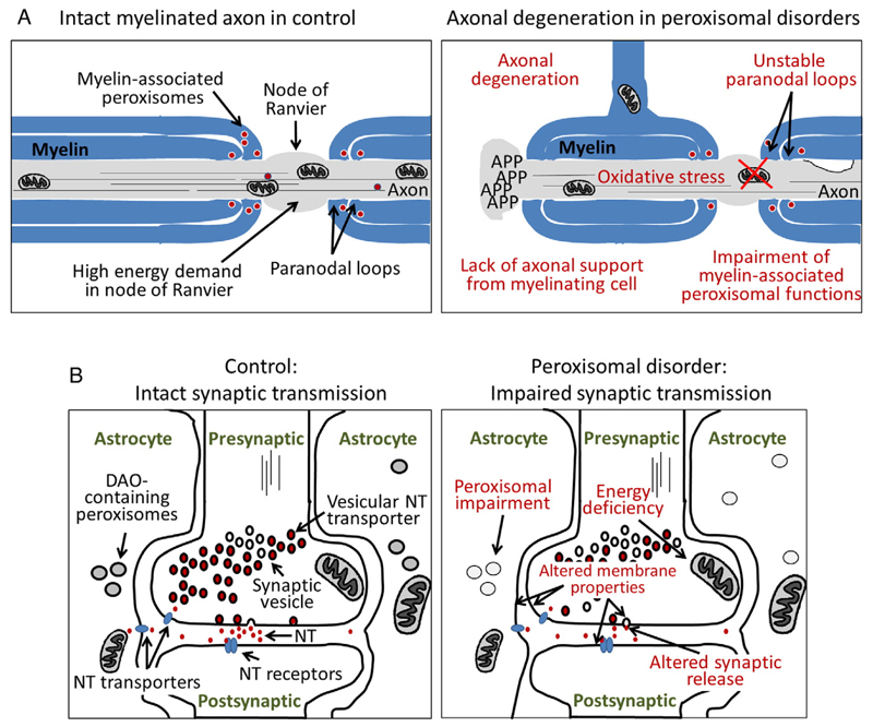
“On the one hand, peroxisomes generate building blocks (intermediates) for the biosynthesis of complex lipids such as ether phospholipids, which are important components of myelin, the membrane processes of oligodendrocytes that ensheath and isolate axons.”
§ Peroxisomes in brain development and function☆ > Brain peroxisomes and how they differ from peroxisomes in other tissues
plaindirect- Asserted: passes — The authors put it forward as true.
- Checkable: passes — It can be checked against evidence.
- Load-bearing: passes — The paper’s conclusions depend on it.
- Based: passes — The paper gives a reason why it holds.
A conclusion the paper draws from its own results — it cites no other source for it, so there is none to check. The quote shows it is in the paper.
In the paper
“This is the reason why in Zellweger spectrum patients, on the cellular level, peroxisomes are either absent or empty (ghosts).”
§ Peroxisomes in brain development and function☆ > Introduction
plaindirect- Asserted: passes — The authors put it forward as true.
- Checkable: passes — It can be checked against evidence.
- Load-bearing: passes — The paper’s conclusions depend on it.
- Based: passes — The paper gives a reason why it holds.
In the paper
“In neurons of human dorsal root ganglia, peroxisomes were readily detected based on immunohistochemistry for ABCD1 [30].”
§ Peroxisomes in brain development and function☆ > Brain peroxisomes and how they differ from peroxisomes in other tissues
What the sources say
- Not checked yetcites [30] Hoftberger R Kunze M Weinhofer I Aboul-Enein F Voigtlander T Oezen I Amann G Bernheimer H Budka H Berger J Distribution and cellular localization of adrenoleukodystrophy protein in human tissues: implications for X-linked adrenoleukodystrophy Neurobiol Dis 2007 28 165 174 17761426 source
plain- Asserted: passes — The authors put it forward as true.
- Checkable: passes — It can be checked against evidence.
- Load-bearing: unclear — It is unclear whether the paper’s conclusions depend on it.
- Based: passes — The paper gives a reason why it holds.
In the paper
“In the rat PNS, peroxisomes were described in Schwann cells, which represent the myelinating cells of the PNS, as well as in dorsal root ganglion satellite cells and, less abundantly, in neuronal somata [29].”
§ Peroxisomes in brain development and function☆ > Brain peroxisomes and how they differ from peroxisomes in other tissues
What the sources say
- Not checked yetcites [29] Citkowitz E Holtzman E Peroxisomes in dorsal root ganglia J Histochem Cytochem 1973 21 34 41 4694538 source
plain- Asserted: passes — The authors put it forward as true.
- Checkable: passes — It can be checked against evidence.
- Load-bearing: unclear — It is unclear whether the paper’s conclusions depend on it.
- Based: passes — The paper gives a reason why it holds.
A conclusion the paper draws from its own results — it cites no other source for it, so there is none to check. The quote shows it is in the paper.
In the paper
“which originate from aberrant development of the brain, demyelination and loss of axonal integrity, neuroinflammation or other neurodegenerative processes”
§ Peroxisomes in brain development and function☆ > Abstract
plaindirect- Asserted: passes — The authors put it forward as true.
- Checkable: passes — It can be checked against evidence.
- Load-bearing: passes — The paper’s conclusions depend on it.
- Based: unclear — It is unclear why the paper says it holds.
- It may state two separate claims as one. 72
- Probably not a substantial claim. 10
- The quote only partly states this claim. 2
In the paper
“In the deep white matter, catalase-positive glia appeared at 31–32 weeks of gestation, their appearance shifting from the deep to the superficial white matter with increasing age [15].”
§ Peroxisomes in brain development and function☆ > Brain peroxisomes and how they differ from peroxisomes in other tissues
What the sources say
- Not checked yetcites [15] Houdou S Kuruta H Hasegawa M Konomi H Takashima S Suzuki Y Hashimoto T Developmental immunohistochemistry of catalase in the human brain Brain Res 1991 556 267 270 1933360
plaincorrelational- Asserted: passes — The authors put it forward as true.
- Checkable: passes — It can be checked against evidence.
- Load-bearing: unclear — It is unclear whether the paper’s conclusions depend on it.
- Based: passes — The paper gives a reason why it holds.
It may state two separate claims as one.
In the paper
“Moreover, the abundance of brain peroxisomes differs between brain areas. Although single membrane-bound structures – detectable with different methods to stain peroxisomes – can be found in most regions, some brain areas were reported to contain only modest numbers of peroxisomes [22].”
§ Peroxisomes in brain development and function☆ > Brain peroxisomes and how they differ from peroxisomes in other tissues
What the sources say
- Not checked yetcites [22] Arnold G Holtzman E Microperoxisomes in the central nervous system of the postnatal rat Brain Res 1978 155 1 17 688002
plaincorrelational- Asserted: passes — The authors put it forward as true.
- Checkable: passes — It can be checked against evidence.
- Load-bearing: unclear — It is unclear whether the paper’s conclusions depend on it.
- Based: passes — The paper gives a reason why it holds.
It may state two separate claims as one.
None of the sources cited for this claim have been checked yet.
In the paper
“In contrast, the mRNAs for the enzymes involved in α-oxidation were not detected during the first postnatal weeks; and the ABCD1 mRNA was most highly expressed in the embryonic brain [26–28].”
§ Peroxisomes in brain development and function☆ > Brain peroxisomes and how they differ from peroxisomes in other tissues
What the sources say
- Not checked yetcites [26] Knoll A Sargueil F Salles J Cassagne C Garbay B Gene expression of peroxisomal beta-oxidation enzymes in rat brain Brain Res Mol Brain Res 1999 74 217 220 10640693
- Not checked yetcites [27] Huyghe S Casteels M Janssen A Meulders L Mannaerts GP Declercq PE Van Veldhoven PP Baes M Prenatal and postnatal development of peroxisomal lipid-metabolizing pathways in the mouse Biochem J 2001 353 673 680 11171065 PMC1221614
- Not checked yetcites [28] Berger J Albet S Bentejac M Netik A Holzinger A Roscher AA Bugaut M Forss-Petter S The four murine peroxisomal ABC-transporter genes differ in constitutive, inducible and developmental expression Eur J Biochem 1999 265 719 727 10504404 source
plain- Asserted: passes — The authors put it forward as true.
- Checkable: passes — It can be checked against evidence.
- Load-bearing: unclear — It is unclear whether the paper’s conclusions depend on it.
- Based: passes — The paper gives a reason why it holds.
It may state two separate claims as one.
In the paper
“In the cerebellum, punctate catalase immunoreactivity (characteristic of a peroxisomal localization) was predominantly observed in Bergmann glia (astrocytes), whereas in Purkinje cells, catalase appeared evenly distributed.”
§ Peroxisomes in brain development and function☆ > Brain peroxisomes and how they differ from peroxisomes in other tissues
What the sources say
- Not checked yetcites [21] Nagase T Shimozawa N Takemoto Y Suzuki Y Komori M Kondo N Peroxisomal localization in the developing mouse cerebellum: implications for neuronal abnormalities related to deficiencies in peroxisomes Biochim Biophys Acta 2004 1671 26 33 15026142
hedged- Asserted: passes — The authors put it forward as true.
- Checkable: passes — It can be checked against evidence.
- Load-bearing: unclear — It is unclear whether the paper’s conclusions depend on it.
- Based: passes — The paper gives a reason why it holds.
It may state two separate claims as one.
In the paper
“In the human brain, catalase-positive neurons emerged early in evolutionary old structures such as the basal ganglia, the thalamus and the cerebellum (about 27–28 weeks of gestation), whereas in the frontal cortex, they appeared later (around 35 weeks of gestation) [15].”
§ Peroxisomes in brain development and function☆ > Brain peroxisomes and how they differ from peroxisomes in other tissues
What the sources say
- Not checked yetcites [15] Houdou S Kuruta H Hasegawa M Konomi H Takashima S Suzuki Y Hashimoto T Developmental immunohistochemistry of catalase in the human brain Brain Res 1991 556 267 270 1933360
plaincorrelational- Asserted: passes — The authors put it forward as true.
- Checkable: passes — It can be checked against evidence.
- Load-bearing: unclear — It is unclear whether the paper’s conclusions depend on it.
- Based: passes — The paper gives a reason why it holds.
It may state two separate claims as one.
In the paper
“Brain peroxisomes in general, and neuronal peroxisomes in particular, are smaller than peroxisomes from other tissues and, thus, were termed microperoxisomes [17].”
§ Peroxisomes in brain development and function☆ > Brain peroxisomes and how they differ from peroxisomes in other tissues
What the sources say
- Not checked yetcites [17] Holtzman E Teichberg S Abrahams SJ Citkowitz E Crain SM Kawai N Peterson ER Notes on synaptic vesicles and related structures, endoplasmic reticulum, lysosomes and peroxisomes in nervous tissue and the adrenal medulla J Histochem Cytochem 1973 21 349 385 4574997
plaindirect- Asserted: passes — The authors put it forward as true.
- Checkable: passes — It can be checked against evidence.
- Load-bearing: unclear — It is unclear whether the paper’s conclusions depend on it.
- Based: passes — The paper gives a reason why it holds.
It may state two separate claims as one.
In the paper
“In mammals, peroxisomes contain around 50 different proteins [2]”
§ Peroxisomes in brain development and function☆ > Introduction
What the sources say
- Not checked yetcites [2] Wiese S Gronemeyer T Ofman R Kunze M Grou CP Almeida JA Eisenacher M Stephan C Hayen H Schollenberger L Korosec T Proteomics characterization of mouse kidney peroxisomes by tandem mass spectrometry and protein correlation profiling Mol Cell Proteomics 2007 6 2045 2057 17768142
plain- Asserted: passes — The authors put it forward as true.
- Checkable: passes — It can be checked against evidence.
- Load-bearing: fails — The paper’s conclusions may not depend on it.
- Based: passes — The paper gives a reason why it holds.
Probably not a substantial claim. The paper’s conclusions may not depend on it.
In the paper
“Comparison of DAO and catalase activity revealed that in the locus coeruleus of the rat brain, peroxisomes that stained positive for catalase activity were found in various cell types, whereas DAO activity-positive peroxisomes were restricted to astrocytes [13].”
§ Peroxisomes in brain development and function☆ > Brain peroxisomes and how they differ from peroxisomes in other tissues
What the sources say
- Not checked yetcites [13] Arnold G Liscum L Holtzman E Ultrastructural localization of D-amino acid oxidase in microperoxisomes of the rat nervous system J Histochem Cytochem 1979 27 735 745 39097
plain- Asserted: passes — The authors put it forward as true.
- Checkable: passes — It can be checked against evidence.
- Load-bearing: unclear — It is unclear whether the paper’s conclusions depend on it.
- Based: passes — The paper gives a reason why it holds.
It may state two separate claims as one.
In the paper
“This fatty acid is enriched in phospholipids including ether phospholipids and, either directly or after enzymatic conversion to a variety of bioactive derivatives, plays an important role in signaling [12].”
§ Peroxisomes in brain development and function☆ > Brain peroxisomes and how they differ from peroxisomes in other tissues
What the sources say
- Not checked yetcites [12] Bazinet RP Laye S Polyunsaturated fatty acids and their metabolites in brain function and disease Nat Rev Neurosci 2014 15 771 785 25387473
plaincorrelational- Asserted: passes — The authors put it forward as true.
- Checkable: passes — It can be checked against evidence.
- Load-bearing: unclear — It is unclear whether the paper’s conclusions depend on it.
- Based: passes — The paper gives a reason why it holds.
It may state two separate claims as one.
In the paper
“This finding was recapitulated in explanted cells from the cerebellum, in which catalase appeared cytosolic (not enriched in peroxisomes) in calbindin-positive Purkinje cells but punctate in astrocytes, whereas the peroxisomal membrane protein PEX14 was found punctate in all cell types [21].”
§ Peroxisomes in brain development and function☆ > Brain peroxisomes and how they differ from peroxisomes in other tissues
What the sources say
- Not checked yetcites [21] Nagase T Shimozawa N Takemoto Y Suzuki Y Komori M Kondo N Peroxisomal localization in the developing mouse cerebellum: implications for neuronal abnormalities related to deficiencies in peroxisomes Biochim Biophys Acta 2004 1671 26 33 15026142
hedged- Asserted: passes — The authors put it forward as true.
- Checkable: passes — It can be checked against evidence.
- Load-bearing: unclear — It is unclear whether the paper’s conclusions depend on it.
- Based: passes — The paper gives a reason why it holds.
It may state two separate claims as one.
In the paper
“By now, mutations in 13 different peroxin (PEX) genes (PEX1, PEX2, PEX3, PEX5, PEX6, PEX10, PEX11b, PEX12, PEX13, PEX14, PEX16, PEX19, PEX26) have been described in patients of the Zellweger spectrum [45].”
§ Peroxisomes in brain development and function☆ > Brain dysfunctions in inherited peroxisomal disorders > Brain pathology under conditions of generalized peroxisome deficiency in man and mice
What the sources say
- Not checked yetcites [45] Steinberg SJ Dodt G Raymond GV Braverman NE Moser AB Moser HW Peroxisome biogenesis disorders Biochim Biophys Acta 2006 1763 1733 1748 17055079
plaincorrelational- Asserted: passes — The authors put it forward as true.
- Checkable: passes — It can be checked against evidence.
- Load-bearing: fails — The paper’s conclusions may not depend on it.
- Based: passes — The paper gives a reason why it holds.
Probably not a substantial claim. The paper’s conclusions may not depend on it.
In the paper
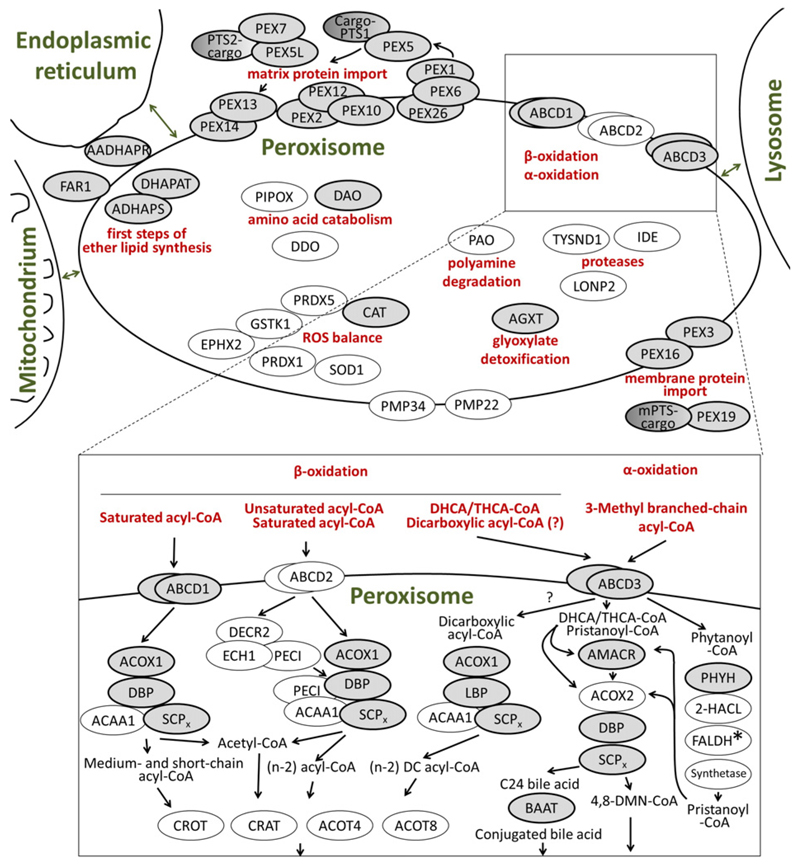
“Peroxisomes are known to house a variety of oxidases generating H2O2 and ROS, but they also enclose various ROS-detoxifying enzymes such as catalase, GSTK1, PRDX5 and SOD1 to limit the detrimental effects of local production [8].”
§ Peroxisomes in brain development and function☆ > Peroxisomes, brain and oxidative stress
What the sources say
- Not checked yetcites [8] Fransen M Nordgren M Wang B Apanasets O Role of peroxisomes in ROS/RNS-metabolism: implications for human disease Biochim Biophys Acta 2012 1822 1363 1373 22178243 source
boosteddirect- Asserted: passes — The authors put it forward as true.
- Checkable: passes — It can be checked against evidence.
- Load-bearing: unclear — It is unclear whether the paper’s conclusions depend on it.
- Based: passes — The paper gives a reason why it holds.
It may state two separate claims as one.
The paper states this without a source or data behind it here, so there is nothing to check it against. The quote shows it is in the paper.
In the paper
“To date, no migration defects have been described in infantile Refsum disease, the least severe form of the Zellweger spectrum.”
§ Peroxisomes in brain development and function☆ > Brain dysfunctions in inherited peroxisomal disorders > Brain pathology under conditions of generalized peroxisome deficiency in man and mice
hedged- Asserted: passes — The authors put it forward as true.
- Checkable: passes — It can be checked against evidence.
- Load-bearing: fails — The paper’s conclusions may not depend on it.
- Based: passes — The paper gives a reason why it holds.
Probably not a substantial claim. The paper’s conclusions may not depend on it.
The paper states this without a source or data behind it here, so there is nothing to check it against. The quote shows it is in the paper.
In the paper
“However, these patients have different clinical symptoms than Zellweger spectrum patients and are clinically not distinguishable from patients suffering from isolated disorders of ether phospholipid synthesis”
§ Peroxisomes in brain development and function☆ > Brain dysfunctions in inherited peroxisomal disorders > Brain pathology under conditions of generalized peroxisome deficiency in man and mice
plain- Asserted: passes — The authors put it forward as true.
- Checkable: passes — It can be checked against evidence.
- Load-bearing: unclear — It is unclear whether the paper’s conclusions depend on it.
- Based: passes — The paper gives a reason why it holds.
It may state two separate claims as one.
In the paper
“and exogenous application of VLCFA to a neuronal cell line induces oxidative stress and mitochondrial damage [35]”
§ Peroxisomes in brain development and function☆ > Peroxisomes, brain and oxidative stress
What the sources say
- Not checked yetcites [35] Zarrouk A Vejux A Nury T El Hajj HI Haddad M Cherkaoui-Malki M Riedinger JM Hammami M Lizard G Induction of mitochondrial changes associated with oxidative stress on very long chain fatty acids (C22:0, C24:0, or C26:0)-treated human neuronal cells (SK-NB-E) Oxidative Med Cell Longev 2012 2012 623257 PMC3420217 22919440
plaindirect- Asserted: passes — The authors put it forward as true.
- Checkable: passes — It can be checked against evidence.
- Load-bearing: unclear — It is unclear whether the paper’s conclusions depend on it.
- Based: passes — The paper gives a reason why it holds.
It may state two separate claims as one.
In the paper
“Artificial local production of ROS inside peroxisomes can induce apoptosis, which can be rescued by ectopic overexpression of peroxisomal detoxifying enzymes [33].”
§ Peroxisomes in brain development and function☆ > Peroxisomes, brain and oxidative stress
What the sources say
- Not checked yetcites [33] Wang B Van Veldhoven PP Brees C Rubio N Nordgren M Apanasets O Kunze M Baes M Agostinis P Fransen M Mitochondria are targets for peroxisome-derived oxidative stress in cultured mammalian cells Free Radic Biol Med 2013 65 882 894 23988789
plaindirect- Asserted: passes — The authors put it forward as true.
- Checkable: passes — It can be checked against evidence.
- Load-bearing: unclear — It is unclear whether the paper’s conclusions depend on it.
- Based: passes — The paper gives a reason why it holds.
It may state two separate claims as one.
The paper states this without a source or data behind it here, so there is nothing to check it against. The quote shows it is in the paper.
In the paper
“The PBD are divided into two types, i) Zellweger spectrum disorders and ii) rhizomelic chondrodysplasia punctata (RCDP) type 1.”
§ Peroxisomes in brain development and function☆ > Brain dysfunctions in inherited peroxisomal disorders > Brain pathology under conditions of generalized peroxisome deficiency in man and mice
plain- Asserted: passes — The authors put it forward as true.
- Checkable: passes — It can be checked against evidence.
- Load-bearing: fails — The paper’s conclusions may not depend on it.
- Based: passes — The paper gives a reason why it holds.
Probably not a substantial claim. The paper’s conclusions may not depend on it.
In the paper
“In our current understanding, the clinical syndromes constituting the Zellweger spectrum (MIM #601539)2 are the Zellweger syndrome, neonatal adrenoleukodystrophy, and infantile Refsum disease, which describe a clinical spectrum of decreasing severity [45].”
§ Peroxisomes in brain development and function☆ > Brain dysfunctions in inherited peroxisomal disorders > Brain pathology under conditions of generalized peroxisome deficiency in man and mice
What the sources say
- Not checked yetcites [45] Steinberg SJ Dodt G Raymond GV Braverman NE Moser AB Moser HW Peroxisome biogenesis disorders Biochim Biophys Acta 2006 1763 1733 1748 17055079
hedged- Asserted: passes — The authors put it forward as true.
- Checkable: passes — It can be checked against evidence.
- Load-bearing: fails — The paper’s conclusions may not depend on it.
- Based: passes — The paper gives a reason why it holds.
Probably not a substantial claim. The paper’s conclusions may not depend on it. It may state two separate claims as one.
In the paper
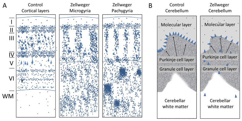
“In the polymicrogyric cortex, typically a fusion of the molecular layers is associated with a modified distribution of medium to large pyramidal cells originating from the deep cortex. This causes a decrease in the numbers of neurons in the outer layers (layer II and layer III) of the cortex; instead, these neurons are located in the deep cortex and within heterotopias of subcortical white matter”
§ Peroxisomes in brain development and function☆ > Brain dysfunctions in inherited peroxisomal disorders > Brain pathology under conditions of generalized peroxisome deficiency in man and mice
What the sources say
- Not checked yetcites [6] Powers JM Moser HW Peroxisomal disorders: genotype, phenotype, major neuropathologic lesions, and pathogenesis Brain Pathol 1998 8 101 120 9458170 PMC8098283 source
plaincorrelational- Asserted: passes — The authors put it forward as true.
- Checkable: passes — It can be checked against evidence.
- Load-bearing: unclear — It is unclear whether the paper’s conclusions depend on it.
- Based: passes — The paper gives a reason why it holds.
It may state two separate claims as one.
None of the sources cited for this claim have been checked yet.
In the paper
“Another striking morphological aberration linked to neuronal migration defects is the heterotopic localization of Purkinje cells in the cerebellar white matter (Fig. 2B) [6,49,50].”
§ Peroxisomes in brain development and function☆ > Brain dysfunctions in inherited peroxisomal disorders > Brain pathology under conditions of generalized peroxisome deficiency in man and mice
What the sources say
- Not checked yetcites [6] Powers JM Moser HW Peroxisomal disorders: genotype, phenotype, major neuropathologic lesions, and pathogenesis Brain Pathol 1998 8 101 120 9458170 PMC8098283 source
- Not checked yetcites [49] Volpe JJ Adams RD Cerebro-hepato-renal syndrome of Zellweger: an inherited disorder of neuronal migration Acta Neuropathol 1972 20 175 198 5043999
- Not checked yetcites [50] Evrard P Caviness VS Jr Prats-Vinas J Lyon G The mechanism of arrest of neuronal migration in the Zellweger malformation: an hypothesis bases upon cytoarchitectonic analysis Acta Neuropathol 1978 41 109 117 636841
plaincorrelational- Asserted: passes — The authors put it forward as true.
- Checkable: passes — It can be checked against evidence.
- Load-bearing: unclear — It is unclear whether the paper’s conclusions depend on it.
- Based: passes — The paper gives a reason why it holds.
The quote only partly states this claim.
The paper states this without a source or data behind it here, so there is nothing to check it against. The quote shows it is in the paper.
In the paper
“Patients with mutations in PEX7 are grouped into PBD because more than one peroxisomal pathway is affected, although the peroxisomal structure remains intact.”
§ Peroxisomes in brain development and function☆ > Brain dysfunctions in inherited peroxisomal disorders > Brain pathology under conditions of generalized peroxisome deficiency in man and mice
plaindirect- Asserted: passes — The authors put it forward as true.
- Checkable: passes — It can be checked against evidence.
- Load-bearing: unclear — It is unclear whether the paper’s conclusions depend on it.
- Based: passes — The paper gives a reason why it holds.
It may state two separate claims as one.
None of the sources cited for this claim have been checked yet.
In the paper
“Other studies reported normal VLCFA levels in plasma of late-onset patients with a PEX2 mutation [59] or normal plasmalogen levels in plasma of patients with PEX16 mutations [58].”
§ Peroxisomes in brain development and function☆ > Brain dysfunctions in inherited peroxisomal disorders > Brain pathology under conditions of generalized peroxisome deficiency in man and mice
What the sources say
- Not checked yetcites [59] Sevin C Ferdinandusse S Waterham HR Wanders RJ Aubourg P Autosomal recessive cerebellar ataxia caused by mutations in the PEX2 gene, Orphanet J Rare Dis 2011 6 8 PMC3064617 21392394
- Not checked yetcites [58] Ebberink MS Csanyi B Chong WK Denis S Sharp P Mooijer PA Dekker CJ Spooner C Ngu LH De Sousa C Wanders RJ Identification of an unusual variant peroxisome biogenesis disorder caused by mutations in the PEX16 gene J Med Genet 2010 47 608 615 20647552
plain- Asserted: passes — The authors put it forward as true.
- Checkable: passes — It can be checked against evidence.
- Load-bearing: passes — The paper’s conclusions depend on it.
- Based: passes — The paper gives a reason why it holds.
It may state two separate claims as one.
In the paper
“In particular, the levels of intermediates of bile acid biosynthesis (DHCA, THCA) and of pipecolic acid declined during the observed time period in many patients, whereas VLCFA and plasmalogen levels normalized only in some.”
§ Peroxisomes in brain development and function☆ > Brain dysfunctions in inherited peroxisomal disorders > Brain pathology under conditions of generalized peroxisome deficiency in man and mice
What the sources say
- Not checked yetcites [47] Berendse K Engelen M Ferdinandusse S Majoie CB Waterham HR Vaz FM Koelman JH Barth PG Wanders RJ Poll-The BT Zellweger spectrum disorders: clinical manifestations in patients surviving into adulthood J Inherit Metab Dis 2015 PMC4710674 26287655
plainn = 19- Asserted: passes — The authors put it forward as true.
- Checkable: passes — It can be checked against evidence.
- Load-bearing: passes — The paper’s conclusions depend on it.
- Based: passes — The paper gives a reason why it holds.
It may state two separate claims as one.
None of the sources cited for this claim have been checked yet.
In the paper
“The number of granule cells was reduced due to defects in their migration from the external to the internal granule cell layer and increased apoptotic cell death.”
§ Peroxisomes in brain development and function☆ > Brain dysfunctions in inherited peroxisomal disorders > Brain pathology under conditions of generalized peroxisome deficiency in man and mice
What the sources say
- Not checked yetcites [68] Faust PL Abnormal cerebellar histogenesis in PEX2 Zellweger mice reflects multiple neuronal defects induced by peroxisome deficiency J Comp Neurol 2003 461 394 413 12746876
- Not checked yetcites [69] Faust PL Banka D Siriratsivawong R Ng VG Wikander TM Peroxisome biogenesis disorders: the role of peroxisomes and metabolic dysfunction in developing brain J Inherit Metab Dis 2005 28 369 383 15868469
plaindirect- Asserted: passes — The authors put it forward as true.
- Checkable: passes — It can be checked against evidence.
- Load-bearing: passes — The paper’s conclusions depend on it.
- Based: passes — The paper gives a reason why it holds.
It may state two separate claims as one.
None of the sources cited for this claim have been checked yet.
In the paper
“In all mice with a global peroxisome deficiency (Pex2, Pex5 and Pex13 deficiency), a reduced thickness of the neocortical plate was observed, which reflects abnormal lamination that has been linked to impaired neuronal migration and increased cellular density in the underlying white matter [60–62].”
§ Peroxisomes in brain development and function☆ > Brain dysfunctions in inherited peroxisomal disorders > Brain pathology under conditions of generalized peroxisome deficiency in man and mice
What the sources say
- Not checked yetcites [60] Baes M Gressens P Baumgart E Carmeliet P Casteels M Fransen M Evrard P Fahimi D Declercq PE Collen D van Veldhoven PP A mouse model for Zellweger syndrome Nat Genet 1997 17 49 57 9288097
- Not checked yetcites [61] Faust PL Hatten ME Targeted deletion of the PEX2 peroxisome assembly gene in mice provides a model for Zellweger syndrome, a human neuronal migration disorder J Cell Biol 1997 139 1293 1305 9382874 PMC2140200
- Not checked yetcites [62] Maxwell M Bjorkman J Nguyen T Sharp P Finnie J Paterson C Tonks I Paton BC Kay GF Crane DI Pex13 inactivation in the mouse disrupts peroxisome biogenesis and leads to a Zellweger syndrome phenotype Mol Cell Biol 2003 23 5947 5957 12897163 PMC166343
plaincorrelational- Asserted: passes — The authors put it forward as true.
- Checkable: passes — It can be checked against evidence.
- Load-bearing: passes — The paper’s conclusions depend on it.
- Based: passes — The paper gives a reason why it holds.
It may state two separate claims as one.
In the paper
“When using mice, which express Cre under the nestin promoter (Nestin-Cre driver mice), inactivation of Pex5 occurs in all neural precursor cells at embryonic stages, but not in the microglia lineage.”
§ Peroxisomes in brain development and function☆ > Brain dysfunctions in inherited peroxisomal disorders > Brain pathology under conditions of generalized peroxisome deficiency in man and mice
What the sources say
- Not checked yetcites [71] Hulshagen L Krysko O Bottelbergs A Huyghe S Klein R Van Veldhoven PP De Deyn PP D’Hooge R Hartmann D Baes M Absence of functional peroxisomes from mouse CNS causes dysmyelination and axon degeneration J Neurosci 2008 28 4015 4027 18400901 PMC6670456
plain- Asserted: passes — The authors put it forward as true.
- Checkable: passes — It can be checked against evidence.
- Load-bearing: passes — The paper’s conclusions depend on it.
- Based: passes — The paper gives a reason why it holds.
It may state two separate claims as one.
In the paper
“During childhood, all 19 patients had a moderate to severe developmental delay as well as a reduction/loss of visual and hearing abilities and seven did not achieve structured speech.”
§ Peroxisomes in brain development and function☆ > Brain dysfunctions in inherited peroxisomal disorders > Brain pathology under conditions of generalized peroxisome deficiency in man and mice
What the sources say
- Not checked yetcites [47] Berendse K Engelen M Ferdinandusse S Majoie CB Waterham HR Vaz FM Koelman JH Barth PG Wanders RJ Poll-The BT Zellweger spectrum disorders: clinical manifestations in patients surviving into adulthood J Inherit Metab Dis 2015 PMC4710674 26287655
plainn = 19- Asserted: passes — The authors put it forward as true.
- Checkable: passes — It can be checked against evidence.
- Load-bearing: unclear — It is unclear whether the paper’s conclusions depend on it.
- Based: passes — The paper gives a reason why it holds.
It may state two separate claims as one.
In the paper
“As the import of peroxisomal proteins is not impaired in this mouse model, no accumulation of VLCFA and only a slight decrease in plasmalogen levels were detected in the brain [65].”
§ Peroxisomes in brain development and function☆ > Brain dysfunctions in inherited peroxisomal disorders > Brain pathology under conditions of generalized peroxisome deficiency in man and mice
What the sources say
- Not checked yetcites [65] Li X Baumgart E Morrell JC Jimenez-Sanchez G Valle D Gould SJ PEX11 beta deficiency is lethal and impairs neuronal migration but does not abrogate peroxisome function Mol Cell Biol 2002 22 4358 4365 12024045 PMC133847
plainSingle case- Asserted: passes — The authors put it forward as true.
- Checkable: passes — It can be checked against evidence.
- Load-bearing: passes — The paper’s conclusions depend on it.
- Based: passes — The paper gives a reason why it holds.
It may state two separate claims as one.
In the paper
“During infancy, four of these patients suffered from hypotonia, five from failure to thrive, 12 had a visual handicap due to retinal degeneration and eight presented with hearing impairment.”
§ Peroxisomes in brain development and function☆ > Brain dysfunctions in inherited peroxisomal disorders > Brain pathology under conditions of generalized peroxisome deficiency in man and mice
What the sources say
- Not checked yetcites [47] Berendse K Engelen M Ferdinandusse S Majoie CB Waterham HR Vaz FM Koelman JH Barth PG Wanders RJ Poll-The BT Zellweger spectrum disorders: clinical manifestations in patients surviving into adulthood J Inherit Metab Dis 2015 PMC4710674 26287655
plainn = 19- Asserted: passes — The authors put it forward as true.
- Checkable: passes — It can be checked against evidence.
- Load-bearing: unclear — It is unclear whether the paper’s conclusions depend on it.
- Based: passes — The paper gives a reason why it holds.
It may state two separate claims as one.
In the paper
“Interestingly, at the time of diagnosis, 17 of these patients had a blood metabolite profile typical of a peroxisomal disorder; but at later time points the concentration of many originally accumulating metabolites had declined and, in some patients, even a complete normalization was observed.”
§ Peroxisomes in brain development and function☆ > Brain dysfunctions in inherited peroxisomal disorders > Brain pathology under conditions of generalized peroxisome deficiency in man and mice
What the sources say
- Not checked yetcites [47] Berendse K Engelen M Ferdinandusse S Majoie CB Waterham HR Vaz FM Koelman JH Barth PG Wanders RJ Poll-The BT Zellweger spectrum disorders: clinical manifestations in patients surviving into adulthood J Inherit Metab Dis 2015 PMC4710674 26287655
plainn = 19- Asserted: passes — The authors put it forward as true.
- Checkable: passes — It can be checked against evidence.
- Load-bearing: passes — The paper’s conclusions depend on it.
- Based: passes — The paper gives a reason why it holds.
It may state two separate claims as one.
The paper states this without a source or data behind it here, so there is nothing to check it against. The quote shows it is in the paper.
In the paper
“As Pex7-deficient mice represent a model for RCDP type 1, but not for Zellweger spectrum disorders, we discuss this model in the context of ether phospholipid deficiency (Section 5.5.).”
§ Peroxisomes in brain development and function☆ > Brain dysfunctions in inherited peroxisomal disorders > Brain pathology under conditions of generalized peroxisome deficiency in man and mice
plain- Asserted: passes — The authors put it forward as true.
- Checkable: passes — It can be checked against evidence.
- Load-bearing: passes — The paper’s conclusions depend on it.
- Based: passes — The paper gives a reason why it holds.
It may state two separate claims as one.
None of the sources cited for this claim have been checked yet.
In the paper
“The phenotype of mice with Pex2, Pex5 and Pex13 deficiency resembles the severe form of human Zellweger syndrome. These mice are born alive, but are growth-retarded and severely hypotonic. Moreover, they do not feed and die within 67#x2013;24 h after birth [60–62].”
§ Peroxisomes in brain development and function☆ > Brain dysfunctions in inherited peroxisomal disorders > Brain pathology under conditions of generalized peroxisome deficiency in man and mice
What the sources say
- Not checked yetcites [60] Baes M Gressens P Baumgart E Carmeliet P Casteels M Fransen M Evrard P Fahimi D Declercq PE Collen D van Veldhoven PP A mouse model for Zellweger syndrome Nat Genet 1997 17 49 57 9288097
- Not checked yetcites [61] Faust PL Hatten ME Targeted deletion of the PEX2 peroxisome assembly gene in mice provides a model for Zellweger syndrome, a human neuronal migration disorder J Cell Biol 1997 139 1293 1305 9382874 PMC2140200
- Not checked yetcites [62] Maxwell M Bjorkman J Nguyen T Sharp P Finnie J Paterson C Tonks I Paton BC Kay GF Crane DI Pex13 inactivation in the mouse disrupts peroxisome biogenesis and leads to a Zellweger syndrome phenotype Mol Cell Biol 2003 23 5947 5957 12897163 PMC166343
plaincorrelational- Asserted: passes — The authors put it forward as true.
- Checkable: passes — It can be checked against evidence.
- Load-bearing: unclear — It is unclear whether the paper’s conclusions depend on it.
- Based: passes — The paper gives a reason why it holds.
It may state two separate claims as one.
None of the sources cited for this claim have been checked yet.
In the paper
“Moreover, mouse models with Pex11α [64] and Pex11β [65] deficiencies have been generated.”
§ Peroxisomes in brain development and function☆ > Brain dysfunctions in inherited peroxisomal disorders > Brain pathology under conditions of generalized peroxisome deficiency in man and mice
What the sources say
- Not checked yetcites [64] Li X Baumgart E Dong GX Morrell JC Jimenez-Sanchez G Valle D Smith KD Gould SJ PEX11alpha is required for peroxisome proliferation in response to 4-phenylbutyrate but is dispensable for peroxisome proliferator-activated receptor alpha-mediated peroxisome proliferation Mol Cell Biol 2002 22 8226 8240 12417726 PMC134051
- Not checked yetcites [65] Li X Baumgart E Morrell JC Jimenez-Sanchez G Valle D Gould SJ PEX11 beta deficiency is lethal and impairs neuronal migration but does not abrogate peroxisome function Mol Cell Biol 2002 22 4358 4365 12024045 PMC133847
plain- Asserted: passes — The authors put it forward as true.
- Checkable: passes — It can be checked against evidence.
- Load-bearing: fails — The paper’s conclusions may not depend on it.
- Based: passes — The paper gives a reason why it holds.
Probably not a substantial claim. The paper’s conclusions may not depend on it.
In the paper
“In these mice, peroxisome-dependent metabolite levels were deranged (increased VLCFA, decreased plasmalogen levels) in the brain at late embryonic stages, but were normal in the liver.”
§ Peroxisomes in brain development and function☆ > Brain dysfunctions in inherited peroxisomal disorders > Brain pathology under conditions of generalized peroxisome deficiency in man and mice
What the sources say
- Not checked yetcites [71] Hulshagen L Krysko O Bottelbergs A Huyghe S Klein R Van Veldhoven PP De Deyn PP D’Hooge R Hartmann D Baes M Absence of functional peroxisomes from mouse CNS causes dysmyelination and axon degeneration J Neurosci 2008 28 4015 4027 18400901 PMC6670456
plain- Asserted: passes — The authors put it forward as true.
- Checkable: passes — It can be checked against evidence.
- Load-bearing: passes — The paper’s conclusions depend on it.
- Based: passes — The paper gives a reason why it holds.
It may state two separate claims as one.
In the paper
“In Acox1−/− mice, marked peroxisome proliferation in the liver was observed and accompanied by an increase in H2O2 concentration, leading to the development of hepatic adenomas and carcinomas at 15 months of age [36]. However, no brain pathology has been described for these mice.”
§ Peroxisomes in brain development and function☆ > Brain dysfunctions in inherited peroxisomal disorders > Brain pathology in peroxisomal β-oxidation disorders in humans and mice
What the sources say
- Not checked yetcites [36] Fan CY Pan J Usuda N Yeldandi AV Rao MS Reddy JK Steatohepatitis, spontaneous peroxisome proliferation and liver tumors in mice lacking peroxisomal fatty acyl-CoA oxidase. Implications for peroxisome proliferator-activated receptor alpha natural ligand metabolism J Biol Chem 1998 273 15639 15645 9624157 source
plaindirect- Asserted: passes — The authors put it forward as true.
- Checkable: passes — It can be checked against evidence.
- Load-bearing: unclear — It is unclear whether the paper’s conclusions depend on it.
- Based: passes — The paper gives a reason why it holds.
It may state two separate claims as one.
In the paper
“However, as both enzymatic steps are essential for peroxisomal β-oxidation, the complete loss of both activities as well as of the individual enzymatic activities causes neurodevelopmental abnormalities and death within the first two years of life [83].”
§ Peroxisomes in brain development and function☆ > Brain dysfunctions in inherited peroxisomal disorders > Brain pathology in peroxisomal β-oxidation disorders in humans and mice
What the sources say
- Not checked yetcites [83] Ferdinandusse S Denis S Mooyer PA Dekker C Duran M Soorani-Lunsing RJ Boltshauser E Macaya A Gartner J Majoie CB Barth PG Clinical and biochemical spectrum of D-bifunctional protein deficiency Ann Neurol 2006 59 92 104 16278854
plaindirect- Asserted: passes — The authors put it forward as true.
- Checkable: passes — It can be checked against evidence.
- Load-bearing: unclear — It is unclear whether the paper’s conclusions depend on it.
- Based: passes — The paper gives a reason why it holds.
It may state two separate claims as one.
The paper states this without a source or data behind it here, so there is nothing to check it against. The quote shows it is in the paper.
In the paper
“Peroxisomal β-oxidation can degrade VLCFA, branched-chain fatty acids, bile acid intermediates, long-chain dicarboxylic acids and polyunsaturated fatty acids like tetracosahexaenoic acid (C24:6), which undergoes one cycle of β-oxidation in peroxisomes to produce DHA (C22:6).”
§ Peroxisomes in brain development and function☆ > Brain dysfunctions in inherited peroxisomal disorders > Brain pathology in peroxisomal β-oxidation disorders in humans and mice
plaindirect- Asserted: passes — The authors put it forward as true.
- Checkable: passes — It can be checked against evidence.
- Load-bearing: unclear — It is unclear whether the paper’s conclusions depend on it.
- Based: passes — The paper gives a reason why it holds.
It may state two separate claims as one.
In the paper
“Interestingly, no developmental defects were observed at birth or after two months although CNPase is expressed in progenitors before myelination as well as in adult oligodendrocytes.”
§ Peroxisomes in brain development and function☆ > Brain dysfunctions in inherited peroxisomal disorders > Brain pathology under conditions of generalized peroxisome deficiency in man and mice
What the sources say
- Not checked yetcites [75] Kassmann CM Lappe-Siefke C Baes M Brugger B Mildner A Werner HB Natt O Michaelis T Prinz M Frahm J Nave KA Axonal loss and neuroinflammation caused by peroxisome-deficient oligodendrocytes Nat Genet 2007 39 969 976 17643102
plain- Asserted: passes — The authors put it forward as true.
- Checkable: passes — It can be checked against evidence.
- Load-bearing: passes — The paper’s conclusions depend on it.
- Based: passes — The paper gives a reason why it holds.
It may state two separate claims as one.
The paper states this without a source or data behind it here, so there is nothing to check it against. The quote shows it is in the paper.
In the paper
“Human peroxisomes harbor two acyl-CoA oxidases, two bifunctional enzymes and two thiolases, whereas murine peroxisomes are equipped with three acyl-CoA oxidases, two bifunctional enzymes and three thiolases.”
§ Peroxisomes in brain development and function☆ > Brain dysfunctions in inherited peroxisomal disorders > Brain pathology in peroxisomal β-oxidation disorders in humans and mice
plain- Asserted: passes — The authors put it forward as true.
- Checkable: passes — It can be checked against evidence.
- Load-bearing: fails — The paper’s conclusions may not depend on it.
- Based: fails — The paper may give no reason why it holds.
Probably not a substantial claim. The paper’s conclusions may not depend on it. The paper may give no reason why it holds. It may state two separate claims as one.
None of the sources cited for this claim have been checked yet.
In the paper
“The phenotype of patients with AMACR deficiency (MIM #614307) often involves adult-onset sensory neuropathy [94] and late-onset cerebellar ataxia [95].”
§ Peroxisomes in brain development and function☆ > Brain dysfunctions in inherited peroxisomal disorders > Brain pathology in peroxisomal β-oxidation disorders in humans and mice
What the sources say
- Not checked yetcites [94] Smith EH Gavrilov DK Oglesbee D Freeman WD Vavra MW Matern D Tortorelli S An adult onset case of alpha-methyl-acyl-CoA racemase deficiency J Inherit Metab Dis 2010 33 Suppl. 3 S349 S353 20821052
- Not checked yetcites [95] Dick D Horvath R Chinnery PF AMACR mutations cause late-onset autosomal recessive cerebellar ataxia Neurology 2011 76 1768 1770 21576695 PMC3100132
plaincorrelational- Asserted: passes — The authors put it forward as true.
- Checkable: passes — It can be checked against evidence.
- Load-bearing: unclear — It is unclear whether the paper’s conclusions depend on it.
- Based: passes — The paper gives a reason why it holds.
It may state two separate claims as one.
In the paper
“Also brain-specific (Nestin-Cre-dependent) inactivation of Pex13 in mice resulted in a similar phenotype with impaired cerebellar development, neuronal cell death, astrogliosis and microgliosis as well as signs of mitochondria-mediated oxidative stress [74].”
§ Peroxisomes in brain development and function☆ > Brain dysfunctions in inherited peroxisomal disorders > Brain pathology under conditions of generalized peroxisome deficiency in man and mice
What the sources say
- Not checked yetcites [74] Muller CC Nguyen TH Ahlemeyer B Meshram M Santrampurwala N Cao S Sharp P Fietz PB Baumgart-Vogt E Crane DI PEX13 deficiency in mouse brain as a model of Zellweger syndrome: abnormal cerebellum formation, reactive gliosis and oxidative stress Dis Model Mech 2011 4 104 119 20959636 PMC3014351
plaindirect- Asserted: passes — The authors put it forward as true.
- Checkable: passes — It can be checked against evidence.
- Load-bearing: passes — The paper’s conclusions depend on it.
- Based: passes — The paper gives a reason why it holds.
It may state two separate claims as one.
None of the sources cited for this claim have been checked yet.
In the paper
“Moreover, Mfp2-deficient mice do not show signs of neurodevelopmental abnormalities such as migration defects at early time points [90] [91]; but later on, these mice develop cerebellar aberrations and axonal loss, which is reflected by motor impairment and lethargy [92].”
§ Peroxisomes in brain development and function☆ > Brain dysfunctions in inherited peroxisomal disorders > Brain pathology in peroxisomal β-oxidation disorders in humans and mice
What the sources say
- Not checked yetcites [90] Huyghe S Schmalbruch H De Gendt K Verhoeven G Guillou F Van Veldhoven PP Baes M Peroxisomal multifunctional protein 2 is essential for lipid homeostasis in Sertoli cells and male fertility in mice Endocrinology 2006 147 2228 2236 16484321
- Not checked yetcites [91] Baes M Dewerchin M Janssen A Collen D Carmeliet P Generation of Pex5-loxP mice allowing the conditional elimination of peroxisomes Genesis 2002 32 177 178 11857813
- Not checked yetcites [92] Huyghe S Schmalbruch H Hulshagen L Veldhoven PV Baes M Hartmann D Peroxisomal multifunctional protein-2 deficiency causes motor deficits and glial lesions in the adult central nervous system Am J Pathol 2006 168 1321 1334 16565505 PMC1606565
plaindirect- Asserted: passes — The authors put it forward as true.
- Checkable: passes — It can be checked against evidence.
- Load-bearing: passes — The paper’s conclusions depend on it.
- Based: passes — The paper gives a reason why it holds.
It may state two separate claims as one.
In the paper
“Marked hypomyelination was detected already during the second to third postnatal week (probably due to insufficient formation of myelin) and was found in all brain regions (later probably also due to demyelination), together with axonal loss, reactive astrocytes as well as activated microglia and macrophages [73].”
§ Peroxisomes in brain development and function☆ > Brain dysfunctions in inherited peroxisomal disorders > Brain pathology under conditions of generalized peroxisome deficiency in man and mice
What the sources say
- Not checked yetcites [73] Bottelbergs A Verheijden S Van Veldhoven PP Just W Devos R Baes M Peroxisome deficiency but not the defect in ether lipid synthesis causes activation of the innate immune system and axonal loss in the central nervous system J Neuroinflammation 2012 9 61 22458306 PMC3419640
plaincorrelational- Asserted: passes — The authors put it forward as true.
- Checkable: passes — It can be checked against evidence.
- Load-bearing: passes — The paper’s conclusions depend on it.
- Based: passes — The paper gives a reason why it holds.
It may state two separate claims as one.
In the paper
“This was surprising, because Pex5 deletion in astrocytes resulted in accumulation of VLCFA as well as reduced plasmalogen levels in the brain.”
§ Peroxisomes in brain development and function☆ > Brain dysfunctions in inherited peroxisomal disorders > Brain pathology under conditions of generalized peroxisome deficiency in man and mice
What the sources say
- Not checked yetcites [76] Bottelbergs A Verheijden S Hulshagen L Gutmann DH Goebbels S Nave KA Kassmann C Baes M Axonal integrity in the absence of functional peroxisomes from projection neurons and astrocytes Glia 2010 58 1532 1543 20578053
plaindirect- Asserted: passes — The authors put it forward as true.
- Checkable: passes — It can be checked against evidence.
- Load-bearing: passes — The paper’s conclusions depend on it.
- Based: passes — The paper gives a reason why it holds.
It may state two separate claims as one.
In the paper
“A mouse model of DBP deficiency (here termed Mfp2 deficiency) has been described, in which VLCFA accumulate specifically in brain, and the degradation of branched-chain fatty acids as well as the maturation of bile acid precursors were disturbed [89]. Mfp2-deficient mice appear quite normal at birth but are severely growth-retarded during the lactation period.”
§ Peroxisomes in brain development and function☆ > Brain dysfunctions in inherited peroxisomal disorders > Brain pathology in peroxisomal β-oxidation disorders in humans and mice
What the sources say
- Not checked yetcites [89] Baes M Huyghe S Carmeliet P Declercq PE Collen D Mannaerts GP Van Veldhoven PP Inactivation of the peroxisomal multifunctional protein-2 in mice impedes the degradation of not only 2-methyl-branched fatty acids and bile acid intermediates but also of very long chain fatty acids J Biol Chem 2000 275 16329 16336 10748062
plaindirect- Asserted: passes — The authors put it forward as true.
- Checkable: passes — It can be checked against evidence.
- Load-bearing: passes — The paper’s conclusions depend on it.
- Based: passes — The paper gives a reason why it holds.
It may state two separate claims as one.
In the paper
“However, young adult mice gradually developed impaired motor function and premature death due to axonal degeneration, progressive subcortical demyelination and neuroinflammation, starting at two to six months of age.”
§ Peroxisomes in brain development and function☆ > Brain dysfunctions in inherited peroxisomal disorders > Brain pathology under conditions of generalized peroxisome deficiency in man and mice
What the sources say
- Not checked yetcites [75] Kassmann CM Lappe-Siefke C Baes M Brugger B Mildner A Werner HB Natt O Michaelis T Prinz M Frahm J Nave KA Axonal loss and neuroinflammation caused by peroxisome-deficient oligodendrocytes Nat Genet 2007 39 969 976 17643102
plaindirect- Asserted: passes — The authors put it forward as true.
- Checkable: passes — It can be checked against evidence.
- Load-bearing: passes — The paper’s conclusions depend on it.
- Based: passes — The paper gives a reason why it holds.
It may state two separate claims as one.
In the paper
“Marked hypomyelination was detected already during the second to third postnatal week (probably due to insufficient formation of myelin) and was found in all brain regions (later probably also due to demyelination), together with axonal loss, reactive astrocytes as well as activated microglia and macrophages [73].”
§ Peroxisomes in brain development and function☆ > Brain dysfunctions in inherited peroxisomal disorders > Brain pathology under conditions of generalized peroxisome deficiency in man and mice
What the sources say
- Not checked yetcites [73] Bottelbergs A Verheijden S Van Veldhoven PP Just W Devos R Baes M Peroxisome deficiency but not the defect in ether lipid synthesis causes activation of the innate immune system and axonal loss in the central nervous system J Neuroinflammation 2012 9 61 22458306 PMC3419640
hedged- Asserted: passes — The authors put it forward as true.
- Checkable: passes — It can be checked against evidence.
- Load-bearing: passes — The paper’s conclusions depend on it.
- Based: passes — The paper gives a reason why it holds.
It may state two separate claims as one.
In the paper
“All known human patients with bifunctional protein deficiency harbor mutations in the HSDI7B4 gene [83], whereas no patients with a mutation in the EHHADH gene, encoding LBP, have yet been identified.”
§ Peroxisomes in brain development and function☆ > Brain dysfunctions in inherited peroxisomal disorders > Brain pathology in peroxisomal β-oxidation disorders in humans and mice
What the sources say
- Not checked yetcites [83] Ferdinandusse S Denis S Mooyer PA Dekker C Duran M Soorani-Lunsing RJ Boltshauser E Macaya A Gartner J Majoie CB Barth PG Clinical and biochemical spectrum of D-bifunctional protein deficiency Ann Neurol 2006 59 92 104 16278854
plain- Asserted: passes — The authors put it forward as true.
- Checkable: passes — It can be checked against evidence.
- Load-bearing: unclear — It is unclear whether the paper’s conclusions depend on it.
- Based: passes — The paper gives a reason why it holds.
It may state two separate claims as one.
A conclusion the paper draws from its own results — it cites no other source for it, so there is none to check. The quote shows it is in the paper.
In the paper
“Because ABCD2 and ABCD3 as well as the peroxisomal β-oxidation enzymes are intact in X-ALD, only saturated straight-chain VLCFA accumulate, but to a variable extent in different cell types and tissues.”
§ Peroxisomes in brain development and function☆ > Brain dysfunctions in inherited peroxisomal disorders > Brain pathology in X-linked adrenoleukodystrophy
plaindirect- Asserted: passes — The authors put it forward as true.
- Checkable: passes — It can be checked against evidence.
- Load-bearing: passes — The paper’s conclusions depend on it.
- Based: passes — The paper gives a reason why it holds.
It may state two separate claims as one.
None of the sources cited for this claim have been checked yet.
In the paper
“It is intriguing that also Abcd1/Abcd2 double-deficient mice do not develop brain inflammation or demyelination [156], in spite of the finding that Abcd1/Abcd2 double-deficient peritoneal macrophages are metabolically much more severely affected than those from single transporter-deficient mice [157]. Also in these mice, the neuropathology is restricted mainly to axonopathy in the spinal cord and,”
§ Peroxisomes in brain development and function☆ > Brain dysfunctions in inherited peroxisomal disorders > Brain pathology in X-linked adrenoleukodystrophy
What the sources say
- Not checked yetcites [156] Pujol A Ferrer I Camps C Metzger E Hindelang C Callizot N Ruiz M Pampols T Giros M Mandel JL Functional overlap between ABCD1 (ALD) and ABCD2 (ALDR) transporters: a therapeutic target for X-adrenoleukodystrophy Hum Mol Genet 2004 13 2997 3006 15489218 source
- Not checked yetcites [157] Muneer Z Wiesinger C Voigtlander T Werner HB Berger J Forss-Petter S Abcd2 Is a strong modifier of the metabolic impairments in peritoneal macrophages of Abcd1-deficient mice PLoS One 2014 9 e108655 25255441 PMC4177892 source
plain- Asserted: passes — The authors put it forward as true.
- Checkable: passes — It can be checked against evidence.
- Load-bearing: unclear — It is unclear whether the paper’s conclusions depend on it.
- Based: passes — The paper gives a reason why it holds.
It may state two separate claims as one.
In the paper
“in case of RCDP type 1, elevated plasma levels of phytanic acid have been detected [186].”
§ Peroxisomes in brain development and function☆ > Brain dysfunctions in inherited peroxisomal disorders > Nervous system pathology in ether phospholipid deficiency in man and mice
What the sources say
- Not checked yetcites [186] Hoefler G Hoefler S Watkins PA Chen WW Moser A Baldwin V McGillivary B Charrow J Friedman JM Rutledge L Biochemical abnormalities in rhizomelic chondrodysplasia punctata J Pediatr 1988 112 726 733 2452243
plaincorrelational- Asserted: passes — The authors put it forward as true.
- Checkable: passes — It can be checked against evidence.
- Load-bearing: fails — The paper’s conclusions may not depend on it.
- Based: passes — The paper gives a reason why it holds.
Probably not a substantial claim. The paper’s conclusions may not depend on it.
None of the sources cited for this claim have been checked yet.
In the paper
“Mutations in the PHYH gene have been established as the genetic cause for classical Refsum disease (MIM #266500) [170, 171]. The 3-methyl branched-chain fatty acid phytanic acid, solely taken up from dietary sources, accumulates in patients with Refsum disease.”
§ Peroxisomes in brain development and function☆ > Brain dysfunctions in inherited peroxisomal disorders > Brain pathology in α-oxidation deficiency in man and mice
What the sources say
- Not checked yetcites [170] Jansen GA Ofman R Ferdinandusse S Ijlst L Muijsers AO Skjeldal OH Stokke O Jakobs C Besley GT Wraith JE Wanders RJ Refsum disease is caused by mutations in the phytanoyl-CoA hydroxylase gene Nat Genet 1997 17 190 193 9326940
- Not checked yetcites [171] Jansen GA Hogenhout EM Ferdinandusse S Waterham HR Ofman R Jakobs C Skjeldal OH Wanders RJ Human phytanoyl-CoA hydroxylase: resolution of the gene structure and the molecular basis of Refsum’s disease Hum Mol Genet 2000 9 1195 1200 10767344
boosteddirect- Asserted: passes — The authors put it forward as true.
- Checkable: passes — It can be checked against evidence.
- Load-bearing: unclear — It is unclear whether the paper’s conclusions depend on it.
- Based: passes — The paper gives a reason why it holds.
It may state two separate claims as one.
None of the sources cited for this claim have been checked yet.
In the paper
“Encouraging results were obtained from a study applying an antioxidant cocktail consisting of vitamin E, N-acetylcystein and lipoic acid to aging Abcd1-deficient mice; this dietary treatment was sufficient to prevent the onset of locomotor disability and axonal damage [163]. In line with these findings, also oral administration of pioglitazone, an agonist of peroxisome proliferator-activated”
§ Peroxisomes in brain development and function☆ > Brain dysfunctions in inherited peroxisomal disorders > Brain pathology in X-linked adrenoleukodystrophy
What the sources say
- Not checked yetcites [163] Lopez-Erauskin J Fourcade S Galino J Ruiz M Schluter A Naudi A Jove M Portero-Otin M Pamplona R Ferrer I Pujol A Antioxidants halt axonal degeneration in a mouse model of X-adrenoleukodystrophy Ann Neurol 2011 70 84 92 21786300 PMC3229843
- Not checked yetcites [164] Morato L Galino J Ruiz M Calingasan NY Starkov AA Dumont M Naudi A Martinez JJ Aubourg P Portero-Otin M Pamplona R Pioglitazone halts axonal degeneration in a mouse model of X-linked adrenoleukodystrophy Brain 2013 136 2432 2443 23794606 PMC4550111
plaindirect- Asserted: passes — The authors put it forward as true.
- Checkable: passes — It can be checked against evidence.
- Load-bearing: passes — The paper’s conclusions depend on it.
- Based: passes — The paper gives a reason why it holds.
It may state two separate claims as one.
None of the sources cited for this claim have been checked yet.
In the paper
“In a classical gene KO model, Acaa1b deficiency showed a very mild phenotype and hardly any accumulation of VLCFA, indicating a compensatory effect from Acaa1a and/or Scpx in mice [103]. In Scpx-deficient mice, methyl-branched-chain fatty acid catabolism is impaired resulting in a mild phenotype under standard conditions [104].”
§ Peroxisomes in brain development and function☆ > Brain dysfunctions in inherited peroxisomal disorders > Brain pathology in peroxisomal β-oxidation disorders in humans and mice
What the sources say
- Not checked yetcites [103] Chevillard G Clemencet MC Latruffe N Nicolas-Frances V Targeted disruption of the peroxisomal thiolase B gene in mouse: a new model to study disorders related to peroxisomal lipid metabolism Biochimie 2004 86 849 856 15589695
- Not checked yetcites [104] Seedorf U Raabe M Ellinghaus P Kannenberg F Fobker M Engel T Denis S Wouters F Wirtz KW Wanders RJ Maeda N Defective peroxisomal catabolism of branched fatty acyl coenzyme A in mice lacking the sterol carrier protein-2/sterol carrier protein-x gene function Genes Dev 1998 12 1189 1201 9553048 PMC316706
hedgeddirect- Asserted: passes — The authors put it forward as true.
- Checkable: passes — It can be checked against evidence.
- Load-bearing: unclear — It is unclear whether the paper’s conclusions depend on it.
- Based: passes — The paper gives a reason why it holds.
It may state two separate claims as one. The quote only partly states this claim.
In the paper
“Although X-ALD does not involve any developmental defect or delay, it is characterized by remarkable clinical heterogeneity. The main phenotypes are adrenomyeloneuropathy (AMN) and cerebral ALD (CALD), the devastating inflammatory and demyelinating form of X-ALD [120].”
§ Peroxisomes in brain development and function☆ > Brain dysfunctions in inherited peroxisomal disorders > Brain pathology in X-linked adrenoleukodystrophy
What the sources say
- Not checked yetcites [120] Kemp S Berger J Aubourg P X-linked adrenoleukodystrophy: Clinical, metabolic, genetic and pathophysiological aspects Biochim Biophys Acta 2012 22483867 source
plain- Asserted: passes — The authors put it forward as true.
- Checkable: passes — It can be checked against evidence.
- Load-bearing: unclear — It is unclear whether the paper’s conclusions depend on it.
- Based: passes — The paper gives a reason why it holds.
It may state two separate claims as one.
The paper states this without a source or data behind it here, so there is nothing to check it against. The quote shows it is in the paper.
In the paper
“However, the extent to which the brain is affected varies remarkably between patients and many of the symptoms are restricted to the severer forms of the disease.”
§ Peroxisomes in brain development and function☆ > Brain dysfunctions in inherited peroxisomal disorders > Nervous system pathology in ether phospholipid deficiency in man and mice
plaincorrelational- Asserted: passes — The authors put it forward as true.
- Checkable: passes — It can be checked against evidence.
- Load-bearing: unclear — It is unclear whether the paper’s conclusions depend on it.
- Based: passes — The paper gives a reason why it holds.
It may state two separate claims as one.
The paper states this without a source or data behind it here, so there is nothing to check it against. The quote shows it is in the paper.
In the paper
“Virtually all male patients with mutations in the ABCD1 gene eventually develop AMN, a slowly progressive myelopathy with typical onset in the third or fourth decade of life.”
§ Peroxisomes in brain development and function☆ > Brain dysfunctions in inherited peroxisomal disorders > Brain pathology in X-linked adrenoleukodystrophy
plaindirect- Asserted: passes — The authors put it forward as true.
- Checkable: passes — It can be checked against evidence.
- Load-bearing: unclear — It is unclear whether the paper’s conclusions depend on it.
- Based: passes — The paper gives a reason why it holds.
It may state two separate claims as one.
In the paper
“The most typical are the eponymous shortening of the proximal long bones (rhizomelia) and epiphyseal stippling (chondrodysplasia punctata) as well as congenital cataracts, joint contractures and growth and developmental retardation [187].”
§ Peroxisomes in brain development and function☆ > Brain dysfunctions in inherited peroxisomal disorders > Nervous system pathology in ether phospholipid deficiency in man and mice
What the sources say
- Not checked yetcites [187] Braverman NE Moser AB Functions of plasmalogen lipids in health and disease Biochim Biophys Acta 2012 1822 1442 1452 22627108
plain- Asserted: passes — The authors put it forward as true.
- Checkable: passes — It can be checked against evidence.
- Load-bearing: unclear — It is unclear whether the paper’s conclusions depend on it.
- Based: passes — The paper gives a reason why it holds.
It may state two separate claims as one.
The paper states this without a source or data behind it here, so there is nothing to check it against. The quote shows it is in the paper.
In the paper
“Mental disability and delayed motor development are hallmarks of RCDP.”
§ Peroxisomes in brain development and function☆ > Brain dysfunctions in inherited peroxisomal disorders > Nervous system pathology in ether phospholipid deficiency in man and mice
plaincorrelational- Asserted: passes — The authors put it forward as true.
- Checkable: passes — It can be checked against evidence.
- Load-bearing: unclear — It is unclear whether the paper’s conclusions depend on it.
- Based: passes — The paper gives a reason why it holds.
It may state two separate claims as one.
In the paper
“Because standard mouse chow is very low in branched-chain fatty acids, Phyh-deficient mice have an unremarkable phenotype. However, dietary supplementation with 0.25% phytol (the precursor of phytanic acid) for three weeks or 0.1% phytol for six weeks caused ataxia, reflecting Purkinje cell loss and astrogliosis in the cerebellum, and peripheral neuropathy, as revealed by nerve conduction velocity”
§ Peroxisomes in brain development and function☆ > Brain dysfunctions in inherited peroxisomal disorders > Brain pathology in α-oxidation deficiency in man and mice
What the sources say
- Not checked yetcites [175] Ferdinandusse S Zomer AW Komen JC van den Brink CE Thanos M Hamers FP Wanders RJ van der Saag PT Poll-The BT Brites P Ataxia with loss of Purkinje cells in a mouse model for Refsum disease Proc Natl Acad Sci U S A 2008 105 17712 17717 19004801 PMC2584743
plaindirect- Asserted: passes — The authors put it forward as true.
- Checkable: passes — It can be checked against evidence.
- Load-bearing: passes — The paper’s conclusions depend on it.
- Based: passes — The paper gives a reason why it holds.
It may state two separate claims as one.
None of the sources cited for this claim have been checked yet.
In the paper
“RCDP type 1 (MIM #215100) is evoked by mutations in the gene encoding PEX7 [176–178], the cytosolic receptor for peroxisomal import of PTS2-containing proteins, whereas RCDP type 2 (MIM #222765) and type 3 (MIM #600121) are caused by mutations in the genes of the first two enzymes for biosynthesis of ether phospholipids, dihydroxyacetone phosphate acyltransferase (DHAPAT, DAPAT; encoded by the GNP”
§ Peroxisomes in brain development and function☆ > Brain dysfunctions in inherited peroxisomal disorders > Nervous system pathology in ether phospholipid deficiency in man and mice
What the sources say
- Not checked yetcites [176] Braverman N Steel G Obie C Moser A Moser H Gould SJ Valle D Human PEX7 encodes the peroxisomal PTS2 receptor and is responsible for rhizomelic chondrodysplasia punctata Nat Genet 1997 15 369 376 9090381
- Not checked yetcites [177] Motley AM Hettema EH Hogenhout EM Brites P ten Asbroek AL Wijburg FA Baas F Heijmans HS Tabak HF Wanders RJ Distel B Rhizomelic chondrodysplasia punctata is a peroxisomal protein targeting disease caused by a non-functional PTS2 receptor Nat Genet 1997 15 377 380 9090382
- Not checked yetcites [178] Purdue PE Zhang JW Skoneczny M Lazarow PB Rhizomelic chondrodysplasia punctata is caused by deficiency of human PEX7, a homologue of the yeast PTS2 receptor Nat Genet 1997 15 381 384 9090383
plaindirect- Asserted: passes — The authors put it forward as true.
- Checkable: passes — It can be checked against evidence.
- Load-bearing: unclear — It is unclear whether the paper’s conclusions depend on it.
- Based: passes — The paper gives a reason why it holds.
It may state two separate claims as one.
In the paper
“In this active region, infiltration of macrophages, CD4+ and CD8 + cytotoxic T cells, as well as activated microglia and astrocytes can be observed [140]. This severe neuroinflammation probably causes the loss of oligodendrocytes, which die by cytolysis rather than by apoptosis [140].”
§ Peroxisomes in brain development and function☆ > Brain dysfunctions in inherited peroxisomal disorders > Brain pathology in X-linked adrenoleukodystrophy
What the sources say
- Not checked yetcites [140] Ito M Blumberg BM Mock DJ Goodman AD Moser AB Moser HW Smith KD Powers JM Potential environmental and host participants in the early white matter lesion of adreno-leukodystrophy: morphologic evidence for CD8 cytotoxic T cells, cytolysis of oligodendrocytes, and CD1-mediated lipid antigen presentation J Neuropathol Exp Neurol 2001 60 1004 1019 11589421 source
plaincorrelational- Asserted: passes — The authors put it forward as true.
- Checkable: passes — It can be checked against evidence.
- Load-bearing: unclear — It is unclear whether the paper’s conclusions depend on it.
- Based: passes — The paper gives a reason why it holds.
It may state two separate claims as one.
None of the sources cited for this claim have been checked yet.
In the paper
“In contrast, patients with a less severe disease course present with only some of the characteristic symptoms and can survive into young adulthood [192–194].”
§ Peroxisomes in brain development and function☆ > Brain dysfunctions in inherited peroxisomal disorders > Nervous system pathology in ether phospholipid deficiency in man and mice
What the sources say
- Not checked yetcites [192] White AL Modaff P Holland-Morris F Pauli RM Natural history of rhizomelic chondrodysplasia punctata Am J Med Genet A 2003 118A 332 342 12687664
- Not checked yetcites [193] Motley AM Brites P Gerez L Hogenhout E Haasjes J Benne R Tabak HF Wanders RJ Waterham HR Mutational spectrum in the PEX7 gene and functional analysis of mutant alleles in 78 patients with rhizomelic chondrodysplasia punctata type 1 Am J Hum Genet 2002 70 612 624 11781871 PMC384941
- Not checked yetcites [194] Poll-The BT Maroteaux P Narcy C Quetin P Guesnu M Wanders RJ Schutgens RB Saudubray JM A new type of chondrodysplasia punctata associated with peroxisomal dysfunction J Inherit Metab Dis 1991 14 361 363 1770792
plaincorrelational- Asserted: passes — The authors put it forward as true.
- Checkable: passes — It can be checked against evidence.
- Load-bearing: unclear — It is unclear whether the paper’s conclusions depend on it.
- Based: passes — The paper gives a reason why it holds.
It may state two separate claims as one.
None of the sources cited for this claim have been checked yet.
In the paper
“Several studies have reported hypomyelination in different brain areas (neocortex, corpus callosum, cerebellum) of ether lipid-deficient mice [217,218], but no progressive demyelination as seen in several mouse models of Zellweger syndrome [73].”
§ Peroxisomes in brain development and function☆ > Brain dysfunctions in inherited peroxisomal disorders > Nervous system pathology in ether phospholipid deficiency in man and mice
What the sources say
- Not checked yetcites [217] Gorgas K Teigler A Komljenovic D Just WW The ether lipid-deficient mouse: tracking down plasmalogen functions Biochim Biophys Acta 2006 1763 1511 1526 17027098
- Not checked yetcites [218] Teigler A Komljenovic D Draguhn A Gorgas K Just WW Defects in myelination, paranode organization and Purkinje cell innervation in the ether lipid-deficient mouse cerebellum Hum Mol Genet 2009 18 1897 1908 19270340 PMC2722228
- Not checked yetcites [73] Bottelbergs A Verheijden S Van Veldhoven PP Just W Devos R Baes M Peroxisome deficiency but not the defect in ether lipid synthesis causes activation of the innate immune system and axonal loss in the central nervous system J Neuroinflammation 2012 9 61 22458306 PMC3419640
plain- Asserted: passes — The authors put it forward as true.
- Checkable: passes — It can be checked against evidence.
- Load-bearing: unclear — It is unclear whether the paper’s conclusions depend on it.
- Based: passes — The paper gives a reason why it holds.
It may state two separate claims as one.
None of the sources cited for this claim have been checked yet.
In the paper
“MR spectroscopy has also revealed elevated levels of mobile lipids, most likely caused by accumulation of long-chain acyl-CoAs, as well as a reduction of choline and the presence of acetate [199,209].”
§ Peroxisomes in brain development and function☆ > Brain dysfunctions in inherited peroxisomal disorders > Nervous system pathology in ether phospholipid deficiency in man and mice
What the sources say
- Not checked yetcites [199] Viola A Confort-Gouny S Ranjeva JP Chabrol B Raybaud C Vintila F Cozzone PJ MR imaging and MR spectroscopy in rhizomelic chondrodysplasia punctata AJNR Am J Neuroradiol 2002 23 480 483 11901023 PMC7975319
- Not checked yetcites [209] Alkan A Kutlu R Yakinci C Sigirci A Aslan M Sarac K Delayed myelination in a rhizomelic chondrodysplasia punctata case: MR spectroscopy findings Magn Reson Imaging 2003 21 77 80 12620550
hedgedcorrelational- Asserted: passes — The authors put it forward as true.
- Checkable: passes — It can be checked against evidence.
- Load-bearing: unclear — It is unclear whether the paper’s conclusions depend on it.
- Based: passes — The paper gives a reason why it holds.
It may state two separate claims as one.
None of the sources cited for this claim have been checked yet.
In the paper
“From a metabolic point of view, MR spectroscopy of the brain has shown increased levels of myo-inositol, a marker for gliosis, in line with previous reports of gliosis in autopsy cases of RCDP [52,208].”
§ Peroxisomes in brain development and function☆ > Brain dysfunctions in inherited peroxisomal disorders > Nervous system pathology in ether phospholipid deficiency in man and mice
What the sources say
- Not checked yetcites [52] Powers JM The pathology of peroxisomal disorders with pathogenetic considerations J Neuropathol Exp Neurol 1995 54 710 719 7666061
- Not checked yetcites [208] Agamanolis DP Novak RW Rhizomelic chondrodysplasia punctata: report of a case with review of the literature and correlation with other peroxisomal disorders Pediatr Pathol Lab Med 1995 15 503 513 8597837
plaincorrelational- Asserted: passes — The authors put it forward as true.
- Checkable: passes — It can be checked against evidence.
- Load-bearing: unclear — It is unclear whether the paper’s conclusions depend on it.
- Based: passes — The paper gives a reason why it holds.
It may state two separate claims as one.
In the paper
“Remarkably, hypo- and dysmyelination in the CNS of ether lipid-deficient animals is accompanied by slight loss of axons and mild astrogliosis in some brain areas, whereas microgliosis, which has been reported in several RCDP cases, and the induction of inflammatory cytokines appear to be less pronounced [73].”
§ Peroxisomes in brain development and function☆ > Brain dysfunctions in inherited peroxisomal disorders > Nervous system pathology in ether phospholipid deficiency in man and mice
What the sources say
- Not checked yetcites [73] Bottelbergs A Verheijden S Van Veldhoven PP Just W Devos R Baes M Peroxisome deficiency but not the defect in ether lipid synthesis causes activation of the innate immune system and axonal loss in the central nervous system J Neuroinflammation 2012 9 61 22458306 PMC3419640
hedgedcorrelational- Asserted: passes — The authors put it forward as true.
- Checkable: passes — It can be checked against evidence.
- Load-bearing: unclear — It is unclear whether the paper’s conclusions depend on it.
- Based: passes — The paper gives a reason why it holds.
It may state two separate claims as one.
None of the sources cited for this claim have been checked yet.
In the paper
“Also myelin of the PNS is affected in ether lipid-deficient mice. Deficiencies in myelination as well as Schwann cell development and differentiation are found [219] resulting in peripheral neuropathy, as judged by reduced motor neuron conduction velocity [220].”
§ Peroxisomes in brain development and function☆ > Brain dysfunctions in inherited peroxisomal disorders > Nervous system pathology in ether phospholipid deficiency in man and mice
What the sources say
- Not checked yetcites [219] da Silva TF Eira J Lopes AT Malheiro AR Sousa V Luoma A Avila RL Wanders RJ Just WW Kirschner DA Sousa MM Peripheral nervous system plasmalogens regulate Schwann cell differentiation and myelination J Clin Invest 2014 124 2560 2570 24762439 PMC4038568
- Not checked yetcites [220] Brites P Mooyer PA El Mrabet L Waterham HR Wanders RJ Plasmalogens participate in very-long-chain fatty acid-induced pathology Brain 2009 132 482 492 19022859
plaindirect- Asserted: passes — The authors put it forward as true.
- Checkable: passes — It can be checked against evidence.
- Load-bearing: unclear — It is unclear whether the paper’s conclusions depend on it.
- Based: passes — The paper gives a reason why it holds.
It may state two separate claims as one.
In the paper
“MRI examination in children with RCDP has revealed varying pathologic features, although some cases, usually with a milder phenotype, with unremarkable MRI results are also found [195].”
§ Peroxisomes in brain development and function☆ > Brain dysfunctions in inherited peroxisomal disorders > Nervous system pathology in ether phospholipid deficiency in man and mice
What the sources say
- Not checked yetcites [195] Bams-Mengerink AM Majoie CB Duran M Wanders RJ Van Hove J Scheurer CD Barth PG Poll-The BT MRI of the brain and cervical spinal cord in rhizomelic chondrodysplasia punctata Neurology 2006 66 798 803 discussion 789 16567694
plaincorrelational- Asserted: passes — The authors put it forward as true.
- Checkable: passes — It can be checked against evidence.
- Load-bearing: unclear — It is unclear whether the paper’s conclusions depend on it.
- Based: passes — The paper gives a reason why it holds.
It may state two separate claims as one.
None of the sources cited for this claim have been checked yet.
In the paper
“Consistently, most reports describe enlargement of ventricles and the subarachnoid space, abnormalities in white matter signal intensity and delayed supratentorial white matter myelination with frequent involvement of the parieto-occipital area [195–200].”
§ Peroxisomes in brain development and function☆ > Brain dysfunctions in inherited peroxisomal disorders > Nervous system pathology in ether phospholipid deficiency in man and mice
What the sources say
- Not checked yetcites [195] Bams-Mengerink AM Majoie CB Duran M Wanders RJ Van Hove J Scheurer CD Barth PG Poll-The BT MRI of the brain and cervical spinal cord in rhizomelic chondrodysplasia punctata Neurology 2006 66 798 803 discussion 789 16567694
- Not checked yetcites [196] Sztriha L Al-Gazali LI Wanders RJ Ofman R Nork M Lestringant GG Abnormal myelin formation in rhizomelic chondrodysplasia punctata type 2 (DHAPAT-deficiency) Dev Med Child Neurol 2000 42 492 495 10972423
- Not checked yetcites [197] Karabayir N Keskindemirci G Adal E Korkmaz O A case of rhizomelic chondrodysplasia punctata in newborn Case Rep Med 2014 2014 879679 24715923 PMC3970257
- Not checked yetcites [198] Sastrowijoto SH Vandenberghe K Moerman P Lauweryns JM Fryns JP Prenatal ultrasound diagnosis of rhizomelic chondrodysplasia punctata in a primigravida Prenat Diagn 1994 14 770 776 7991519
- Not checked yetcites [199] Viola A Confort-Gouny S Ranjeva JP Chabrol B Raybaud C Vintila F Cozzone PJ MR imaging and MR spectroscopy in rhizomelic chondrodysplasia punctata AJNR Am J Neuroradiol 2002 23 480 483 11901023 PMC7975319
- Not checked yetcites [200] Goh S Neuroimaging features in a neonate with rhizomelic chondrodysplasia punctata Pediatr. Neurol 2007 37 382 384 17950430
plaincorrelational- Asserted: passes — The authors put it forward as true.
- Checkable: passes — It can be checked against evidence.
- Load-bearing: unclear — It is unclear whether the paper’s conclusions depend on it.
- Based: passes — The paper gives a reason why it holds.
It may state two separate claims as one.
In the paper
“However, neuroinflammation with marked microglia activation was observed upon combined deficiency of Pex7 and Abcd1 in 11-months-old mice of mixed background (Swiss Webster and C57BL/6J:129S1) [220] suggesting that ether lipid deficiency has little effect on immune cell activation under basal conditions but more drastic consequences in the presence of an additional immunostimulatory factor.”
§ Peroxisomes in brain development and function☆ > Brain dysfunctions in inherited peroxisomal disorders > Nervous system pathology in ether phospholipid deficiency in man and mice
What the sources say
- Not checked yetcites [220] Brites P Mooyer PA El Mrabet L Waterham HR Wanders RJ Plasmalogens participate in very-long-chain fatty acid-induced pathology Brain 2009 132 482 492 19022859
hedgedconditional- Asserted: passes — The authors put it forward as true.
- Checkable: passes — It can be checked against evidence.
- Load-bearing: passes — The paper’s conclusions depend on it.
- Based: passes — The paper gives a reason why it holds.
It may state two separate claims as one.
None of the sources cited for this claim have been checked yet.
In the paper
“Also epileptic seizures are very common but non-specific; seizure type and frequency vary considerably and even affected children with multiple seizure types have been reported [191,192].”
§ Peroxisomes in brain development and function☆ > Brain dysfunctions in inherited peroxisomal disorders > Nervous system pathology in ether phospholipid deficiency in man and mice
What the sources say
- Not checked yetcites [191] Bams-Mengerink AM Koelman JH Waterham H Barth PG Poll-The BT The neurology of rhizomelic chondrodysplasia punctata Orphanet J Rare Dis 2013 8 174 24172221 PMC4228450
- Not checked yetcites [192] White AL Modaff P Holland-Morris F Pauli RM Natural history of rhizomelic chondrodysplasia punctata Am J Med Genet A 2003 118A 332 342 12687664
plain- Asserted: passes — The authors put it forward as true.
- Checkable: passes — It can be checked against evidence.
- Load-bearing: unclear — It is unclear whether the paper’s conclusions depend on it.
- Based: passes — The paper gives a reason why it holds.
It may state two separate claims as one.
None of the sources cited for this claim have been checked yet.
In the paper
“Currently, gene KO mouse models exist for RCDP types 1–3 (Pex 7, Gnpat and Agps KO mice, respectively) [210–212], of which the first two models have been extensively characterized.”
§ Peroxisomes in brain development and function☆ > Brain dysfunctions in inherited peroxisomal disorders > Nervous system pathology in ether phospholipid deficiency in man and mice
What the sources say
- Not checked yetcites [210] Brites P Motley AM Gressens P Mooyer PA Ploegaert I Everts V Evrard P Carmeliet P Dewerchin M Schoonjans L Duran M Impaired neuronal migration and endochondral ossification in Pex7 knockout mice: a model for rhizomelic chondrodysplasia punctata Hum Mol Genet 2003 12 2255 2267 12915479
- Not checked yetcites [211] Rodemer C Thai TP Brugger B Kaercher T Werner H Nave KA Wieland F Gorgas K Just WW Inactivation of ether lipid biosynthesis causes male infertility, defects in eye development and optic nerve hypoplasia in mice Hum Mol Genet 2003 12 1881 1895 12874108
- Not checked yetcites [212] Liegel RP Ronchetti A Sidjanin DJ Alkylglycerone phosphate synthase (AGPS) deficient mice: models for rhizomelic chondrodysplasia punctate type 3 (RCDP3) malformation syndrome Mol Genet Metab Rep 2014 1 299 311 25197626 PMC4151185
plain- Asserted: passes — The authors put it forward as true.
- Checkable: passes — It can be checked against evidence.
- Load-bearing: unclear — It is unclear whether the paper’s conclusions depend on it.
- Based: passes — The paper gives a reason why it holds.
It may state two separate claims as one.
In the paper
“Microscopy studies revealed structural abnormalities in the innervation of Purkinje cells by parallel fibers and climbing fibers as well as axonal swellings with accumulation of smooth ER-like structures in Purkinje cells [218].”
§ Peroxisomes in brain development and function☆ > Brain dysfunctions in inherited peroxisomal disorders > Nervous system pathology in ether phospholipid deficiency in man and mice
What the sources say
- Not checked yetcites [218] Teigler A Komljenovic D Draguhn A Gorgas K Just WW Defects in myelination, paranode organization and Purkinje cell innervation in the ether lipid-deficient mouse cerebellum Hum Mol Genet 2009 18 1897 1908 19270340 PMC2722228
plain- Asserted: passes — The authors put it forward as true.
- Checkable: passes — It can be checked against evidence.
- Load-bearing: unclear — It is unclear whether the paper’s conclusions depend on it.
- Based: passes — The paper gives a reason why it holds.
It may state two separate claims as one.
None of the sources cited for this claim have been checked yet.
In the paper
“Neuronal migration defects never reach the extent of those observed in Zellweger syndrome (see Fig. 2A), but several cases with dysplastic olivary bodies have been reported [202,206].”
§ Peroxisomes in brain development and function☆ > Brain dysfunctions in inherited peroxisomal disorders > Nervous system pathology in ether phospholipid deficiency in man and mice
What the sources say
- Not checked yetcites [202] Powers JM Kenjarski TP Moser AB Moser HW Cerebellar atrophy in chronic rhizomelic chondrodysplasia punctata: a potential role for phytanic acid and calcium in the death of its Purkinje cells Acta Neuropathol 1999 98 129 134 10442551
- Not checked yetcites [206] Poulos A Sheffield L Sharp P Sherwood G Johnson D Beckman K Fellenberg AJ Wraith JE Chow CW Usher S Rhizomelic chondrodysplasia punctata: clinical, pathologic, and biochemical findings in two patients J Pediatr 1988 113 685 690 3171792
plain- Asserted: passes — The authors put it forward as true.
- Checkable: passes — It can be checked against evidence.
- Load-bearing: unclear — It is unclear whether the paper’s conclusions depend on it.
- Based: passes — The paper gives a reason why it holds.
It may state two separate claims as one.
None of the sources cited for this claim have been checked yet.
In the paper
“Also, a migration defect of granule cell precursors [218] and increased numbers of apoptotic cells in the external granule layer [225] were found.”
§ Peroxisomes in brain development and function☆ > Brain dysfunctions in inherited peroxisomal disorders > Nervous system pathology in ether phospholipid deficiency in man and mice
What the sources say
- Not checked yetcites [218] Teigler A Komljenovic D Draguhn A Gorgas K Just WW Defects in myelination, paranode organization and Purkinje cell innervation in the ether lipid-deficient mouse cerebellum Hum Mol Genet 2009 18 1897 1908 19270340 PMC2722228
- Not checked yetcites [225] Krysko O Bottelbergs A Van Veldhoven P Baes M Combined deficiency of peroxisomal beta-oxidation and ether lipid synthesis in mice causes only minor cortical neuronal migration defects but severe hypotonia Mol Genet Metab 2010 100 71 76 20202875
plaincorrelational- Asserted: passes — The authors put it forward as true.
- Checkable: passes — It can be checked against evidence.
- Load-bearing: passes — The paper’s conclusions depend on it.
- Based: passes — The paper gives a reason why it holds.
It may state two separate claims as one.
None of the sources cited for this claim have been checked yet.
In the paper
“Furthermore, hypoplasia of the optic nerve, microphtalmia, a persistent hyaloid artery [211] and abnormal vascularization [229] were detected in Gnpat KO mice.”
§ Peroxisomes in brain development and function☆ > Brain dysfunctions in inherited peroxisomal disorders > Nervous system pathology in ether phospholipid deficiency in man and mice
What the sources say
- Not checked yetcites [211] Rodemer C Thai TP Brugger B Kaercher T Werner H Nave KA Wieland F Gorgas K Just WW Inactivation of ether lipid biosynthesis causes male infertility, defects in eye development and optic nerve hypoplasia in mice Hum Mol Genet 2003 12 1881 1895 12874108
- Not checked yetcites [229] Saab S Buteau B Leclere L Bron AM Creuzot-Garcher CP Bretillon L Acar N Involvement of plasmalogens in post-natal retinal vascular development PLoS One 2014 9 e101076 24963632 PMC4071069
plaincorrelational- Asserted: passes — The authors put it forward as true.
- Checkable: passes — It can be checked against evidence.
- Load-bearing: unclear — It is unclear whether the paper’s conclusions depend on it.
- Based: passes — The paper gives a reason why it holds.
It may state two separate claims as one.
In the paper
“Remarkably, although similar techniques were used, no such alterations could be detected in the brains of Gnpat KO mice [225], pointing towards a role of phytanic acid in the development of these migration defects or potential differences in the background strains of the mice used in the different studies.”
§ Peroxisomes in brain development and function☆ > Brain dysfunctions in inherited peroxisomal disorders > Nervous system pathology in ether phospholipid deficiency in man and mice
What the sources say
- Not checked yetcites [225] Krysko O Bottelbergs A Van Veldhoven P Baes M Combined deficiency of peroxisomal beta-oxidation and ether lipid synthesis in mice causes only minor cortical neuronal migration defects but severe hypotonia Mol Genet Metab 2010 100 71 76 20202875
hedged- Asserted: passes — The authors put it forward as true.
- Checkable: passes — It can be checked against evidence.
- Load-bearing: unclear — It is unclear whether the paper’s conclusions depend on it.
- Based: passes — The paper gives a reason why it holds.
It may state two separate claims as one.
In the paper
“This goes along with decreased ATP content and an inability of synaptosomal respiration to adapt to the higher energy requirements of depolarization, thereby offering a potential explanation for the defects in synaptosomal neurotransmitter release [40] (Fig. 3B).”
§ Peroxisomes in brain development and function☆ > Brain dysfunctions in inherited peroxisomal disorders > Nervous system pathology in ether phospholipid deficiency in man and mice
What the sources say
- Not checked yetcites [40] Brodde A Teigler A Brugger B Lehmann WD Wieland F Berger J Just WW Impaired neurotransmission in ether lipid-deficient nerve terminals Hum Mol Genet 2012 21 2713 2724 22403185 PMC4880048
hedgedconditional- Asserted: passes — The authors put it forward as true.
- Checkable: passes — It can be checked against evidence.
- Load-bearing: passes — The paper’s conclusions depend on it.
- Based: passes — The paper gives a reason why it holds.
It may state two separate claims as one.
None of the sources cited for this claim have been checked yet.
In the paper
“Furthermore, it has been suggested that deficiency in plasmalogens impairs membrane rafts (formerly termed lipid rafts) [211], small membrane domains that organize various cellular processes [236,237] and are enriched in plasmalogens [238].”
§ Peroxisomes in brain development and function☆ > Brain dysfunctions in inherited peroxisomal disorders > Nervous system pathology in ether phospholipid deficiency in man and mice
What the sources say
- Not checked yetcites [211] Rodemer C Thai TP Brugger B Kaercher T Werner H Nave KA Wieland F Gorgas K Just WW Inactivation of ether lipid biosynthesis causes male infertility, defects in eye development and optic nerve hypoplasia in mice Hum Mol Genet 2003 12 1881 1895 12874108
- Not checked yetcites [236] Pike LJ Rafts defined: a report on the Keystone Symposium on Lipid Rafts and Cell Function J Lipid Res 2006 47 1597 1598 16645198
- Not checked yetcites [237] Simons K Ikonen E Functional rafts in cell membranes Nature 1997 387 569 572 9177342
- Not checked yetcites [238] Pike LJ Han X Chung KN Gross RW Lipid rafts are enriched in arachidonic acid and plasmenylethanolamine and their composition is independent of caveolin-1 expression: a quantitative electrospray ionization/mass spectrometric analysis Biochemistry 2002 41 2075 2088 11827555
hedgedconditional- Asserted: passes — The authors put it forward as true.
- Checkable: passes — It can be checked against evidence.
- Load-bearing: fails — The paper’s conclusions may not depend on it.
- Based: passes — The paper gives a reason why it holds.
Probably not a substantial claim. The paper’s conclusions may not depend on it.
In the paper
“Recently, a defect in the activation and downstream signaling of AKT was proposed to be responsible for the myelination deficit in the PNS of ether lipid-deficient animals [219].”
§ Peroxisomes in brain development and function☆ > Brain dysfunctions in inherited peroxisomal disorders > Nervous system pathology in ether phospholipid deficiency in man and mice
What the sources say
- Not checked yetcites [219] da Silva TF Eira J Lopes AT Malheiro AR Sousa V Luoma A Avila RL Wanders RJ Just WW Kirschner DA Sousa MM Peripheral nervous system plasmalogens regulate Schwann cell differentiation and myelination J Clin Invest 2014 124 2560 2570 24762439 PMC4038568
hedgedconditional- Asserted: passes — The authors put it forward as true.
- Checkable: passes — It can be checked against evidence.
- Load-bearing: fails — The paper’s conclusions may not depend on it.
- Based: passes — The paper gives a reason why it holds.
Probably not a substantial claim. The paper’s conclusions may not depend on it.
The paper relies on this without support.
Observations in mouse models of peroxisomal disorders are informative about human brain development and function.
“with a specific focus on human physiology and pathophysiology supplemented by observations made in various mouse models”
“genetically engineered mouse models with tissue- or cell type-specific inactivation of peroxisome biogenesis demonstrate the importance of peroxisomes for the different brain cell types”
The paper relies on this without support.
The brain pathologies observed in inherited peroxisomal disorders reveal the normal functions of peroxisomes in brain development and maintenance.
“correlating peroxisomal properties with a compilation of pathologies observed in human patients and mouse models lacking all or individual peroxisomal functions”
“the observed pathology cannot be attributed to individual peroxisomal metabolic pathways but rather reflects the importance of the entire organelle for brain development and maintenance”
Claims other papers make about this one
Each is checked against this paper’s own text: does it say what the citing paper says it does?
In the citing paper
“However, when the hyperphosphorylated Tau protein is overexpressed (which is a pathophysiological indicator of AD) a depletion of these organelles has been evidenced, suggesting it is the cause of H2O2 and ROS increase [72,108,126].”
From Multi-Target Mechanisms of Phytochemicals in Alzheimer’s Disease: Effects on Oxidative Stress, Neuroinflammation and Protein Aggregation (2022) · cites this paper as [108]
In the citing paper
“Although AD has complex multifactorial, effects OS is key to the initiation of the pathophysiological process in cholinergic neurons [107,108,109].”
From Multi-Target Mechanisms of Phytochemicals in Alzheimer’s Disease: Effects on Oxidative Stress, Neuroinflammation and Protein Aggregation (2022) · cites this paper as [108]
In the citing paper
“Hydrogen peroxide seems to be the relevant actor in promoting excessive oxidation in neuronal cells.”
From Multi-Target Mechanisms of Phytochemicals in Alzheimer’s Disease: Effects on Oxidative Stress, Neuroinflammation and Protein Aggregation (2022) · cites this paper as [108]
Self-check
Percentages, sums and reported differences re-computed from the paper’s own numbers.
Rated minor: it would not touch a result — a value no result rests on, a p-value that stays on the same side of the significance level, or a conflict that is clerical, resolved elsewhere in the paper, or outside its own findings.
In the paper
“About 60% of male X-ALD patients develop CALD, the fatal cerebral demyelinating form of the disease. This can occur either in childhood, most commonly between 5 and 10 years of age, before onset of AMN (about 35%) or later in adolescence or adulthood, often on the background of AMN (35%).”
§ Peroxisomes in brain development and function☆ > Brain dysfunctions in inherited peroxisomal disorders > Brain pathology in X-linked adrenoleukodystrophy
Reported 60 · recomputed 70
Citations
Cites
The Metabolic and Molecular Bases of Inherited Disease
Scriver, R. C. (1995) · Medical Entomology and Zoology · Cited by 12,101
Functional rafts in cell membranes
Simons, K., & Ikonen, E. (1997) · Nature · doi:10.1038/42408 · Cited by 9,757 · cited 1×
“[211], small membrane domains that organize various cellular processes [236,237] and are enriched in plasmalogens [238].”
BackgroundThe metabolic and molecular bases of inherited disease
Chen, Y.-T. (2001) · Medical Entomology and Zoology · Cited by 6,928
Mitochondrial Reactive Oxygen Species (ROS) and ROS-Induced ROS Release
Zorov, D. B., Juhaszova, M., & Sollott, S. J. (2014) · Physiological Reviews · doi:10.1152/physrev.00026.2013 · Cited by 5,383 · cited 11×
“Evidence for the role of oxidative stress in plasma of X-ALD patients comes from an increased level of thiobarbituric acid reactive species (TBA-RS) reflecting induction of lipid peroxidation, aswell as a decrease of plasma total antioxidant reactivity, indicating a deficient capacity to rapidly handle an increase of…”
BackgroundInfluentialMucke, L., Masliah, E., Yu, G.-Q., Mallory, M., Rockenstein, E. M., Tatsuno, G., Hu, K., Kholodenko, D., Johnson-Wood, K., & McConlogue, L. (2000) · The Journal of Neuroscience · doi:10.1523/jneurosci.20-11-04050.2000 · Cited by 1,856 · cited 1×
“Moreover, the neuron-specific ectopic expression of IDE in a mouse model for AD (APPSwe/Ind) [301] was sufficient to relieve the burden of amyloid plaque-related pathology [302].”
BackgroundPolyunsaturated fatty acids and their metabolites in brain function and disease
Bazinet, R. P., & Layé, S. (2014) · Nature reviews. Neuroscience · doi:10.1038/nrn3820 · Cited by 1,703 · cited 1×
“This fatty acid is enriched in phospholipids including ether phospholipids and, either directly or after enzymatic conversion to a variety of bioactive derivatives, plays an important role in signaling [12].”
BackgroundFarris, W., Mansourian, S., Chang, Y., Lindsley, L., Eckman, E. A., Frosch, M. P., Eckman, C. B., Tanzi, R. E., Selkoe, D. J., & Guénette, S. Y. (2003) · Proceedings of the National Academy of Sciences · doi:10.1073/pnas.0230450100 · Cited by 1,526
Hematopoietic Stem Cell Gene Therapy with a Lentiviral Vector in X-Linked Adrenoleukodystrophy
Cartier, N., Hacein-Bey-Abina, S., Bartholomae, C. C., Veres, G., Schmidt, M., Kutschera, I., Vidaud, M., Abel, U., Dal-Cortivo, L., Caccavelli, L., et al. (2009) · Science · doi:10.1126/science.1171242 · Cited by 1,508
Rafts defined: a report on the Keystone symposium on lipid rafts and cell function
PIKE, L. J. (2006) · Journal of Lipid Research · doi:10.1194/jlr.e600002-jlr200 · Cited by 1,500 · cited 1×
“[211], small membrane domains that organize various cellular processes [236,237] and are enriched in plasmalogens [238].”
BackgroundPutative X-linked adrenoleukodystrophy gene shares unexpected homology with ABC transporters
Mosser, J., Douar, A.-M., Sarde, C.-O., Kioschis, P., Feil, R., Moser, H., Poustka, A.-M., Mandel, J.-L., & Aubourg, P. (1993) · Nature · doi:10.1038/361726a0 · Cited by 1,202 · cited 2×
“member 1 (ABCD1) X-linked adrenoleukodystrophy 300100 [108]”
Background
Research works printed but not linked: 1
- Nave, 2006, Oligodendrocytes and Schwann cells support axon function and survival independent of their role in myelination: implications for multiple sclerosis, J. Neuroimmunol., 178, 36-36
Cited by
Sorted by citations
The peroxisome: an update on mysteries 2.0
Islinger, M., Völkl, A., Fahimi, H. D., & Schrader, M. (2018) · Histochemistry and Cell Biology · doi:10.1007/s00418-018-1722-5 · Cited by 296
“ders is the often severe neuropathological phenotype represented by developmental alterations in neuronal migration, a progressive demyelination of neurons or inflammatory activation of microglia (Berger et al. 2016).”
BackgroundInfluentialThe physiological functions of human peroxisomes
Wanders, R. J. A., Baes, M., Ribeiro, D., Ferdinandusse, S., & Waterham, H. R. (2023) · Physiological Reviews · doi:10.1152/physrev.00051.2021 · Cited by 228
Peroxisomal Hydrogen Peroxide Metabolism and Signaling in Health and Disease
Lismont, C., Revenco, I., & Fransen, M. (2019) · International Journal of Molecular Sciences · doi:10.3390/ijms20153673 · Cited by 218
“Interestingly, patients suffering from a peroxisomal deficiency also typically develop severe neurological deficits [171].”
BackgroundHypoxia and Selective Autophagy in Cancer Development and Therapy
Daskalaki, I., Gkikas, I., & Tavernarakis, N. (2018) · Frontiers in Cell and Developmental Biology · doi:10.3389/fcell.2018.00104 · Cited by 188
“Their key functions are oxygen-dependent and related to lipid synthesis, ROS metabolism and the degradation of both polyunsaturated fatty acids (PUFAs) and very long fatty acids (VLCFAs), among others (Berger et al., 2016).”
BackgroundDorninger, F., Forss‐Petter, S., & Berger, J. (2017) · FEBS Letters · doi:10.1002/1873-3468.12788 · Cited by 165
“Inborn ether lipid deficiency is also a secondary feature of peroxisome biogenesis disorders of the Zellweger syndrome spectrum, in which all peroxisomal functions are impaired due to deficient biogenesis of the whole organelle and whose nervous system phenotype has been reviewed extensively elsewhere [27].”
Self-citation
Metadata
- Authors
- Berger, J., Dorninger, F., Forss-Petter, S., & Kunze, M.
- Affiliations
- Medical University of Vienna (AT)
- Corresponding author
- Berger, Johannes
- Venue
- Biochimica et Biophysica Acta (BBA) - Molecular Cell Research
- Publisher
- Elsevier BV
- Published
- 2016-05
- Volume
- 1863
- Issue
- 5
- Pages
- 934-955
- ISSN
- 0167-4889 (ISSN-L)
- PMC
- PMC4880039
- OpenAlex
- W2194234530
- Semantic Scholar
- 68bd48b50547368a06d2cb6adc013ae54ddad4f5
- Cited by
- 194
- References
- 352
- Citations by year
- 2026: 13 · 2025: 20 · 2024: 19 · 2023: 16 · 2022: 27 · 2021: 10 · 2020: 22 · 2019: 26 · 2018: 16 · 2017: 16 · 2016: 9
- FWCI
- 11.30
- Type
- journal article
- Language
- en
- Open access
- hybrid
- doi.org
- Locations
- Biochimica et Biophysica Acta (BBA) - Molecular Cell Research (publishedVersion, cc-by)
- PubMed (publishedVersion)
- Europe PMC (PubMed Central) (acceptedVersion, cc-by)
- PubMed Central (acceptedVersion, cc-by)
- Funders
- Austrian Science Fund (FWF) (P26112-B19,, P24843-B24, I2738-B26)
- Topics
- Life Sciences › Biochemistry, Genetics and Molecular Biology › Clinical Biochemistry › Metabolism and Genetic Disorders
- Health Sciences › Medicine › Genetics (clinical) › Lysosomal Storage Disorders Research
- Life Sciences › Neuroscience › Neurology › Neurological diseases and metabolism
- Keywords
- peroxisomes, peroxisome biogenesis disorders, brain development, demyelination, axonal integrity, neuroinflammation, neurodegeneration, Alzheimer's disease, autism spectrum disorder, amyotrophic lateral sclerosis, neurological disorders
- MeSH terms
- ATPases Associated with Diverse Cellular Activities, Animals, Brain, Disease Models, Animal, Gene Expression Regulation, Humans, Membrane Proteins, Mutation, Synaptic Transmission, Peroxisomal Disorders, Protein Isoforms, Peroxisomes, Protein Transport, Mice, Metabolic Networks and Pathways
About these checks
Registry, retraction, Cites and statistics results are computed, not judged, with one exception: a printed reference that no index lists is read once by a model before it counts as a citation error. Claims are extracted by a model and kept only when their quote appears verbatim in the text; each cited source is then checked, with a model’s help, for whether it says what the claim relies on. Those verdicts can be wrong — read the claim’s quote and the cited source before relying on one.
A language model drew the claims from the paper on 2026-10-10, under version 3 of the claim definition (model deepseek-v4.1-flash, prompt extract-v3).
A second model labelled each claim on 2026-10-10 (model jev-1.13-20260917, definition revision gold-guideline-v0.2).
Statistics and arithmetic are recomputed, not judged.
Confidence bands are shown only for fields calibrated on our annotated test set. None is calibrated yet, so no band is shown.
What counts as a claim ›How we measure confidence ›How we check this ›
Are you an author, or do you see a problem with this page? Request a review
