

The DOI matches its Crossref record; 68 of the 68 references it cites were found in a registry or an index.
Hubs in the human fetal brain network
Open accessAbstract
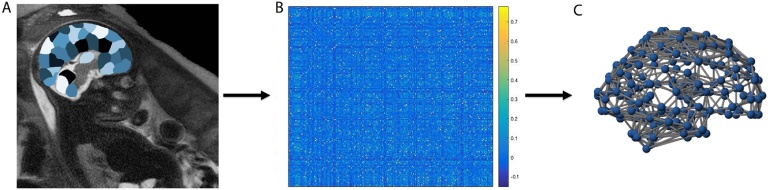
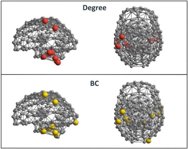
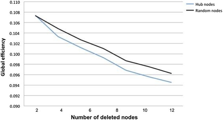
Advances in neuroimaging and network analyses have lead to discovery of highly connected regions, or hubs, in the connectional architecture of the human brain. Whether these hubs emerge in utero, has yet to be examined. The current study addresses this question and aims to determine the location of neural hubs in human fetuses. Fetal resting-state fMRI data (N = 105) was used to construct connectivity matrices for 197 discrete brain regions. We discovered that within the connectional functional organization of the human fetal brain key hubs are emerging. Consistent with prior reports in infants, visual and motor regions were identified as emerging hub areas, specifically in cerebellar areas. We also found evidence for network hubs in association cortex, including areas remarkably close to the adult fusiform facial and Wernicke areas. Functional significance of hub structure was confirmed by computationally deleting hub versus random nodes and observing that global efficiency decreased significantly more when hubs were removed (p < .001). Taken together, we conclude that both primary and association brain regions demonstrate centrality in network organization before birth. While fetal hubs may be important for facilitating network communication, they may also form potential points of vulnerability in fetal brain development.
Overview
The claims and statistics of this paper haven’t been checked yet.
Run the check — free, no account needed. It reads the open-access text and checks the sources it cites.
Integrity signals
Registry
Cites
Statistics
Figures
Verification
Claims the paper makes
The claims and statistics of this paper haven’t been checked yet.
Run the check — free, no account needed. It reads the open-access text and checks the sources it cites.
Claims other papers make about this one
Each is checked against this paper’s own text: does it say what the citing paper says it does?
The cited source doesn’t support the claim as written.
In the citing paper
“Most hubs are localized in the cerebellum, while some are in the primary and association cortices [71].”
From Brain structural and functional outcomes in the offspring of women experiencing psychological distress during pregnancy (2024) · cites this paper as [71]
Checked by a reasoning model — may be wrong.
Experimental — accuracy not yet measured
Self-check
Percentages, sums and reported differences re-computed from the paper’s own numbers.
The self-check needs the paper’s full text.
Citations
Cites
Complex network measures of brain connectivity: Uses and interpretations
Rubinov, M., & Sporns, O. (2010) · NeuroImage · doi:10.1016/j.neuroimage.2009.10.003 · Cited by 11,522 · cited 4×
“From these matrices, measures of network centrality, degree and betweenness centrality (BC), were computed for every region by means of graph theoretical analysis, using the brain connectivity toolbox for Matlab (Rubinov and Sporns, 2010).”
InfluentialFunctional connectivity in the resting brain: a network analysis of the default mode hypothesis
Greicius, M. D., Krasnow, B., Reiss, A. L., & Menon, V. (2003) · Proceedings of the National Academy of Sciences · doi:10.1073/pnas.0135058100 · Cited by 6,358 · cited 3×
“…the ventral and dorsal precuneus, posterior and anterior cingulate gyrus, ventromedial frontal cortex, and interior parietal brain regions (Zuo et al., 2012; Tomasi and Volkow, 2011), brain areas with considerable overlap with sub regions of the default mode network (DMN) (Greicius et al., 2003).”
A report: the definition and classification of cerebral palsy April 2006
Rosenbaum, P., Paneth, N., Leviton, A., Goldstein, M., BAX, M. C. O., Damiano, D. L., Dan, B., & Jacobsson, B. (2007) · Developmental Medicine & Child Neurology · doi:10.1111/j.1469-8749.2007.tb12610.x · Cited by 5,028
Conn : A Functional Connectivity Toolbox for Correlated and Anticorrelated Brain Networks
Whitfield-Gabrieli, S., & Nieto-Castanon, A. (2012) · Brain Connectivity · doi:10.1089/brain.2012.0073 · Cited by 4,885 · cited 2×
“The CONN-fMRI Toolbox (Whitfield-Gabrieli and Nieto Castanon, 2012) was used to compute functional connectivity (FC) matrices.”
MethodA component based noise correction method (CompCor) for BOLD and perfusion based fMRI
Behzadi, Y., Restom, K., Liau, J., & Liu, T. T. (2007) · NeuroImage · doi:10.1016/j.neuroimage.2007.04.042 · Cited by 4,502 · cited 1×
“The anatomical component correction (aCompCor) method of estimating and removing noise (Behzadi et al., 2007; Chai et al., 2012) was applied.”
The economy of brain network organization
Bullmore, E., & Sporns, O. (2012) · Nature Reviews Neuroscience · doi:10.1038/nrn3214 · Cited by 3,284 · cited 4×
“Given prior assertions that the centrality of hubs increase their susceptibility to insults (Crossley et al., 2014; Aerts et al., 2016; Bullmore and Sporns, 2012), and given prior reports of the importance of the cerebellum for early development, our results provide new data suggesting the…”
InfluentialWhy do many psychiatric disorders emerge during adolescence?
Paus, T., Keshavan, M., & Giedd, J. N. (2008) · Nature Reviews Neuroscience · doi:10.1038/nrn2513 · Cited by 3,123 · cited 2×
“…notion, the origins of neurodevelopmental disorders are increasingly attributed to alterations occurring during the prenatal period (Di Martino et al., 2014; Paneth et al., 2005; Rosenbaum et al., 2007) despite onsets in symptomatology between the 3rd and 20th years of life (Paus et al., 2008).”
Rice, D., & Barone, S. (2000) · Environmental Health Perspectives · doi:10.1289/ehp.00108s3511 · Cited by 2,944 · cited 2×
“Insight into hub emergence before birth may be of great importance, since the fetal period is a critical time in brain development (Rice and Barone, 2000; Keunen et al., 2017) in which seemingly small disturbances, insults or exposures can produce lifelong changes in neurological and mental…”
Rich-club organization of the human connectome
van den Heuvel, M. P., & Sporns, O. (2011) · The Journal of Neuroscience · doi:10.1523/jneurosci.3539-11.2011 · Cited by 2,354
Network hubs in the human brain
van den Heuvel, M. P., & Sporns, O. (2013) · Trends in Cognitive Sciences · doi:10.1016/j.tics.2013.09.012 · Cited by 2,123 · cited 6×
“Advances in neuroimaging and network analyses have revealed the existence of sets of highly connected regions, often called “hubs”, that are critically important for enabling efficient neuronal signaling and communication [1, 2].”
Influential
Cited by
Sorted by citations
The development of brain network hubs
Oldham, S., & Fornito, A. (2018) · Developmental Cognitive Neuroscience · doi:10.1016/j.dcn.2018.12.005 · Cited by 257
Functional Connectome of the Fetal Brain
Turk, E., van den Heuvel, M. I., Benders, M. J., de Heus, R., Franx, A., Manning, J. H., Hect, J. L., Hernandez-Andrade, E., Hassan, S. S., Romero, R., et al. (2019) · The Journal of Neuroscience · doi:10.1523/jneurosci.2891-18.2019 · Cited by 176
Self-citationFunctional brain connectivity changes across the human life span: From fetal development to old age
Edde, M., Leroux, G., Altena, E., & Chanraud, S. (2021) · Journal of Neuroscience Research · doi:10.1002/jnr.24669 · Cited by 143
“Yet, caution should be exercised because all studies, but one (van den Heuvel et al., 2018), were based on small sample sizes which is a strong bias during FC analysis.”
BackgroundPremature Birth and Developmental Programming: Mechanisms of Resilience and Vulnerability
Lammertink, F., Vinkers, C. H., Tataranno, M. L., & Benders, M. J. N. L. (2021) · Frontiers in Psychiatry · doi:10.3389/fpsyt.2020.531571 · Cited by 145
“Both the temporal lobe (97) and the insular cortex (76, 98) were found to be already highly connected before birth.”
BackgroundSex differences in functional connectivity during fetal brain development
Wheelock, M. D., Hect, J. L., Hernandez-Andrade, E., Hassan, S. S., Romero, R., Eggebrecht, A. T., & Thomason, M. E. (2019) · Developmental Cognitive Neuroscience · doi:10.1016/j.dcn.2019.100632 · Cited by 126
“Pycluster (Craddock et al., 2012) was used to generate 197 spatially contiguous, similarly sized regions of interest (ROI) in a spatially normalized fetal atlas space, that have been previously described (van den Heuvel et al., 2018), and are available for download at www. brainnexus.com.”
BackgroundMethodInfluentialSelf-citationWu, Y., De Asis-Cruz, J., & Limperopoulos, C. (2024) · Molecular Psychiatry · doi:10.1038/s41380-024-02449-0 · Cited by 124
Kostović, I. (2020) · Progress in Neurobiology · doi:10.1016/j.pneurobio.2020.101883 · Cited by 105
Development of Brain Networks In Utero: Relevance for Common Neural Disorders
Thomason, M. E. (2020) · Biological Psychiatry · doi:10.1016/j.biopsych.2020.02.007 · Cited by 110
Self-citationAssociation of Prenatal Maternal Anxiety With Fetal Regional Brain Connectivity
De Asis-Cruz, J., Krishnamurthy, D., Zhao, L., Kapse, K., Vezina, G., Andescavage, N., Quistorff, J., Lopez, C., & Limperopoulos, C. (2020) · JAMA Network Open · doi:10.1001/jamanetworkopen.2020.22349 · Cited by 81
van den Heuvel, M. I., Hect, J. L., Smarr, B. L., Qawasmeh, T., Kriegsfeld, L. J., Barcelona, J., Hijazi, K. E., & Thomason, M. E. (2021) · Scientific Reports · doi:10.1038/s41598-021-81681-y · Cited by 76
Self-citationThomason, M. E., Hect, J. L., Waller, R. E., & Curtin, P. C. P. (2021) · Neuropsychopharmacology · doi:10.1038/s41386-021-01066-7 · Cited by 86
Self-citationMahadevan, A. S., Tooley, U. A., Bertolero, M. A., Mackey, A. P., & Bassett, D. S. (2021) · NeuroImage · doi:10.1016/j.neuroimage.2021.118408 · Cited by 67
Functional brain connectivity in ex utero premature infants compared to in utero fetuses
De Asis-Cruz, J., Kapse, K., Basu, S. K., Said, M., Scheinost, D., Murnick, J., Chang, T., du Plessis, A., & Limperopoulos, C. (2020) · NeuroImage · doi:10.1016/j.neuroimage.2020.117043 · Cited by 63
“Frames with translational motion >1 mm and rotational motion >1.5 were considered high motion; these cut-offs were based on thresholds used in recently published fetal resting state studies (Li et al., 2019; Thomason et al., 2019, 2017; van den Heuvel et al., 2018; Wheelock et al., 2019).”
MethodInfluentialCook, K. M., Asis‐Cruz, J. D., Kim, J., Basu, S. K., Andescavage, N. N., Murnick, J. G., Spoehr, E., Liggett, M., du Plessis, A. J., & Limperopoulos, C. (2023) · BMC Medicine · doi:10.1186/s12916-023-03141-w · Cited by 62
“Pearson correlations were computed between all ROIs and z -transformed using a Fischer transformation (Additional file 1: SI 2) [25, 58, 59].”
BackgroundMethodAutomated Brain Masking of Fetal Functional MRI with Open Data
Rutherford, S. E., Sturmfels, P., Angstadt, M., Hect, J. L., Wiens, J., van den Heuvel, M. I., Scheinost, D., Sripada, C., & Thomason, M. E. (2021) · Neuroinformatics · doi:10.1007/s12021-021-09528-5 · Cited by 46
Self-citationThomason, M. E., Palopoli, A. C., Jariwala, N. N., Werchan, D. M., Chen, A., Adhikari, S., Espinoza‐Heredia, C., Brito, N. H., & Trentacosta, C. J. (2021) · Developmental Cognitive Neuroscience · doi:10.1016/j.dcn.2021.101000 · Cited by 43
Self-citationGlobal Network Organization of the Fetal Functional Connectome
De Asis-Cruz, J., Andersen, N., Kapse, K., Khrisnamurthy, D., Quistorff, J., Lopez, C., Vezina, G., & Limperopoulos, C. (2021) · Cerebral Cortex · doi:10.1093/cercor/bhaa410 · Cited by 40
“…range of neuroscience studies involving different species (Watts and Strogatz 1998; Oh et al. 2014; van den Heuvel et al. 2016; Rilling and van den Heuvel 2018) at different scales, using different imaging modalities (e.g., diffusion tensor imaging and EEG) (Ferri et al. 2007) including in vitro…”
BackgroundAge-Related Alterations in EEG Network Connectivity in Healthy Aging
Javaid, H., Kumarnsit, E., & Chatpun, S. (2022) · Brain Sciences · doi:10.3390/brainsci12020218 · Cited by 39
“The structural and functional systems of the brain have the characteristics of a complex network, such as network modularity, highly connected hubs, and small world topology [1,2].”
BackgroundIndividual variability in functional organization of the neonatal brain
Molloy, M. F., & Saygin, Z. M. (2022) · NeuroImage · doi:10.1016/j.neuroimage.2022.119101 · Cited by 42
Rotem‐Kohavi, N., Williams, L. J., Muller, A. M., Abdi, H., Virji‐Babul, N., Björnson, B., Brain, U. M., Werker, J. F., GRUNAU, R. E., Miller, S. P., et al. (2019) · Depression and Anxiety · doi:10.1002/da.22906 · Cited by 34
“…to characterize the functional topography of the human brain from the fetal period through to adulthood (Bathelt, O’Reilly, Clayden, Cross, & De Haan, 2013; De Asis‐Cruz, Bouyssi‐Kobar, Evangelou, Vezina, & Limperopoulos, 2015; Gao et al., 2015; Thomason et al., 2017; van den Heuvel et al., 2018).”
BackgroundResultRobust sex differences in functional brain connectivity are present in utero
Cook, K. M., De Asis-Cruz, J., Lopez, C., Quistorff, J., Kapse, K., Andersen, N., Vezina, G., & Limperopoulos, C. (2023) · Cerebral Cortex · doi:10.1093/cercor/bhac218 · Cited by 31
Early and late development of hub connectivity in the human brain
Oldham, S., Ball, G., & Fornito, A. (2021) · Current Opinion in Psychology · doi:10.1016/j.copsyc.2021.10.010 · Cited by 30
Prenatal Programming of Neurodevelopment: Structural and Functional Changes
Demers, C. H., Aran, Ö., Glynn, L. M., & Davis, E. P. (2021) · Springer eBooks · doi:10.1007/978-3-030-60159-1_9 · Cited by 25
van den Heuvel, M. I., Monk, C. E., Hendrix, C. L., Hect, J. L., Lee, S., Feng, T., & Thomason, M. E. (2023) · Journal of the American Academy of Child & Adolescent Psychiatry · doi:10.1016/j.jaac.2023.03.020 · Cited by 26
Self-citationTrajectories of human brain functional connectome maturation across the birth transition
Ji, L., Menu, I., Majbri, A., Bhatia, T., Trentacosta, C. J., & Thomason, M. E. (2024) · PLoS Biology · doi:10.1371/journal.pbio.3002909 · Cited by 29
Self-citationAutism-related shifts in the brain’s information processing hierarchy
Bernhardt, B. C., Valk, S. L., Hong, S., Soulières, I., & Mottron, L. (2025) · Trends in Cognitive Sciences · doi:10.1016/j.tics.2025.04.008 · Cited by 29
Karolis, V. R., Fitzgibbon, S. P., Cordero‐Grande, L., Farahibozorg, S.-R., Price, A. N., Hughes, E., Fetit, A. E., Kyriakopoulou, V., Pietsch, M., Rutherford, M., et al. (2023) · Communications Biology · doi:10.1038/s42003-023-04969-x · Cited by 26
Graph Theory Measures and Their Application to Neurosurgical Eloquence
Tanglay, O., Dadario, N. B., Chong, E. H. N., Tang, S. J., Young, I. M., & Sughrue, M. E. (2023) · Cancers · doi:10.3390/cancers15020556 · Cited by 24
“Some clues may be derived from developmental studies examining hub node formation, though results of these have so far been conflicting [70,71].”
BackgroundVasilkovska, T., Adhikari, M. H., Audekerke, J. V., Salajeghe, S., Pustina, D., Cachope, R., Tang, H., Liu, L., Muñoz-Sanjuán, I., der Linden, A. V., et al. (2023) · Neurobiology of Disease · doi:10.1016/j.nbd.2023.106095 · Cited by 22
“Spontaneous fluctuations in the low frequency (0.01–0.1 Hz) BOLD signal contain information about the connectivity between distinct brain regions and reveal the brain’s functional architecture, organized in resting-state networks (RSN) (van den Heuvel et al., 2018; Fransson, 2005).”
BackgroundInfluentialSubcortico-Cortical Functional Connectivity in the Fetal Brain: A Cognitive Development Blueprint
Canini, M., Cavoretto, P. I., Scifo, P., Pozzoni, M., Petrini, A., Iadanza, A., Pontesilli, S., Scotti, R., Candiani, M., Falini, A., et al. (2020) · Cerebral Cortex Communications · doi:10.1093/texcom/tgaa008 · Cited by 22
He, Z., Du, L., Huang, Y., Jiang, X., Lv, J., Guo, L., Zhang, S., & Zhang, T. (2021) · Cerebral Cortex · doi:10.1093/cercor/bhab420 · Cited by 19
Asis‐Cruz, J. D., & Limperopoulos, C. (2022) · Biological Psychiatry · doi:10.1016/j.biopsych.2022.11.019 · Cited by 22
Motion correction and volumetric reconstruction for fetal functional magnetic resonance imaging data
Sobotka, D., Ebner, M., Schwartz, E., Nenning, K.-H., Taymourtash, A., Vercauteren, T., Ourselin, S., Kasprian, G., Prayer, D., Langs, G., et al. (2022) · NeuroImage · doi:10.1016/j.neuroimage.2022.119213 · Cited by 20
“In recent fetal studies [22, 23, 24, 25], motion censoring or scrubbing instead of motion correction is used where datasets with high motion are excluded, i.”
BackgroundMethodImaging Developmental and Interventional Plasticity Following Perinatal Stroke
Craig, B. T., Hilderley, A. J., Kirton, A., & Carlson, H. L. (2020) · Canadian Journal of Neurological Sciences / Journal Canadien des Sciences Neurologiques · doi:10.1017/cjn.2020.166 · Cited by 24
“for primary sensory functions (vision, audition, and sensorimotor) appear to develop very early, before birth.(163) Throughout childhood, networks strengthen and follow a local-to-global pattern of development.”
BackgroundRawls, E., Kummerfeld, E., Mueller, B. A., Ma, S., & Zilverstand, A. (2022) · NeuroImage · doi:10.1016/j.neuroimage.2022.119211 · Cited by 24
Fetal behavior during MRI changes with age and relates to network dynamics
Ji, L., Majbri, A., Hendrix, C. L., & Thomason, M. E. (2022) · Human Brain Mapping · doi:10.1002/hbm.26167 · Cited by 18
Self-citationThill, B. (2023) · Frontiers in Pain Research · doi:10.3389/fpain.2023.1128530 · Cited by 26
Afacan, O., Estroff, J. A., Yang, E., Barnewolt, C. E., Connolly, S. A., Parad, R. B., Mulkern, R. V., Warfield, S. K., & Gholipour, A. (2019) · Topics in Magnetic Resonance Imaging · doi:10.1097/rmr.0000000000000219 · Cited by 20
Developmental Pathoconnectomics and Advanced Fetal MRI
András, J. (2019) · Topics in Magnetic Resonance Imaging · doi:10.1097/rmr.0000000000000220 · Cited by 19
“All rights reserved. ules begin to overlap with known functional systems, and the www.topicsinmri.com | 279 posterior cingulate cortex in the default mode network shows increased negative functional connectivity with other brain areas.80 The fetal connectome is also characterized by an increasing proportion of brain r…”
ResultDesrosiers, J., Caron-Desrochers, L., René, A., Gaudet, I., Pincivy, A., Paquette, N., & Gallagher, A. (2024) · Neuroscience & Biobehavioral Reviews · doi:10.1016/j.neubiorev.2024.105778 · Cited by 23
Millevert, C., Vidas-Guscic, N., Vanherp, L., Jonckers, E., Verhoye, M., Staelens, S. G., Bertoglio, D., & Weckhuysen, S. (2023) · Journal of Neuroscience · doi:10.1523/jneurosci.1043-23.2023 · Cited by 17
Early development of the functional brain network in newborns
Nazari, R., & Salehi, M. (2023) · Brain Structure and Function · doi:10.1007/s00429-023-02681-4 · Cited by 16
A Neuroscience Perspective on Emotional Development
Morales, S., & Fox, N. A. (2019) · Springer eBooks · doi:10.1007/978-3-030-17332-6_4 · Cited by 14
Empirical evaluation of human fetal fMRI preprocessing steps
Ji, L., Hendrix, C. L., & Thomason, M. E. (2022) · Network Neuroscience · doi:10.1162/netn_a_00254 · Cited by 15
“…connectivity patterns in utero precede and may guide functional selectivity of certain brain regions, such as the fusiform face area (van den Heuvel et al., 2018), and that macroscale characteristics of fetal functional networks share significant overlap with adult networks (Turk et al.,…”
BackgroundSelf-citationAn integrative perspective on the role of touch in the development of intersubjectivity
Plinio, S. D., Scalabrini, A., & Ebisch, S. J. H. (2022) · Brain and Cognition · doi:10.1016/j.bandc.2022.105915 · Cited by 15
Functional connectivity of the pediatric brain
Argyropoulou, M. I., Xydis, V. G., & Astrakas, L. G. (2024) · Neuroradiology · doi:10.1007/s00234-024-03453-5 · Cited by 13
“Further studies characterized the maturation of the fetal functional connectome, revealing features such as small-worldness, modularity, and hubs [ 83, 84], along with gradient organization of connectivity within RSNs [ 31].”
Jang, Y. H., Kim, H., Lee, J. Y., Ahn, J., Chung, A. W., & Lee, H. J. (2022) · Cerebral Cortex · doi:10.1093/cercor/bhac438 · Cited by 11
Li, Y., Yang, B., Wang, Z., Huang, R., Lu, X., Bi, X., & Zhou, S. (2023) · Frontiers in Neuroscience · doi:10.3389/fnins.2023.1133834 · Cited by 10
“For functional brain networks, researchers have proposed many coupling methods, such as correlation-based method, partial order correlation-based method, and sparse method (Bullmore and Sporns, 2012; Sporns, 2013; Li et al., 2015; van den Heuvel et al., 2018).”
MethodAdvances in Fetal Brain Imaging
Calixto, C. A., Taymourtash, A., Karimi, D., Snoussi, H., Velasco‐Annis, C., Jaimes, C. E., & Gholipour, A. (2024) · Magnetic Resonance Imaging Clinics of North America · doi:10.1016/j.mric.2024.03.004 · Cited by 11
RS-FetMRI: a MATLAB-SPM Based Tool for Pre-processing Fetal Resting-State fMRI Data
Pecco, N., Canini, M., Mosser, K. H. H., Caglioni, M., Scifo, P., Castellano, A., Cavoretto, P. I., Candiani, M., Baldoli, C., Falini, A., et al. (2022) · Neuroinformatics · doi:10.1007/s12021-022-09592-5 · Cited by 11
“…Milan, Italy 4 Vita-Salute San Raffaele University, Milan, Italy 1 3 to accommodate motion in a fetal rs-fMRI time-series (Ferrazzi et al., 2014; Van den Heuvel et al., 2018; Thomason et al., 2018; Van Dijk et al., 2012) and on spatial normalization of the functional time-series to…”
BackgroundJunaković, A., Kopić, J., Duque, A., RAKIC, P. T., Krsnik, Ž., & Kostović, I. (2023) · Brain Structure and Function · doi:10.1007/s00429-022-02606-7 · Cited by 12
Funcmasker-flex: An Automated BIDS-App for Brain Segmentation of Human Fetal Functional MRI data
Nichols, E. S., Correa, S., Dyken, P. C. V., Kai, J., Kuehn, T. K., de Ribaupierre, S., Duerden, E. G., & KHAN, A. R. (2023) · Neuroinformatics · doi:10.1007/s12021-023-09629-3 · Cited by 10
“In the past, brain extraction of fetal fMRI data was performed by manually (Thomason et al., 2014, 2015) or semi-automatically (Thomason et al., 2013; van den Heuvel et al., 2018) segmenting the brain from the surrounding tissue.”
MethodLi, X., Hect, J. L., Thomason, M. E., & Zhu, D. (2020) · IEEE International Symposium on Biomedical Imaging (ISBI) · doi:10.1109/isbi45749.2020.9098606 · Cited by 10
“For fetal neurodevelopmental studies, existing works focus on analysis of the functional connectivity in fetal brain, such as discoveries of functional networks and identification of highly connected hubs [4].”
BackgroundSelf-citationCook, K. M., Asis‐Cruz, J. D., Sitrin, C., Barnett, S. D., Krishnamurthy, D., & Limperopoulos, C. (2024) · The Journal of Pediatrics · doi:10.1016/j.jpeds.2024.114201 · Cited by 9
Kaiser, M. (2020) · The MIT Press eBooks · doi:10.7551/mitpress/11980.001.0001 · Cited by 25
The developing Human Connectome Project fetal functional MRI release: Methods and data structures
Karolis, V. R., Cordero‐Grande, L., Price, A. N., Hughes, E., Fitzgibbon, S. P., Kyriakopoulou, V., Uus, A. U., Harper, N., Prokopenko, D. A., Bridglal, D., et al. (2025) · Imaging Neuroscience · doi:10.1162/imag_a_00512 · Cited by 11
“…et al., 2023; Sch ö pf et al., 2012, 2014; Seshamani et al., 2016; Sobotka et al., 2022; Taymourtash et al., 2021, 2023; Thomason et al., 2015; van den Heuvel et al., 2018), fetal fMRI remains a relatively novel field, where pre-processing standards are yet to be established and which until…”
Feng, S., Huang, Y., Lu, H., Li, H., Zhou, S., Lu, H., Feng, Y., Ning, Y., Han, W., Chang, Q., et al. (2024) · Journal of Psychiatric Research · doi:10.1016/j.jpsychires.2024.03.007 · Cited by 7
Overview of the Development of the Human Brain and Spinal Cord
ten Donkelaar, H. J., Takakuwa, T., Vasung, L., Yamada, S., Shiota, K., & van der Vliet, T. (2023) · Springer eBooks · doi:10.1007/978-3-031-26098-8_1 · Cited by 8
Uberos, J., Nieto-Ruíz, A., Contreras‐Chova, F., Carrasco-Solís, M. E., Ruiz‐López, A., Fernández‐Marin, E., Láynez-Rubio, C., & Campos‐Martínez, A. (2024) · The Pediatric Infectious Disease Journal · doi:10.1097/inf.0000000000004262 · Cited by 6
Reproducibility of Functional Connectivity Estimates in Motion Corrected Fetal fMRI
Sobotka, D., Licandro, R., Ebner, M., Schwartz, E., Vercauteren, T. K., Sebastien Ourselin, A., Kasprian, G. J., Prayer, D., & Langs, G. (2019) · Lecture notes in computer science · doi:10.1007/978-3-030-32875-7_14 · Cited by 6
Moore, J. W., Wilson, S., Oldehinkel, M., Cordero‐Grande, L., Uus, A. U., Kyriakopoulou, V., Duff, E. P., O’Muircheartaigh, J., Rutherford, M., Andreae, L. C., et al. (2023) · doi:10.7554/elife.90536.1 · Cited by 6
Cook, K. M., Asis‐Cruz, J. D., Basu, S. K., Andescavage, N. N., Murnick, J. G., Spoehr, E., du Plessis, A. J., & Limperopoulos, C. (2023) · Frontiers in Neuroscience · doi:10.3389/fnins.2023.1214080 · Cited by 5
Schirmer, M. D., & Chung, A. W. (2019) · DZNE Pub · Cited by 5
Dai, Y., He, Q., Wang, S., Cao, T., Chai, X., Wang, N., Dong, Y., Wong, P., He, J., Duan, F., et al. (2024) · Frontiers in Psychology · doi:10.3389/fpsyg.2024.1458339 · Cited by 5
Utilizing functional neuroimaging to study early language development
Olson, H. A., Day, T. K. M., Hiersche, K. J., Manning, B. L., Bradley, H., Choi, S. J., Abdurokhmonova, G., McCormick, S. A., Conrad, A. L., Scheinost, D., et al. (2025) · Developmental Cognitive Neuroscience · doi:10.1016/j.dcn.2025.101641 · Cited by 4
Fetal functional connectivity prospectively associates with autistic traits in toddlerhood
Chen, B., Menu, I., Ji, L., Trentacosta, C. J., & Thomason, M. E. (2025) · NeuroImage Clinical · doi:10.1016/j.nicl.2025.103938 · Cited by 4
Self-citationMorphological development of the human fetal striatum during the second trimester
Tian, M., Xu, F., Xia, Q., Tang, Y., Zhang, Z., Lin, X., Meng, H., Feng, L., & Liu, S. (2021) · Cerebral Cortex · doi:10.1093/cercor/bhab532 · Cited by 4
Zhang, S., Wang, R., Han, Z., Yu, S., Gao, H. B., Jiang, X., & Zhang, T. (2022) · Chaos Solitons & Fractals · doi:10.1016/j.chaos.2022.112018 · Cited by 4
Li, B., Li, W., Guo, Y., Wang, Y., Xu, L., Yang, Y., Xu, S.-G., Tan, Z., Mei, Y., & Wang, K.-Y. (2023) · Frontiers in Neurology · doi:10.3389/fneur.2023.1266167 · Cited by 4
“It also reveals that these are essential compensatory brain regions that enable integration and coupling across the whole brain at the acute phase of sTBI (20).”
BackgroundKipping, J., Margulies, D. S., Eickhoff, S. B., Lee, A., & Qiu, A. (2018) · NeuroImage · doi:10.1016/j.neuroimage.2018.04.067 · Cited by 4
Correa, S., Nichols, E. S., Mueller, M. E., de Vrijer, B., Eagleson, R. A., McKenzie, C. A., de Ribaupierre, S., & Duerden, E. G. (2023) · Cerebral Cortex · doi:10.1093/cercor/bhad190 · Cited by 5
Hayat, T. T. A., & Sami, M. B. (2020) · Development and Psychopathology · doi:10.1017/s0954579420000218 · Cited by 2
“Furthermore. of the dominant hubs, there appears to be age-related variation in some frontal and temporal hubs (van den Heuvel et al., 2018).”
BackgroundLes stress pendant les 1 000 premiers jours de la vie quand tout commence
Verney, C., & Vitalis, T. (2023) · médecine/sciences · doi:10.1051/medsci/2023124 · Cited by 3
Schirmer, M. D., & Chung, A. W. (2019) · Lecture notes in computer science · doi:10.1007/978-3-030-32391-2_6 · Cited by 6
“Such hubs are the first to develop and present at birth, with strong similarity to adults [14, 12].”
BackgroundYorita, A., Kawayama, T., Inoue, M., Kinoshita, T., Oda, H., Tokunaga, Y., Tateishi, T., Shoji, Y., Uchimura, N., Abe, T., et al. (2024) · Journal of Clinical Medicine · doi:10.3390/jcm13092556 · Cited by 3
“To investigate the possibility that the secondary visual cortex functions as a hub, we analysed the degree and betweenness centralities using graph theoretical analysis [54].”
MethodIndividual Topological Analysis of Synchronization-Based Brain Connectivity
Lombardi, A., Amoroso, N., Diacono, D., Monaco, A., Tangaro, S., & Bellotti, R. (2020) · Applied Sciences · doi:10.3390/app10093275 · Cited by 2
Im, K. (2021) · Advances in magnetic resonance technology and applications · doi:10.1016/b978-0-12-816633-8.00004-1 · Cited by 1
The Representation of Nociception and Pain in the Developing Brain
Lorenzo Fabrizi, D., & Fitzgerald, M. (2025) · Annual Review of Physiology · doi:10.1146/annurev-physiol-040125-112145 · Cited by 1
Early functional organization of the anterior and posterior hippocampus in the fetal brain
Nichols, E. S., Al‐Saoud, S., Fang, M., Eagleson, R. A., de Vrijer, B., McKenzie, C. A., de Ribaupierre, S., & Duerden, E. G. (2025) · Cerebral Cortex · doi:10.1093/cercor/bhaf327 · Cited by 2
“Although the presence of functional connectivity networks has been well-established in the fetal brain (van den Heuvel and Thomason 2016; van den Heuvel et al. 2018; Thomason 2020), the third trimester covers a three-month period in which large developmental changes occur (Clouchoux et al. 2012;…”
Li, X., Hect, J. L., Thomason, M. E., & Zhu, D. (2019) · arXiv (Cornell University) · doi:10.48550/arxiv.1906.03691 · Cited by 1
“For fetal neurodevelopmental studies, existing works focus on analysis of the functional connectivity in fetal brain, such as discoveries of functional networks and identification of highly connected hubs [4].”
BackgroundSelf-citationAutomated Brain Masking of Fetal Functional MRIPreprint
Rutherford, S. E., Sturmfels, P., Angstadt, M., Hect, J. L., Wiens, J., van den Heuval, M., Scheinost, D., Thomason, M. E., & Sripada, C. (2019) · bioRxiv (Cold Spring Harbor Laboratory) · doi:10.1101/525386 · Cited by 1
Self-citationMoore, J. W., Wilson, S., Oldehinkel, M., Cordero‐Grande, L., Uus, A. U., Kyriakopoulou, V., Duff, E. P., O’Muircheartaigh, J., Rutherford, M., Andreae, L. C., et al. (2023) · bioRxiv (Cold Spring Harbor Laboratory) · doi:10.1101/2023.06.26.546607 · Cited by 1
Moore, J. W., Wilson, S., Oldehinkel, M., Cordero‐Grande, L., Uus, A. U., Kyriakopoulou, V., Duff, E. P., O’Muircheartaigh, J., Rutherford, M., Andreae, L. C., et al. (2023) · doi:10.7554/elife.90536 · Cited by 2
Characterizing Large-Scale Human Circuit Development with In Vivo NeuroimagingPreprint
Arichi, T. (2024) · Cold Spring Harbor Perspectives in Biology · doi:10.1101/cshperspect.a041496 · Cited by 1
Early Childhood Brain Development and Schizophrenia: An Imaging Perspective
Gilmore, J. H. (2020) · Springer eBooks · doi:10.1007/978-3-030-35206-6_15
Gountas, J. (2020) · Scholarly Journal of Psychology and Behavioral Sciences · doi:10.32474/sjpbs.2020.03.000166
“Amalric [39] found that blind mathematicians process advanced mathematics in similar brain networks as sighted without the visual experience.”
BackgroundA blueprint of the developing mind
Turk, E. (2022) · Utrecht University Repository (Utrecht University) · doi:10.33540/627
Self-citationVasilkovska, T., Adhikari, M. H., Audekerke, J. V., Pustina, D., Cachope, R., Tang, H., Liu, L., Muñoz-Sanjuán, I., der Linden, A. V., & Verhoye, M. (2022) · bioRxiv (Cold Spring Harbor Laboratory) · doi:10.1101/2022.11.19.517166
Wu, Z., Yu, J., Chen, X., Shen, J. F., Xie, S., & Zeng, Y. (2023) · Multimedia Tools and Applications · doi:10.1007/s11042-023-17918-x
“Our results revealed that infant brain structural network exhibited sets of hub nodes in limbic subsystem, superior frontal, insula, fusiform, and lingual gyrus, which is in line with the findings in [38, 42].”
Biological basis of neurodevelopmental disabilities
Kielty, K., Risen, S., & Melendez-Zaidi, A. E. (2024) · Elsevier eBooks · doi:10.1016/b978-0-12-824060-1.00003-1 · Cited by 1
Wasserman, T. H., & Wasserman, L. D. (2024) · Springer eBooks · doi:10.1007/978-3-031-78732-4_5
Wasserman, T. H., & Wasserman, L. D. (2024) · Springer eBooks · doi:10.1007/978-3-031-78732-4_10
Connectivité fonctionnelle du cerveau fœtal et néonatal : de la technique à l’application
Argyropoulou, M. I., Lopes, R., & Astrakas, L. G. (2025) · Bulletin de l Académie Nationale de Médecine · doi:10.1016/j.banm.2024.11.025
Evaluating the effects of volume censoring on fetal functional connectivity
Kim, J., Asis‐Cruz, J. D., Cook, K. M., & Limperopoulos, C. (2025) · Scientific Reports · doi:10.1038/s41598-025-96538-x
Maternal Prenatal Psychological Stress and Iron Levels in the Fetal Brain
Chen, B., Ji, L., Yoon, Y. B., Duffy, M., Menu, I., Trentacosta, C. J., & Thomason, M. E. (2025) · Developmental Psychobiology · doi:10.1002/dev.70062
Self-citationSun, H., Toyonaga, T., Mehta, S., Liu, H., Camp, C., Rossano, S. M., Fowles, K., Martins, S., Spann, M. N., Groman, S., et al. (2025) · bioRxiv (Cold Spring Harbor Laboratory) · doi:10.1101/2025.09.03.674103
“For example, major white-matter tracts are present in the third trimester 2 , leading to the establishment of highly connected hub regions and an adult-like network organization by birth 3 .”
Charting the Normal Development of Structural Brain Connectivity in Utero Using Diffusion MRI
Karimi, D., Li, B., Taymourtash, A., Jaimes, C. E., Grant, P. E., & Warfield, S. K. (2026) · Human Brain Mapping · doi:10.1002/hbm.70609
Trajectories of functional connectivity in brain maturation
Liang, X., Sun, L., Cao, M., & Yong He, A. (2025) · Elsevier eBooks · doi:10.1016/b978-0-443-19099-5.00009-x
Cook, K. M., Asis‐Cruz, J. D., Kim, J.-H., Basu, S. K., Andescavage, N. N., Murnick, J., Spoehr, E., Liggett, M., du Plessis, A. J., & Limperopoulos, C. (2023) · Figshare · doi:10.6084/m9.figshare.24558050
Chen, B., Ji, L., Menu, I., Taylor, A., Trentacosta, C. J., & Thomason, M. E. (2026) · Neuroimage Reports · doi:10.1016/j.ynirp.2026.100326
“…sensory networks (e.g., visual, somatosensory) and thalamocortical pathways warrant further investigation, as these connections are among the earliest to form and mature and serve as functional hubs in the fetal brain (Sch ¨ opf et al., 2012; Taymourtash et al., 2023; van den Heuvel et al., 2018).”
Self-citation
Metadata
- Authors
- van den Heuvel, M. I., Turk, E., Manning, J. H., Hect, J., Hernandez-Andrade, E., Hassan, S. S., Romero, R., van den Heuvel, M. P., & Thomason, M. E.
- Affiliations
- University Medical Center Utrecht (NL)
- National Institutes of Health (US)
- Wayne State University (US)
- United States Department of Health and Human Services (US)
- Venue
- Developmental Cognitive Neuroscience
- Publisher
- Elsevier BV
- Published
- 2018-04
- Volume
- 30
- Pages
- 108-115
- ISSN
- 1878-9293 (ISSN-L)
- PMC
- PMC5963507
- OpenAlex
- W2793015464
- Cited by
- 108
- References
- 68
- Citations by year
- 2026: 9 · 2025: 11 · 2024: 14 · 2023: 21 · 2022: 14 · 2021: 11 · 2020: 14 · 2019: 12 · 2018: 2
- FWCI
- 5.80
- Type
- journal article
- Language
- en
- Open access
- gold
- doi.org
- Licence
- Locations
- Developmental Cognitive Neuroscience (publishedVersion, cc-by-nc-nd)
- Europe PMC (PubMed Central) (publishedVersion, cc-by-nc-nd)
- PubMed Central (publishedVersion, cc-by-nc-nd)
- PubMed (publishedVersion)
- Pure Amsterdam UMC (publishedVersion, other-oa)
- Research portal (Tilburg University) (submittedVersion, cc-by-nc-nd)
- Data Archiving and Networked Services (DANS) (submittedVersion, cc-by-nc-nd)
- DOAJ (DOAJ: Directory of Open Access Journals) (submittedVersion, cc-by-sa)
- Utrecht University Repository (Utrecht University) (submittedVersion, other-oa)
- Amsterdam UMC (VU Amsterdam) - Institutional Repository (submittedVersion, other-oa)
- Data Archiving and Networked Services (DANS) (submittedVersion, other-oa)
- Funders
- Nederlandse Organisatie voor Wetenschappelijk Onderzoek (452-16-015)
- U.S. Department of Health and Human Services
- National Institutes of Health (ES020957, ES026022, MH110793)
- National Alliance for Research on Schizophrenia and Depression
- Eunice Kennedy Shriver National Institute of Child Health and Human Development
- Topics
- Life Sciences › Neuroscience › Cognitive Neuroscience › Functional Brain Connectivity Studies
- Health Sciences › Medicine › Radiology, Nuclear Medicine and Imaging › Advanced Neuroimaging Techniques and Applications
- Physical Sciences › Physics and Astronomy › Statistical and Nonlinear Physics › Complex Network Analysis Techniques
- Keywords
- human fetal brain, brain network development, resting-state functional connectivity, cerebellum, visual cortex, motor cortex, association cortex, fusiform face area, Wernicke's area, global efficiency, fetal brain development
- MeSH terms
- Adult, Brain, Female, Humans, Male, Neural Pathways, Pregnancy, Young Adult
About these checks
Registry, retraction, Cites and statistics results are computed, not judged, with one exception: a printed reference that no index lists is read once by a model before it counts as a citation error. Claims are extracted by a model and kept only when their quote appears verbatim in the text; each cited source is then checked, with a model’s help, for whether it says what the claim relies on. Those verdicts can be wrong — read the claim’s quote and the cited source before relying on one.
Statistics and arithmetic are recomputed, not judged.
Confidence bands are shown only for fields calibrated on our annotated test set. None is calibrated yet, so no band is shown.
What counts as a claim ›How we measure confidence ›How we check this ›
Are you an author, or do you see a problem with this page? Request a review
