

The DOI matches its Crossref record; 51 of the 52 references it cites were found in a registry or an index.
Q493K and Q498H substitutions in Spike promote adaptation of SARS-CoV-2 in mice
Open accessAbstract
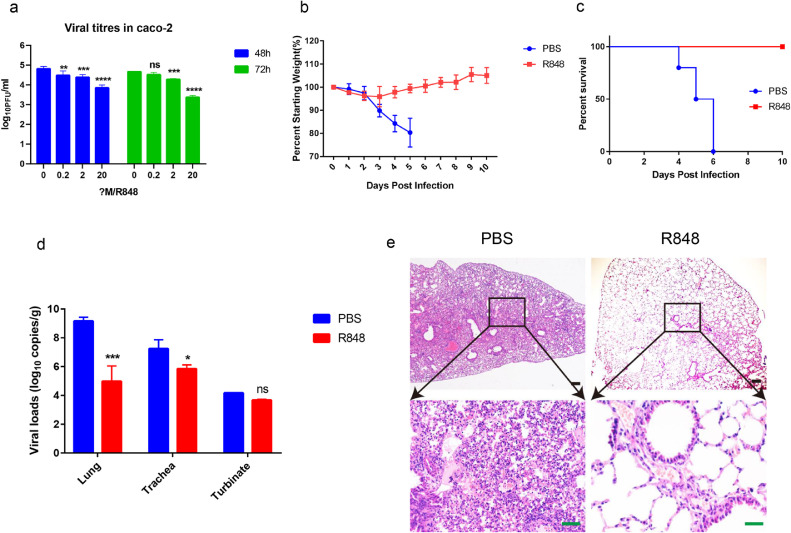
BACKGROUND: An ideal animal model to study SARS-coronavirus 2 (SARS-CoV-2) pathogenesis and evaluate therapies and vaccines should reproduce SARS-CoV-2 infection and recapitulate lung disease like those seen in humans. The angiotensin-converting enzyme 2 (ACE2) is a functional receptor for SARS-CoV-2, but mice are resistant to the infection because their ACE2 is incompatible with the receptor-binding domain (RBD) of the SARS-CoV-2 spike protein . METHODS: SARS-CoV-2 was passaged in BALB/c mice to obtain mouse-adapted virus strain. Complete genome deep sequencing of different generations of viruses was performed to characterize the dynamics of the adaptive mutations in SARS-CoV-2. Indirect immunofluorescence analysis and Biolayer interferometry experiments determined the binding affinity of mouse-adapted SARS-CoV-2 WBP-1 RBD to mouse ACE2 and human ACE2. Finally, we tested whether TLR7/8 agonist Resiquimod (R848) could also inhibit the replication of WBP-1 in the mouse model. FINDINGS: The mouse-adapted strain WBP-1 showed increased infectivity in BALB/c mice and led to severe interstitial pneumonia. We characterized the dynamics of the adaptive mutations in SARS-CoV-2 and demonstrated that Q493K and Q498H in RBD significantly increased its binding affinity towards mouse ACE2. Additionally, the study tentatively found that the TLR7/8 agonist Resiquimod was able to protect mice against WBP-1 challenge. Therefore, this mouse-adapted strain is a useful tool to investigate COVID-19 and develop new therapies. INTERPRETATION: We found for the first time that the Q493K and Q498H mutations in the RBD of WBP-1 enhanced its interactive affinities with mACE2. The mouse-adapted SARS-CoV-2 provides a valuable tool for the evaluation of novel antiviral and vaccine strategies. This study also tentatively verified the antiviral activity of TLR7/8 agonist Resiquimod against SARS-CoV-2 in vitro and in vivo. FUNDING: This research was funded by the National Key Research and Development Program of China (2020YFC0845600) and Emergency Science and Technology Project of Hubei Province (2020FCA046) and Robert A. Welch Foundation (C-1565).
Overview
The claims and statistics of this paper haven’t been checked yet.
Run the check — free, no account needed. It reads the open-access text and checks the sources it cites.
Integrity signals
Registry
Cites
Statistics
Figures
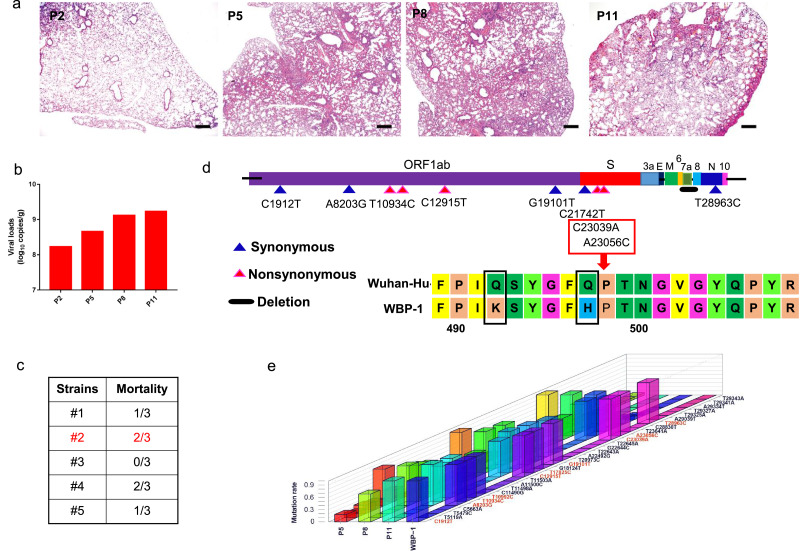
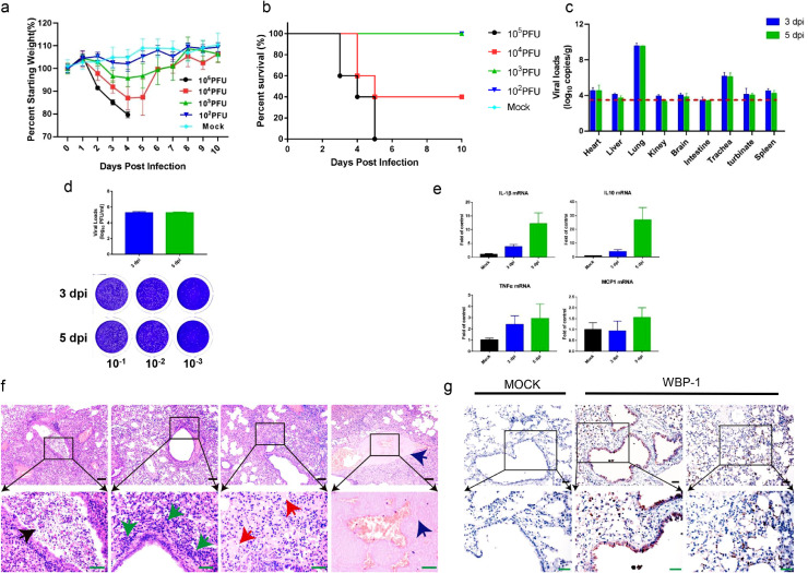
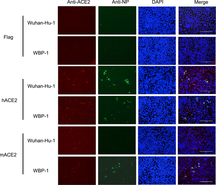
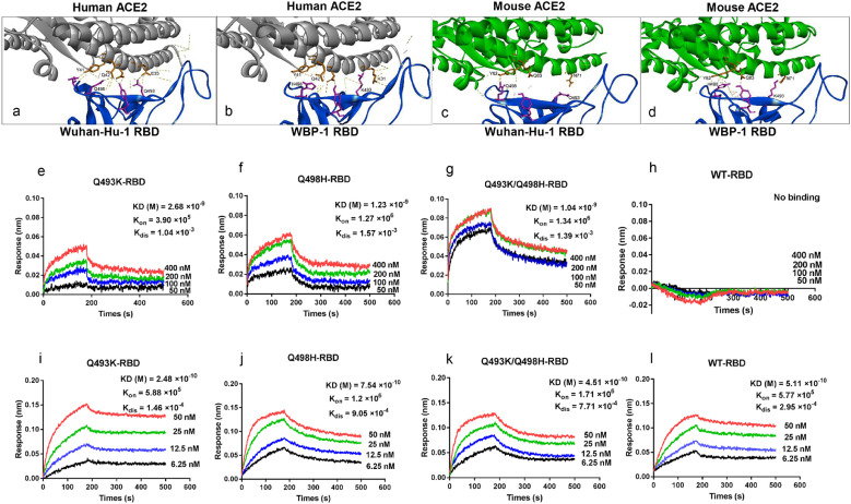
Verification
Claims the paper makes
The claims and statistics of this paper haven’t been checked yet.
Run the check — free, no account needed. It reads the open-access text and checks the sources it cites.
Claims other papers make about this one
Each is checked against this paper’s own text: does it say what the citing paper says it does?
In the citing paper
“The efficient usage of murine ACE2 is in keeping with the presence of the mutations K417N and N501Y, which can also emerge upon SARS-CoV-2 adaptation to experimentally infected mice”
From The Omicron variant is highly resistant against antibody-mediated neutralization: Implications for control of the COVID-19 pandemic (2022) · cites this paper as Huang et al., 2021
In the citing paper
“In addition, the Omicron spike harbors mutations Q493R and Q498R, which are related to exchanges Q493K and Q498H, which were also detected in mouse-adapted SARS-CoV-2”
From The Omicron variant is highly resistant against antibody-mediated neutralization: Implications for control of the COVID-19 pandemic (2022) · cites this paper as Huang et al., 2021
Self-check
Percentages, sums and reported differences re-computed from the paper’s own numbers.
The self-check needs the paper’s full text.
Citations
Cites
Fast gapped-read alignment with Bowtie 2
Langmead, B., & Salzberg, S. L. (2012) · Nature Methods · doi:10.1038/nmeth.1923 · Cited by 56,400
Clinical features of patients infected with 2019 novel coronavirus in Wuhan, China
Huang, C., Wang, Y., Li, X., Ren, L., Zhao, J., Hu, Y., Zhang, L., Fan, G., Xu, J., Gu, X., et al. (2020) · The Lancet · doi:10.1016/s0140-6736(20)30183-5 · Cited by 48,884
Clinical characteristics of coronavirus disease 2019 in China
Guan, W.-J., Ni, Z.-Y., Hu, Y., Liang, W.-H., Ou, C.-Q., He, J.-X., Liu, L., Shan, H., Lei, C.-L., Hui, D. S. C., et al. (2020) · New England Journal of Medicine · doi:10.1056/nejmoa2002032 · Cited by 30,246
Self-citationA novel coronavirus from patients with pneumonia in China, 2019
Zhu, N., Zhang, D., Wang, W., Li, X., Yang, B., Song, J., Zhao, X., Huang, B., Shi, W., Lu, R., et al. (2020) · New England Journal of Medicine · doi:10.1056/nejmoa2001017 · Cited by 28,244
A pneumonia outbreak associated with a new coronavirus of probable bat origin
Zhou, P., Yang, X.-L., Wang, X.-G., Hu, B., Zhang, L., Zhang, W., Si, H.-R., Zhu, Y., Li, B., Huang, C.-L., et al. (2020) · Nature · doi:10.1038/s41586-020-2012-7 · Cited by 21,969
Self-citationPulmonary Vascular Endothelialitis, Thrombosis, and Angiogenesis in Covid-19
Ackermann, M., Verleden, S. E., Kuehnel, M., Haverich, A., Welte, T., Laenger, F., Vanstapel, A., Werlein, C., Stark, H., Tzankov, A., et al. (2020) · New England Journal of Medicine · doi:10.1056/nejmoa2015432 · Cited by 5,613
Isolation and characterization of a bat SARS-like coronavirus that uses the ACE2 receptor
Ge, X.-Y., Li, J.-L., Yang, X.-L., Chmura, A. A., Zhu, G., Epstein, J. H., Mazet, J. K., Hu, B., Zhang, W., Peng, C., et al. (2013) · Nature · doi:10.1038/nature12711 · Cited by 1,778 · cited 1×
“Several mouse-adapted SARS-CoV-2 strains, such as MASCp6 (N501Y), MA10 (Q493K) and HRB26 (Q498H), increase virulence or replication in mice likely attributed to one of the mutations in the S protein [15,26,45,46].”
Self-citationWilm, A., Aw, P. P. K., Bertrand, D., Yeo, G. H. T., Ong, S. H., Wong, C. H., Khor, C. C., Petric, R., Hibberd, M. L., & Nagarajan, N. (2012) · Nucleic Acids Research · doi:10.1093/nar/gks918 · Cited by 1,370
Pathogenesis and transmission of SARS-CoV-2 in golden hamsters
Sia, S. F., Yan, L.-M., Chin, A. W. H., Fung, K., Choy, K.-T., Wong, A. Y. L., Kaewpreedee, P., Perera, R. A. P. M., Poon, L. L. M., Nicholls, J. M., et al. (2020) · Nature · doi:10.1038/s41586-020-2342-5 · Cited by 1,342
The pathogenicity of SARS-CoV-2 in hACE2 transgenic mice
Bao, L., Deng, W., Huang, B., Gao, H., Liu, J., Ren, L., Wei, Q., Yu, P., Xu, Y., Qi, F., et al. (2020) · Nature · doi:10.1038/s41586-020-2312-y · Cited by 1,126
Chan, J. F., Zhang, J., Yuan, S., Poon, V. K., Chan, C. C., Lee, A. C.-Y., Chan, W.-M., Fan, Z., Tsoi, H.-W., Wen, L., et al. (2020) · Clinical Infectious Diseases · doi:10.1093/cid/ciaa325 · Cited by 1,048
Self-citationSyrian hamsters as a small animal model for SARS-CoV-2 infection and countermeasure development
Imai, M., Iwatsuki‐Horimoto, K., Hatta, M., Loeber, S. J., Halfmann, P., Nakajima, N., Watanabe, T., Ujie, M., Takahashi, K., Ito, M., et al. (2020) · Proceedings of the National Academy of Sciences · doi:10.1073/pnas.2009799117 · Cited by 994
Evasion of Type I Interferon by SARS-CoV-2
Xia, H., Cao, Z., Xie, X., Zhang, X., Chen, J. Y.-C., Wang, H., Menachery, V. D., Rajsbaum, R., & Shi, P.-Y. (2020) · Cell Reports · doi:10.1016/j.celrep.2020.108234 · Cited by 957
Comparative pathogenesis of COVID-19, MERS, and SARS in a nonhuman primate model
Rockx, B., Kuiken, T., Herfst, S., Bestebroer, T., Lamers, M. M., Oude Munnink, B. B., de Meulder, D., van Amerongen, G., van den Brand, J., Okba, N. M. A., et al. (2020) · Science · doi:10.1126/science.abb7314 · Cited by 901
Infection and Rapid Transmission of SARS-CoV-2 in Ferrets
Kim, Y., Kim, S.-G., Kim, S.-M., Kim, E.-H., Park, S.-J., Yu, K.-M., Chang, J.-H., Kim, E. J., Lee, S., Casel, M. A. B., et al. (2020) · Cell Host & Microbe · doi:10.1016/j.chom.2020.03.023 · Cited by 883
SARS-CoV-2 infection protects against rechallenge in rhesus macaques
Chandrashekar, A., Liu, J., Martinot, A. J., McMahan, K., Mercado, N. B., Peter, L., Tostanoski, L. H., Yu, J., Maliga, Z., Nekorchuk, M., et al. (2020) · Science · doi:10.1126/science.abc4776 · Cited by 816
Adaptation of SARS-CoV-2 in BALB/c mice for testing vaccine efficacy
Gu, H., Chen, Q., Yang, G., He, L., Fan, H., Deng, Y.-Q., Wang, Y., Teng, Y., Zhao, Z., Cui, Y., et al. (2020) · Science · doi:10.1126/science.abc4730 · Cited by 762
Molecular Evolution of Human Coronavirus Genomes
Forni, D., Cagliani, R., Clerici, M., & Sironi, M. (2016) · Trends in Microbiology · doi:10.1016/j.tim.2016.09.001 · Cited by 751
Is a “Cytokine Storm” Relevant to COVID-19?
Sinha, P., Matthay, M. A., & Calfee, C. S. (2020) · JAMA Internal Medicine · doi:10.1001/jamainternmed.2020.3313 · Cited by 724
A mouse-adapted model of SARS-CoV-2 to test COVID-19 countermeasures
Dinnon, K. H., Leist, S. R., Schäfer, A., Edwards, C. E., Martinez, D. R., Montgomery, S. A., West, A., Yount, B. L., Hou, Y. J., Adams, L. E., et al. (2020) · Nature · doi:10.1038/s41586-020-2708-8 · Cited by 675
A Mouse Model of SARS-CoV-2 Infection and Pathogenesis
Sun, S.-H., Chen, Q., Gu, H.-J., Yang, G., Wang, Y.-X., Huang, X.-Y., Liu, S.-S., Zhang, N.-N., Li, X.-F., Xiong, R., et al. (2020) · Cell Host & Microbe · doi:10.1016/j.chom.2020.05.020 · Cited by 617
A Mouse-Adapted SARS-CoV-2 Induces Acute Lung Injury and Mortality in Standard Laboratory Mice
Leist, S. R., Dinnon, K. H., Schäfer, A., Tse, L. V., Okuda, K., Hou, Y. J., West, A., Edwards, C. E., Sanders, W., Fritch, E. J., et al. (2020) · Cell · doi:10.1016/j.cell.2020.09.050 · Cited by 613
Angelini, M. M., Akhlaghpour, M., Neuman, B. W., & Buchmeier, M. J. (2013) · mBio · doi:10.1128/mbio.00524-13 · Cited by 539
SARS-CoV-2 nsp13, nsp14, nsp15 and orf6 function as potent interferon antagonists
Yuen, C., Lam, J.-Y., Wong, W., Mak, L., Wang, X., Chu, H., Cai, J., Jin, D., To, K. K., Chan, J. F., et al. (2020) · Emerging Microbes & Infections · doi:10.1080/22221751.2020.1780953 · Cited by 525
A Mouse-Adapted SARS-Coronavirus Causes Disease and Mortality in BALB/c Mice
Roberts, A., Deming, D., Paddock, C. D., Cheng, A., Yount, B. L., Vogel, L., Herman, B. D., Sheahan, T., Heise, M. T., Genrich, G. L., et al. (2007) · PLoS Pathogens · doi:10.1371/journal.ppat.0030005 · Cited by 447
Generation of a Broadly Useful Model for COVID-19 Pathogenesis, Vaccination, and Treatment
Sun, J., Zhuang, Z., Zheng, J. H., Li, K., Wong, R. L.-Y., Liu, D., Huang, J., He, J., Zhu, A., Zhao, J., et al. (2020) · Cell · doi:10.1016/j.cell.2020.06.010 · Cited by 440
Young, B. E., Fong, S., Chan, Y., Mak, T. M., Ang, L. W., Anderson, D. E., Lee, C. Y., Amrun, S. N., Lee, B. T. K., Goh, Y. S., et al. (2020) · The Lancet · doi:10.1016/s0140-6736(20)31757-8 · Cited by 437
Su, Y. C. F., Anderson, D. E., Young, B. E., Linster, M., Zhu, F., Jayakumar, J., Zhuang, Y., Kalimuddin, S., Low, J. G. H., Tan, C. W., et al. (2020) · mBio · doi:10.1128/mbio.01610-20 · Cited by 313
SARS-CoV 3CL protease cleaves its C-terminal autoprocessing site by novel subsite cooperativity
Muramatsu, T., Takemoto, C., Kim, Y.-T., Wang, H., Nishii, W., Terada, T., Shirouzu, M., & Yokoyama, S. (2016) · Proceedings of the National Academy of Sciences · doi:10.1073/pnas.1601327113 · Cited by 268
Benvenuto, D., Angeletti, S., Giovanetti, M., Bianchi, M., Pascarella, S., Cauda, R., Ciccozzi, M., & Cassone, A. (2020) · Journal of Infection · doi:10.1016/j.jinf.2020.03.058 · Cited by 257
The nsp9 Replicase Protein of SARS-Coronavirus, Structure and Functional Insights
Sutton, G. C., Fry, E. E., Carter, L. G., Sainsbury, S., Walter, T., Nettleship, J. E., Berrow, N. S., Owens, R. J., Gilbert, R. J. C., Davidson, A. D., et al. (2004) · Structure · doi:10.1016/j.str.2004.01.016 · Cited by 256
Infection with novel coronavirus (SARS-CoV-2) causes pneumonia in Rhesus macaques
Shan, C., Yao, Y., Yang, X., Zhou, Y., Gao, G., Peng, Y., Yang, L., Hu, X., Xiong, J., Jiang, R.-D., et al. (2020) · Cell Research · doi:10.1038/s41422-020-0364-z · Cited by 241
Immune suppression in the early stage of COVID-19 disease
Tian, W., Zhang, N., Jin, R., Feng, Y., Wang, S., Gao, S., Gao, R., Wu, G., Tian, D., Tan, W., et al. (2020) · Nature Communications · doi:10.1038/s41467-020-19706-9 · Cited by 231
Vascular Disease and Thrombosis in SARS-CoV-2-Infected Rhesus Macaques
Aid, M., Busman-Sahay, K., Vidal, S. J., Maliga, Z., Bondoc, S., Starke, C., Terry, M., Jacobson, C. A., Wrijil, L., Ducat, S., et al. (2020) · Cell · doi:10.1016/j.cell.2020.10.005 · Cited by 215
A mouse model for MERS coronavirus-induced acute respiratory distress syndrome
Cockrell, A. S., Yount, B. L., Scobey, T., Jensen, K., Douglas, M. G., Beall, A. E., Tang, X.-C., Marasco, W. A., Heise, M. T., & Baric, R. S. (2016) · Nature Microbiology · doi:10.1038/nmicrobiol.2016.226 · Cited by 209
Lau, S. K. P., Feng, Y., Chen, H., Luk, H. K. H., Yang, W.-H., Li, K. S. M., Zhang, Y.-Z., Huang, Y. P., Song, Z.-Z., Chow, F. W., et al. (2015) · Journal of Virology · doi:10.1128/jvi.01048-15 · Cited by 191
Self-citationCrystal Structure of the SARS-CoV-2 Non-structural Protein 9, Nsp9
Littler, D. R., Gully, B. S., Colson, R. N., & Rossjohn, J. (2020) · iScience · doi:10.1016/j.isci.2020.101258 · Cited by 191
Oostra, M., Hagemeijer, M. C., van Gent, M., Bekker, C. P. J., Lintelo, E. G. T., Rottier, P. J. M., & de Haan, C. A. M. (2008) · Journal of Virology · doi:10.1128/jvi.01219-08 · Cited by 172
Intranasal Treatment with Poly(I·C) Protects Aged Mice from Lethal Respiratory Virus Infections
Zhao, J., Wohlford-Lenane, C., Zhao, J., Fleming, E., Lane, T. E., McCray, P. B., & Perlman, S. M. (2012) · Journal of Virology · doi:10.1128/jvi.01410-12 · Cited by 145
Frieman, M. B., Yount, B. L., Agnihothram, S., Page, C., Donaldson, E., Roberts, A., Vogel, L., Woodruff, B., Scorpio, D. G., Subbarao, K., et al. (2011) · Journal of Virology · doi:10.1128/jvi.05957-11 · Cited by 141
Robust SARS-CoV-2 Infection in Nasal Turbinate Despite Systemic Neutralizing Antibody
Zhou, D., Chan, J. F., Zhou, B., Zhou, R., Li, S., Shan, S., Liu, L., Zhang, J., Chen, S. J., Chan, C. C., et al. (2020) · Cell Host & Microbe · doi:10.1016/j.chom.2021.02.019 · Cited by 101
Self-citationA mouse-adapted SARS-CoV-2 model for the evaluation of COVID-19 medical countermeasuresPreprint
Dinnon, K. H., Leist, S. R., Schäfer, A., et al. (2020) · bioRxiv (Cold Spring Harbor Laboratory) · doi:10.1101/2020.05.06.081497 · Cited by 90
Wang, J., Shuai, L., Wang, C., Liu, R., He, X., Zhang, X., Sun, Z., Shan, D., Ge, J., Wang, X., et al. (2020) · Protein & Cell · doi:10.1007/s13238-020-00767-x · Cited by 89
Vanwalscappel, B., Tada, T., & Landau, N. R. (2018) · Virology · doi:10.1016/j.virol.2018.07.014 · Cited by 80
Mutational Frequencies of SARS-CoV-2 Genome during the Beginning Months of the Outbreak in USA
Kaushal, N., Gupta, Y., Goyal, M., Khaiboullina, S. F., Baranwal, M., & Verma, S. C. (2020) · Pathogens · doi:10.3390/pathogens9070565 · Cited by 61
Novel Pyrimidine Toll-like Receptor 7 and 8 Dual Agonists to Treat Hepatitis B Virus
McGowan, D. C., Herschke, F., Pauwels, F., Stoops, B., Last, S., Pieters, S., Scholliers, A., Thoné, T., Schoubroeck, B. V., Pooter, D. D., et al. (2016) · Journal of Medicinal Chemistry · doi:10.1021/acs.jmedchem.6b00747 · Cited by 43
Tracing SARS-Coronavirus Variant with Large Genomic Deletion
Chiu, R. W. K., Chim, S. S. C., Tong, Y., Fung, K. S. C., Chan, P. K. S., Zhao, G., & Lo, Y. M. D. (2005) · Emerging infectious diseases · doi:10.3201/eid1101.040544 · Cited by 40
The Large 386‐nt Deletion in SARS‐Associated Coronavirus: Evidence for Quasispecies?
Tang, J. W.-T., Cheung, J. L. K., Chu, I. M., Sung, J. J. �., Peiris, M. J. S., & Chan, P. K. S. (2006) · The Journal of Infectious Diseases · doi:10.1086/507044 · Cited by 30
TLR7/8 agonist induces a post-entry SAMHD1-independent block to HIV-1 infection of monocytes
Hofmann, H., Vanwalscappel, B., Bloch, N., & Landau, N. R. (2016) · Retrovirology · doi:10.1186/s12977-016-0316-3 · Cited by 21
Karadimou, G., Gisterå, A., Gallina, A. L., Caravaca, A. S., Centa, M., Salagianni, M. L., Andreakos, E., Hansson, G. K., Malin, S., Olofsson, P. S., et al. (2020) · Journal of Internal Medicine · doi:10.1111/joim.13085 · Cited by 15
General Principles of the Laboratory Diagnosis of Viral Infections
Cheever, F. S. (1947) · New England Journal of Medicine · doi:10.1056/nejm194710162371604 · Cited by 5
Research works printed but not linked: 2
- Dinnon, 2020, A mouse-adapted model of SARS-CoV-2 to test COVID-19 countermeasures, Nature, 586, 560, 10.1038/s41586-020-2708-8 · DOI · Paper page
- Zhao, 2012, Intranasal treatment with poly(I*C) protects aged mice from lethal respiratory virus infections, J Virol, 86, 11416, 10.1128/JVI.01410-12 · DOI
Cited by
Sorted by citations
Hoffmann, M., Krüger, N., Schulz, S., Cossmann, A., Rocha, C., Kempf, A., Nehlmeier, I., Graichen, L., Moldenhauer, A.-S., Winkler, M. S., et al. (2022) · Cell · doi:10.1016/j.cell.2021.12.032 · Cited by 889
Bat coronaviruses related to SARS-CoV-2 and infectious for human cells
Temmam, S., Vongphayloth, K., Baquero, E., Munier, S., Bonomi, M., Regnault, B., Douangboubpha, B., Karami, Y., Chrétien, D., Sanamxay, D., et al. (2022) · Nature · doi:10.1038/s41586-022-04532-4 · Cited by 553
Receptor binding and complex structures of human ACE2 to spike RBD from omicron and delta SARS-CoV-2
Han, P., Li, L., Liu, S., Wang, Q., Zhang, D., Xu, Z., Han, P., Li, X., Peng, Q., Su, C., et al. (2022) · Cell · doi:10.1016/j.cell.2022.01.001 · Cited by 485
Understanding COVID-19-associated coagulopathy
Conway, E. M., Mackman, N., Warren, R. Q., Wolberg, A. S., Mosnier, L. O., Campbell, R. A., Gralinski, L. E., Rondina, M. T., van de Veerdonk, F. L., Hoffmeister, K. M., et al. (2022) · Nature reviews. Immunology · doi:10.1038/s41577-022-00762-9 · Cited by 380
Evidence for a mouse origin of the SARS-CoV-2 Omicron variant
Wei, C., Shan, K.-J., Wang, W., Zhang, S., Huan, Q., & Qian, W. (2021) · Journal of genetics and genomics/Journal of Genetics and Genomics · doi:10.1016/j.jgg.2021.12.003 · Cited by 278
Self-citation
Metadata
- Authors
- Huang, K., Zhang, Y., Hui, X., Zhao, Y., Gong, W., Wang, T., Zhang, S., Yang, Y., Deng, F., Zhang, Q., Chen, X., Yang, Y., Sun, X., Chen, H., Tao, Y. J., Zou, Z., & Jin, M.
- Affiliations
- Huazhong Agricultural University (CN)
- Rice University (US)
- Corresponding author
- Zou, Zhong
- Jin, Meilin
- ORCID
- Venue
- EBioMedicine
- Publisher
- Elsevier BV
- Published
- 2021-05
- Volume
- 67
- Pages
- 103381
- ISSN
- 2352-3964 (ISSN-L)
- PMC
- PMC8118724
- OpenAlex
- W3162133013
- Cited by
- 119
- References
- 52
- Citations by year
- 2026: 5 · 2025: 8 · 2024: 20 · 2023: 17 · 2022: 55 · 2021: 14
- FWCI
- 8.55
- Type
- journal article
- Language
- en
- Open access
- gold
- doi.org
- Licence
- Locations
- EBioMedicine (publishedVersion, cc-by-nc-nd)
- PubMed Central (publishedVersion, cc-by-nc-nd)
- PubMed (publishedVersion)
- DOAJ (DOAJ: Directory of Open Access Journals) (submittedVersion, cc-by-sa)
- Rice Research Repository (Rice University) (submittedVersion, cc-by-nc-nd)
- Funders
- Welch Foundation (C-1565)
- Chinese Academy of Sciences
- Hubei Province Department of Science and Technology (2020FCA046)
- State Key Laboratory of Virology
- National Key Research and Development Program of China (2020YFC0845600)
- Topics
- Health Sciences › Medicine › Infectious Diseases › SARS-CoV-2 and COVID-19 Research
- Life Sciences › Agricultural and Biological Sciences › Animal Science and Zoology › Animal Virus Infections Studies
- Health Sciences › Medicine › Infectious Diseases › COVID-19 Clinical Research Studies
- Keywords
- SARS-CoV-2, spike protein, receptor binding domain, mouse-adapted virus, ACE2 receptor, receptor binding affinity, interstitial lung disease, TLR7/8 agonist, resiquimod, antiviral activity, adaptive mutation
- MeSH terms
- Whole Genome Sequencing, Angiotensin-Converting Enzyme 2, COVID-19, SARS-CoV-2, Adaptation, Physiological, Animals, Binding Sites, Chlorocebus aethiops, Disease Models, Animal, Female, Humans, Imidazoles, Mice, Inbred BALB C, Serial Passage, Vero Cells, Virus Replication, Caco-2 Cells, Amino Acid Substitution, Mice, High-Throughput Nucleotide Sequencing, Spike Glycoprotein, Coronavirus
About these checks
Registry, retraction, Cites and statistics results are computed, not judged, with one exception: a printed reference that no index lists is read once by a model before it counts as a citation error. Claims are extracted by a model and kept only when their quote appears verbatim in the text; each cited source is then checked, with a model’s help, for whether it says what the claim relies on. Those verdicts can be wrong — read the claim’s quote and the cited source before relying on one.
Statistics and arithmetic are recomputed, not judged.
Confidence bands are shown only for fields calibrated on our annotated test set. None is calibrated yet, so no band is shown.
What counts as a claim ›How we measure confidence ›How we check this ›
Are you an author, or do you see a problem with this page? Request a review
